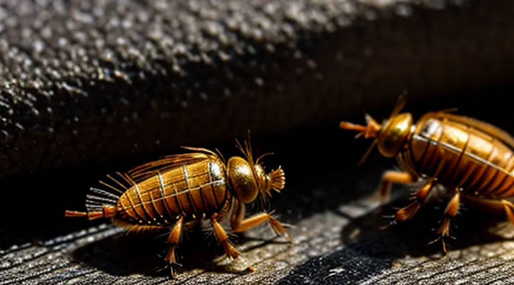
Size and Shape «An Overview»
Length and Width «Typical Measurements»
Human fleas (Pulex irritans) are small, laterally flattened insects whose size is a primary identifying characteristic.
- Length: 1.5 mm to 3.0 mm (approximately 0.06 in to 0.12 in) when unfed; up to 4 mm (0.16 in) after a blood meal.
- Width: 0.5 mm to 0.8 mm (0.02 in to 0.03 in) at the thorax; slightly broader at the abdomen after engorgement.
Males generally occupy the lower end of the length range, while females reach the upper limits, especially when engorged. Size variation correlates with feeding status rather than geographic origin.
Body Segmentation «Head, Thorax, Abdomen»
Human fleas (Pulex irritans) are tiny, laterally flattened insects measuring 1–3 mm in length. Their bodies are divided into three distinct sections that facilitate feeding, movement, and reproduction.
- Head – Compact capsule bearing large, compound eyes and short antennae with sensory pits. Mouthparts form a piercing‑sucking stylet adapted for blood extraction from the host’s skin.
- Thorax – Central segment comprising three pairs of short, stout legs equipped with claws that grip hair shafts. The thorax supports the legs and provides attachment points for the wing‑less musculature.
- Abdomen – Elongated, segmented abdomen containing the digestive tract, reproductive organs, and a flexible exoskeleton that expands after blood meals. The dorsal surface bears fine setae that aid in respiration.
External Features and Their Functions
Exoskeleton «Protective Covering»
Human fleas (Pulex irritans) are small, laterally compressed insects measuring 1.5–3 mm in length. Their bodies exhibit a reddish‑brown color that darkens after a blood meal. The outermost layer is a rigid exoskeleton composed of chitin and protein, forming a protective covering that defines the flea’s shape and preserves internal organs.
The exoskeleton consists of articulated sclerites linked by flexible membranes. Dorsal plates protect the thorax and abdomen, while ventral plates support the legs and mouthparts. Cuticular layers are impregnated with waxes that reduce desiccation, and the surface bears microscopic setae that enhance grip on host hair.
Key characteristics of the protective covering include:
- Hardness: sclerotized plates resist mechanical damage.
- Water retention: waxy cuticle limits evaporative loss.
- Flexibility: membrane joints permit rapid jumping motions.
- Attachment: setae and microstructures enable firm anchorage to mammalian fur.
Coloration «Pigmentation and Appearance»
Human fleas (Pulex irritans) are small, laterally flattened insects measuring 1.5–3 mm in length. Their exoskeleton exhibits a uniform pigmentation that ranges from reddish‑brown to dark brown, sometimes appearing almost black in mature specimens. The coloration results from a thin layer of sclerotized cuticle containing melanin pigments, which provides protection against desiccation and UV exposure.
Key visual characteristics of the cuticular hue include:
- Base color: reddish‑brown in newly emerged adults; darkens to deep brown or black after several molts.
- Abdominal shading: lighter ventral surface contrasting with a darker dorsal plate.
- Leg and antennae: same pigment as the body, occasionally showing a slightly paler tone near joints.
- Eyes: absent; the head appears uniformly colored, lacking distinct markings.
The overall appearance is a smooth, glossy surface that reflects light, giving the flea a subtle metallic sheen when viewed under magnification. This consistent pigmentation distinguishes human fleas from other ectoparasites that may display striped or spotted patterns.
Head Structure «Antennae and Mouthparts»
The human flea (Pulex irritans) is a small, laterally compressed insect, typically 1.5–3 mm long, dark brown to reddish‑brown, with a distinctive head that houses specialized sensory and feeding structures.
The antennae consist of six short, compact segments. The basal segments are robust, while the distal flagellum bears numerous sensilla that detect host heat, carbon dioxide, and movement. Antennae are concealed beneath the head capsule when the flea is at rest, giving the appearance of a smooth, rounded head.
Mouthparts form a piercing‑sucking apparatus adapted for rapid blood extraction. Key components include:
- Labrum forming a shallow groove that guides the food canal.
- Paired maxillae and mandibles reduced to slender stylets that penetrate the host’s skin.
- A central labium that houses the dorsal and ventral stylets, forming a tube through which blood is drawn.
- A muscular salivary pump located in the thorax but operatively linked to the head, generating suction.
These structures enable the flea to locate a human host, pierce the epidermis, and ingest blood within seconds, contributing to the insect’s recognizable silhouette when viewed under magnification.
Antennae «Sensory Organs»
Human fleas are small, laterally compressed insects measuring roughly 1.5–3 mm in length. Their bodies are dark brown to reddish‑brown, with a hard exoskeleton that gives a glossy appearance. The head is proportionally large, bearing compound eyes and a pair of short, segmented antennae that serve as primary sensory organs.
The antennae consist of three distinct segments: a basal scape, a pedicel, and a multi‑flagellated club. Each segment is covered with fine setae that increase surface area for detecting chemical and tactile stimuli. The scape attaches directly to the head capsule, while the pedicel contains the Johnston’s organ, a mechanoreceptive structure sensitive to air currents. The terminal club bears numerous sensilla specialized for olfactory reception.
Antennae enable fleas to locate a human host by perceiving:
- Carbon dioxide gradients emitted from breathing.
- Body heat and humidity cues.
- Volatile compounds present on skin and clothing.
These sensory capabilities allow the flea to initiate rapid jumping behavior when a suitable host is identified. The morphology of the antennae, combined with the flea’s compact body, contributes to the insect’s overall appearance and effectiveness as an ectoparasite.
Mouthparts «Piercing and Sucking»
Human fleas (Pulex irritans) are laterally flattened, dark brown insects measuring 1.5–3 mm in length. Their bodies are covered with microscopic spines that give a slightly rough texture, and the abdomen expands after a blood meal, appearing engorged and more translucent.
The feeding apparatus is a specialized piercing‑sucking structure. It consists of a rigid labrum that forms the outer sheath, two slender mandibles that act as cutting blades, and a pair of maxillae that function as supportive stylets. These components converge into a narrow canal through which saliva and anticoagulant enzymes are introduced before blood is drawn upward by the muscular pharynx.
Key elements of the mouthparts:
- Labrum: protective covering, guides the stylet assembly.
- Mandibles: sharp, curved, cut host skin.
- Maxillae: reinforce the piercing assembly, aid in maintaining a stable channel.
- Stylet canal: transports fluids, prevents clotting.
- Salivary glands: release anticoagulant substances during feeding.
The combined action of cutting and suction enables rapid extraction of host blood, a process completed within seconds. This morphology distinguishes human fleas from other ectoparasites that employ chewing or sponging mouthparts.
Thorax and Legs «Locomotion and Jumping»
Human fleas (Pulex irritans) are small, laterally flattened insects measuring 1.5–3 mm in length, dark brown to reddish‑black, with a hard, chitinous exoskeleton divided into head, thorax, and abdomen.
The thorax forms a compact, convex capsule covering the attachment points for the three pairs of legs. It is covered by a dense layer of short setae that aid in sensory detection and reduce friction during rapid movement. Sclerotized dorsal plates (pronotum and mesonotum) provide rigidity, while internal musculature connects directly to the leg coxae, enabling swift force transmission for jumps.
Legs exhibit several adaptations for propulsion:
- Three pairs, each with a robust coxa linking to the thorax.
- Enlarged femora filled with resilin, a rubber‑like protein that stores elastic energy.
- Tibiae bearing curved spines that grip host fur.
- Tarsi ending in claws and pulvilli for temporary adhesion during take‑off.
Locomotion relies on a catapult mechanism. Muscles contract the femoral resilin, compressing it against the thoracic cuticle. Upon release, stored energy converts to kinetic energy, propelling the flea upward and forward. Jumps reach 10–15 cm, approximately 30–50 times the insect’s body length, with launch acceleration exceeding 100 g. The thorax‑leg architecture thus directly determines the flea’s ability to move quickly between hosts and navigate the host’s fur.
Legs «Adaptations for Movement»
Human fleas (Pulex irritans) possess six legs that function as highly specialized locomotor organs. Each leg is divided into three main segments—coxa, femur, and tarsus—followed by a terminal claw. The tarsus contains a pair of elongated, curved claws that grip the host’s hair and skin, preventing dislodgement during rapid movement.
Key adaptations for movement include:
- Resilin pads in the femur‑tibia joint store elastic energy, enabling the flea to launch up to 18 mm in a single jump, equivalent to 100 times its body length.
- Sensory setae on the coxa and tibia detect vibrations and temperature changes, guiding the flea toward a suitable host.
- Articulated joints provide a wide range of motion, allowing the flea to navigate dense fur and tight skin folds.
- Reduced tarsal segments minimize weight while maintaining grip strength, essential for maintaining contact during high‑speed jumps.
The combination of powerful elastic mechanisms, precise sensory input, and compact claw architecture equips human fleas with the ability to locate, latch onto, and rapidly traverse a host’s surface. This leg morphology distinguishes them from other ectoparasites that rely on crawling rather than jumping for host acquisition.
Claws «Grip on Host»
Human fleas (Pulex irritans) possess three pairs of curved, sclerotized claws located at the distal end of each tarsus. Each claw measures approximately 0.10–0.15 mm in length, with a tapered tip that penetrates the host’s skin or hair shaft. The claws are arranged in a triangular pattern, providing a stable tripod that distributes gripping force evenly across the attachment point.
The claw surface is covered with microscopic ridges and setae that increase friction against keratinized structures. When the flea jumps onto a human host, muscular contraction of the tibial flexor muscles pulls the claws into the substrate, locking them in place. This mechanism enables the flea to maintain attachment during rapid movements of the host, such as walking or scratching.
Key morphological features that enhance grip include:
- Curvature: pronounced bend that matches the contour of hair or skin folds.
- Angle of insertion: approximately 30° relative to the tarsal axis, allowing deep penetration.
- Micro‑texturing: fine cuticular ridges that create interlocking contact with host fibers.
These adaptations ensure that the flea remains anchored while feeding, facilitating blood extraction and reducing the likelihood of dislodgement.
Abdomen «Reproduction and Digestion»
The abdomen of the human‑associated flea is a compact, segmented structure housing the systems responsible for nutrient processing and egg production. Its dorsal cuticle is smooth, while the ventral side bears the opening of the digestive tract and the genital aperture.
Digestive function concentrates in the mid‑abdomen. After a blood meal, the ingested blood pools in the crop, then moves to the midgut where proteolytic enzymes break down hemoglobin. Nutrient absorption occurs across the midgut epithelium, and waste material is expelled through the hindgut and anus at the posterior end of the abdomen.
Reproductive activity is confined to the posterior abdomen. Female fleas possess a paired oviduct that opens near the genital pore. After a blood meal, vitellogenesis accelerates, leading to the production of several eggs within a few days. Fertilized eggs travel through the oviducts and are deposited in the environment through the genital opening. The abdomen expands slightly during egg development, a change visible as a modest bulge in the posterior segments.
Key abdominal components:
- Crop: temporary storage for blood.
- Midgut: enzymatic digestion and nutrient absorption.
- Hindgut: waste elimination.
- Oviducts and genital pore: egg passage and deposition.
- Muscular sheath: facilitates expansion during feeding and oviposition.
Distinguishing Human Fleas from Other Species
Key Differences «Human Flea vs. Cat/Dog Flea»
The human flea (Pulex irritans) is a laterally flattened, wing‑less insect measuring 1.5–3 mm in length. Its body is dark brown to reddish‑black, with a smooth dorsal surface lacking the prominent combs (genal and pronotal) seen on cat and dog fleas. The head is small, rounded, and positioned low on the thorax, giving the flea a compact silhouette. Legs are relatively short, limiting jump distance to about 5–10 cm.
Cat and dog fleas (Ctenocephalides felis and C. canis) range from 2–4 mm, slightly larger on average. Their coloration varies from reddish‑brown to tan, often with a lighter abdominal band. Distinctive features include:
- Pronounced genal and pronotal combs (rows of fine spines) used for attachment to host hair.
- Elongated, powerful hind legs that enable jumps up to 30 cm.
- A more tapered abdomen, especially in engorged females.
- Slightly flattened body, but with visible dorsal setae giving a slightly fuzzy appearance.
Host specificity also differs. Pulex irritans prefers humans and other mammals but is less adapted to dense fur, whereas Ctenocephalides species thrive on the thick coats of cats and dogs, exploiting the combs for secure anchorage. These morphological distinctions allow reliable identification under a microscope or with a hand lens.
Identifying Characteristics «Unique Traits»
Human fleas are tiny, laterally flattened insects measuring approximately 1.5–3 mm in length. Their bodies exhibit a reddish‑brown hue that darkens after a blood meal. The thorax and abdomen are fused, giving a smooth, streamlined silhouette without distinct segmentation. Six legs end in enlarged, backward‑pointing tibial spines that function as levers for rapid jumps. Antennae are short, concealed beneath the head capsule, and eyes are absent, rendering the flea reliant on sensory hairs for navigation.
Key identifying traits include:
- Body compression: extreme lateral flattening allows movement through hair and clothing fibers.
- Jumping apparatus: powerful femoral muscles and a resilin pad enable leaps up to 150 mm, a distance far exceeding body length.
- Comb‑like claws: serrated tarsal claws grip host hair, preventing dislodgement during motion.
- Blood‑filled abdomen: after feeding, the abdomen expands and adopts a darker, engorged appearance.
- Lack of wings: wings are absent, distinguishing fleas from other ectoparasitic insects such as lice.
These characteristics together create a distinctive profile that separates human fleas from other arthropod parasites.
Microscopic Examination «What You Can't See with the Naked Eye»
Bristles and Spines «Sensory and Locomotor Aids»
Human fleas (Pulex irritans) are small, laterally compressed insects measuring 1–3 mm in length. Their bodies are covered with a dense array of microscopic bristles and spines that serve both sensory and locomotor functions.
The bristles are slender, flexible setae distributed across the dorsal thorax and abdomen. They detect air currents and surface textures, allowing the flea to locate a host and maintain orientation while moving through hair or clothing. Each seta terminates in a sensory pore connected to mechanoreceptors that transmit tactile information to the central nervous system.
Spines are short, rigid cuticular extensions found primarily on the legs and the ventral surface of the abdomen. Their roles include:
- Grip enhancement: Spines interlock with host hair or fabric fibers, providing traction during rapid jumps and sustained crawling.
- Stability: Ventral spines prevent slippage on smooth skin surfaces, enabling the flea to remain attached while feeding.
- Anchorage: During blood meals, spines anchor the flea’s mouthparts, reducing the risk of dislodgement.
Together, bristles and spines form an integrated system that compensates for the flea’s diminutive size, ensuring effective host detection, secure attachment, and efficient locomotion.
Sensory Organs «Beyond the Antennae»
Human fleas (Pulex irritans) are laterally flattened, 1–3 mm long, dark brown to reddish, with hard‑sclerotized exoskeleton and pronounced genal and pronotal combs. Their compact body houses a suite of sensory structures that operate independently of the prominent antennae.
Beyond the antennae, fleas rely on:
- Compound eyes: small, laterally positioned lenses provide motion detection and low‑light vision essential for host localization.
- Mouthpart sensilla: mechanoreceptive hairs on the labrum and maxillae detect pressure changes when penetrating skin, ensuring precise feeding.
- Tarsal receptors: dense arrays of chemosensory setae on the fore‑tarsi sense host odorants and temperature gradients, guiding the flea toward warm blood sources.
- Vibrissae on the head capsule: tactile bristles detect airflow and substrate vibration, alerting the insect to potential disturbances.
These organs integrate multimodal signals through the central nervous system, allowing rapid orientation toward a host despite the flea’s minute size. The coordination of visual, thermal, chemical, and mechanical inputs compensates for the limited resolution of the antennae, enabling effective parasitic behavior.
