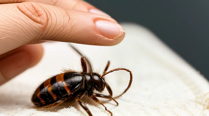
Preparing for Tick Removal
What You'll Need
Tools for Safe Removal
When a tiny tick attaches to the skin, precise instruments reduce the risk of pathogen transmission and minimize tissue damage. The removal method should grasp the parasite as close to the surface as possible, applying steady pressure without crushing the body.
• Fine‑point tweezers – stainless‑steel tips, narrow enough to clasp the tick’s head.
• Tick‑removal device («tick extractor») – curved slot designed to slide under the mouthparts.
• Fine‑tipped forceps – useful for ticks embedded in hair or difficult angles.
• Protective gloves – prevent direct contact with saliva or bodily fluids.
• Magnifying glass – enhances visibility of the attachment point.
• Antiseptic solution – cleans the bite site after extraction.
• Sealed disposal container – ensures safe discarding of the tick for later identification if needed.
Personal Protection
Personal protection against tick bites begins with preventive measures before exposure. Wear long sleeves and trousers, tuck pant legs into socks, and choose light-colored clothing to spot attached arthropods. Apply an EPA‑registered repellent containing 20 % DEET, picaridin, or IR3535 to exposed skin and clothing. Perform a thorough body inspection after outdoor activities; remove any attached tick immediately.
When removal is necessary, follow a precise technique. Use fine‑pointed, non‑slipping tweezers or a specialized tick‑removal tool. Grasp the tick as close to the skin surface as possible, pulling upward with steady, even pressure. Avoid twisting or jerking, which can leave mouthparts embedded. After extraction, clean the bite area with antiseptic and wash hands thoroughly.
Post‑removal care includes monitoring the site for signs of infection. If redness, swelling, or a rash develops within two weeks, seek medical evaluation. Maintaining a personal protection routine—protective clothing, appropriate repellents, diligent skin checks, and correct removal methods—significantly reduces the risk of disease transmission from small ticks.
The Tick Removal Process
Locating the Tick
Visual Inspection
Visual inspection provides the first line of defense when addressing a small tick attached to skin. Accurate identification of the parasite’s position and condition guides safe removal and minimizes the risk of disease transmission.
To conduct a thorough visual examination, follow these steps:
- Use a well‑lit area or a magnifying device to enhance clarity.
- Part hair around the suspected site with a fine‑toothed comb or disposable gloves.
- Scan the skin surface for the tick’s shape, noting any engorgement that indicates prolonged attachment.
- Confirm that the tick is attached by observing its mouthparts penetrating the epidermis.
Key observations include:
- Size: early‑stage ticks measure less than 3 mm; larger dimensions suggest a later feeding stage.
- Location: ticks often cluster in warm, moist regions such as the scalp, armpits, groin, and behind the knees.
- Attachment depth: visible legs or a clear outline of the body imply a superficial attachment, while a swollen abdomen may hide the mouthparts.
After removal, re‑examine the bite area within 24 hours. Persistent redness, swelling, or a bullseye rash warrants medical consultation. Document the tick’s appearance with a photograph if possible; this record assists healthcare providers in assessing potential pathogen exposure.
Palpation for Hidden Ticks
Palpation is essential for locating ticks that are not immediately visible. The method relies on systematic pressure applied to the skin to reveal concealed arthropods without causing them to embed deeper.
A practical approach includes:
- Use the pads of the thumb and index finger to press gently but firmly along the length of each limb, especially around the ankles, wrists, and neck.
- Apply a rolling motion while maintaining constant pressure; a tick will feel like a small, firm nodule distinct from surrounding tissue.
- Examine areas of hair or fur by parting strands with a fine-toothed comb; the tick often becomes apparent as a dark speck.
- After detection, isolate the tick with a pair of fine-tipped tweezers, grasping as close to the skin as possible to avoid crushing the body.
Following detection, immediate removal minimizes the risk of pathogen transmission. Ensure the surrounding skin is clean, and avoid squeezing the tick’s abdomen to prevent release of infectious fluids. The entire process should be performed in a well‑lit environment to enhance visual confirmation after palpation.
Step-by-Step Removal
Grasping the Tick Correctly
Grasping the tick properly prevents the head from breaking off and reduces the risk of infection.
- Use fine‑pointed tweezers or a specialized tick‑removal tool.
- Position the instrument as close to the skin as possible, gripping the tick’s mouthparts, not the body.
- Apply steady, even pressure and pull upward in a straight line; avoid twisting or jerking motions.
- Release the tick once it separates from the skin, then place it in a sealed container for identification if needed.
- Clean the bite area with antiseptic solution and wash hands thoroughly.
- Observe the site for several days; seek medical advice if redness, swelling, or flu‑like symptoms develop.
Pulling Technique
The pulling technique provides a reliable method for removing a small tick without increasing the risk of disease transmission.
To execute the procedure correctly, follow these steps:
- Select fine‑pointed tweezers; avoid blunt instruments.
- Grasp the tick as close to the skin surface as possible, securing the head rather than the body.
- Apply steady, upward pressure; do not twist or jerk the tick.
- Maintain traction until the entire mouth‑parts detach from the skin.
- Place the tick in a sealed container for identification if needed.
- Disinfect the bite area with an antiseptic solution.
- Clean the tweezers with alcohol or a disinfectant after use.
After removal, observe the site for several days. Any signs of redness, swelling, or fever warrant medical evaluation. The technique minimizes the chance that the tick’s mouthparts remain embedded, which can introduce pathogens. Using proper tools and a controlled pull ensures safe elimination of the parasite.
After Removal Care
Cleaning the Bite Area
After a tick is removed, the bite site requires prompt decontamination to prevent infection and reduce irritation. First, wash the area with mild soap and running water for at least 20 seconds, ensuring that all residual saliva and debris are cleared. Rinse thoroughly and pat dry with a clean disposable towel.
Next, apply a broad‑spectrum antiseptic such as povidone‑iodine or chlorhexidine. Allow the solution to remain on the skin for the manufacturer‑recommended contact time before wiping away excess. If the antiseptic is unavailable, an alcohol‑based swab can serve as a temporary measure, though it may cause mild stinging.
A thin layer of over‑the‑counter antibiotic ointment (e.g., bacitracin or neomycin) may be spread over the cleaned wound, then covered with a sterile adhesive bandage. Change the dressing daily, or sooner if it becomes wet or contaminated.
Monitor the bite area for signs of complications:
- Redness extending beyond the immediate margin
- Swelling or heat accumulation
- Pus formation or foul odor
- Development of a target‑shaped rash (potential early Lyme disease indicator)
If any of these symptoms appear, seek medical evaluation promptly. Maintaining a clean, protected bite site significantly lowers the risk of secondary infection while the skin heals naturally.
Disposing of the Tick
After a tick is extracted, immediate containment prevents accidental reattachment. Place the specimen in a small, sealable plastic bag or a puncture‑resistant container.
To render the arthropod harmless, employ one of the following methods:
- Submerge the bag in isopropyl alcohol (≥70 %) for at least five minutes.
- Freeze the sealed bag at –20 °C or lower for 24 hours.
- Immerse the tick in a solution of 10 % bleach for a minimum of ten minutes.
Once the tick is dead, discard the sealed bag in a regular waste bin. Do not crush the insect, as saliva may contain pathogens.
Finally, clean the tweezers or extraction tool with alcohol or soap and water, then wash hands thoroughly with soap for at least 20 seconds. This sequence eliminates the risk of disease transmission while complying with safe disposal practices.
Monitoring for Symptoms
After a tick has been removed, careful observation for emerging health signs is essential. Early detection of infection reduces the risk of complications and guides timely medical intervention.
Key indicators to monitor include:
- Redness or expanding rash at the bite site, especially a bullseye‑shaped lesion.
- Fever, chills, or sweats without an obvious cause.
- Headache, neck stiffness, or facial muscle weakness.
- Joint pain, swelling, or stiffness, particularly in the knees or elbows.
- Fatigue, muscle aches, or malaise persisting beyond a few days.
Symptoms may appear within a few days or develop weeks after exposure. Document any changes promptly, noting onset time, severity, and accompanying factors. If any of the listed signs emerge, seek professional evaluation without delay. Early treatment for conditions such as «Lyme disease» or other tick‑borne illnesses improves outcomes and minimizes long‑term effects.
When to Seek Medical Attention
Signs of Infection
After a tick is removed, monitor the bite site for indications that an infection is developing. Early detection prevents complications and guides timely medical intervention.
Typical signs include:
- Redness expanding beyond the immediate area of the bite.
- Swelling that increases in size or becomes painful to touch.
- Warmth localized around the wound.
- Pus or other fluid discharge.
- Fever, chills, or unexplained fatigue.
- Headache, muscle aches, or joint pain accompanying the skin changes.
If any of these symptoms appear within a few days of removal, seek professional evaluation. Prompt antibiotic therapy may be required to address bacterial infection and reduce the risk of systemic involvement. Regular cleaning of the area with mild soap and antiseptic solution supports healing, but does not replace medical assessment when infection signs emerge.
Concerns About Tick-Borne Diseases
Tick bites present a direct pathway for pathogens that cause serious illness. Common agents include «Lyme disease», «Rocky Mountain spotted fever», «Anaplasmosis», and «Ehrlichiosis». Even a diminutive tick can harbor these microorganisms, and infection risk rises with the duration of attachment.
Monitoring after removal is essential. Look for the following signs within days to weeks:
- Fever or chills
- Headache or neck stiffness
- Muscle or joint pain
- Fatigue or malaise
- Rash, especially a bull’s‑eye pattern
Any appearance of these symptoms warrants immediate medical evaluation, even if the tick was extracted promptly.
After the tick is removed, follow a strict protocol: grasp the mouthparts with fine‑point tweezers, pull upward with steady pressure, avoid crushing the body, then cleanse the bite area with antiseptic. Preserve the specimen in a sealed container for possible laboratory identification. Contact a healthcare professional to discuss prophylactic treatment options and to arrange testing if symptoms develop.
Incomplete Tick Removal
Incomplete removal of a small tick leaves mouthparts embedded in the skin, creating a portal for pathogens and increasing inflammation. The residual fragment can detach spontaneously, but prompt action reduces complications.
- Grasp the tick as close to the skin as possible with fine‑point tweezers.
- Apply steady, downward pressure without twisting.
- Maintain traction until the entire body separates from the host.
If the mouthparts remain after initial extraction, follow these steps. Clean the area with antiseptic solution, then use a sterilized needle or fine‑point tweezers to lift the exposed portion. Gently pull the fragment out, keeping the motion linear and avoiding compression of surrounding tissue. Do not dig or crush the embedded parts.
After removal, disinfect the site with iodine or alcohol and cover with a sterile bandage. Observe the bite for signs of redness, swelling, or a rash within the next 24‑48 hours. Document the date of the bite and the tick’s appearance, as this information assists healthcare providers.
Seek medical evaluation if the wound enlarges, develops a bullseye rash, or if fever, headache, or joint pain appear. Professional assessment may include antibiotic therapy or serologic testing to rule out tick‑borne diseases.
