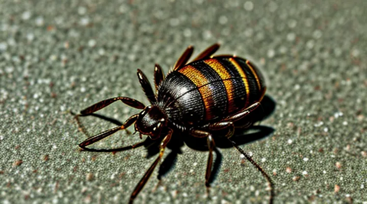
Understanding Tick-Borne Diseases
Importance of Testing Ticks
Testing ticks provides essential data for disease surveillance, species distribution, and risk assessment. Laboratory analysis confirms pathogen presence, informs treatment decisions, and guides public‑health interventions. Even ticks that are no longer alive retain diagnostic value because DNA, RNA, and protein markers remain stable when preserved correctly.
Key reasons for submitting deceased specimens:
- Pathogen detection – Molecular assays identify bacteria, viruses, and parasites despite the tick’s death.
- Species verification – Morphological features and genetic sequencing verify tick identity, which influences disease probability.
- Epidemiological mapping – Aggregated results reveal geographic hotspots and seasonal trends.
- Quality control – Testing known dead samples validates laboratory protocols and assay sensitivity.
Acceptable submission conditions include immediate placement in a sealed container, storage at low temperature, or immersion in ethanol. Laboratories typically reject specimens that have decomposed, been exposed to high heat, or lack proper labeling. Proper documentation of collection site, date, and host enhances the utility of the analysis.
Accurate testing of both live and dead ticks sustains reliable surveillance networks, reduces diagnostic delays, and supports targeted prevention strategies.
Common Pathogens Tested
Submitting a deceased tick for laboratory analysis is acceptable for most molecular diagnostics. Viability is not required when the goal is to detect pathogen DNA or RNA; preservation in cold storage or ethanol suffices. Live specimens are only necessary for culture‑based methods or for maintaining pathogen viability in vector competence studies.
Typical pathogens screened in tick specimens include:
- Borrelia burgdorferi complex (Lyme disease agents)
- Anaplasma phagocytophilum (human granulocytic anaplasmosis)
- Ehrlichia spp. (e.g., E. chaffeensis, E. ewingii)
- Rickettsia spp. (spotted fever group)
- Babesia spp. (e.g., B. microti)
- Powassan virus and other tick‑borne flaviviruses
- Coxiella burnetii (occasionally detected in ticks)
Diagnostic platforms rely on polymerase chain reaction (PCR) or reverse transcription PCR (RT‑PCR) for nucleic‑acid detection, followed by sequencing or probe‑based confirmation. Serological assays are uncommon for tick samples because the tick does not produce antibodies.
Optimal specimen handling: place the dead tick in a sterile tube, add 70 % ethanol or keep at –20 °C, and ship with cold packs. Avoid formalin or other fixatives that degrade nucleic acids. Laboratories receiving the sample will process it according to the requested pathogen panel and report results in a standard format.
Submitting a Dead Tick for Analysis
Feasibility of Testing Dead Ticks
What Happens to Pathogens After Tick Death
When a tick dies, the microorganisms it carried are subject to rapid physiological changes. Cellular structures of bacteria, viruses, and protozoa begin to break down as the tick’s internal environment loses homeostasis. Enzymatic activity, pH shifts, and loss of nutrient supply accelerate degradation, reducing the likelihood that pathogens remain viable for extended periods.
Key factors influencing pathogen persistence after tick mortality include:
- Time since death: Viability declines sharply within hours; some spirochetes may survive up to 24 hours, while most viruses lose infectivity within minutes.
- Temperature: Warm conditions (20‑30 °C) hasten decay; refrigeration slows enzymatic breakdown and can preserve nucleic acids for several days.
- Tick species and pathogen type: Hard ticks (Ixodidae) often retain Borrelia DNA longer than soft ticks; intracellular protozoa such as Babesia are more resistant than extracellular bacteria.
- Preservation method: Immediate placement in ethanol, RNAlater, or dry ice maintains genetic material for molecular assays, though live cultures become unrecoverable.
For diagnostic laboratories, the presence of pathogen DNA or RNA in a deceased tick remains sufficient for molecular testing, provided the specimen is collected promptly and stored under appropriate conditions. Cultivation of live organisms generally requires the tick to be alive at the time of collection; otherwise, culture attempts will fail. Consequently, submission of a dead tick is acceptable for PCR‑based identification, serological antigen detection, and sequencing, but not for attempts to isolate viable pathogens.
Impact of Decomposition on Test Results
Submitting a deceased tick for laboratory analysis introduces variables that can compromise the reliability of diagnostic outcomes. Decomposition initiates biochemical and microbial processes that alter the specimen’s composition, affecting both morphological and molecular assessments.
During the post‑mortem interval, the cuticle softens, obscuring key identification features such as scutum shape and leg segmentation. This degradation reduces the accuracy of species determination, which is essential for evaluating disease risk.
Molecular assays are particularly sensitive to nucleic‑acid decay. Enzymatic breakdown of DNA and RNA leads to fragmentation, lowering amplification efficiency in PCR‑based tests. Consequently, pathogen detection rates decline, and false‑negative results become more likely.
Microbial proliferation on a dead tick can introduce contaminant DNA, creating background noise in sequencing runs and potentially yielding misleading pathogen profiles.
The following points summarize the principal effects of decomposition on test results:
- Morphological distortion: loss of defining structures, hindering visual identification.
- Nucleic‑acid degradation: reduced quantity and quality of genetic material, impairing PCR and sequencing.
- Contamination risk: growth of opportunistic microbes that may confound pathogen detection.
- Variable preservation: inconsistent environmental conditions cause unpredictable rates of decay, complicating result interpretation.
To mitigate these impacts, specimens should be collected promptly, stored in cold conditions, and preserved in ethanol or RNA‑stabilizing solutions whenever possible. Immediate processing minimizes degradation, preserving the integrity required for accurate identification and pathogen detection.
Optimal Preservation Methods for Deceased Ticks
Storage Recommendations
When a tick has died before laboratory submission, proper preservation is essential to maintain morphological integrity and DNA quality. Immediate action after collection determines whether the specimen remains viable for identification, pathogen detection, or research purposes.
- Place the tick in a sealed, airtight container such as a screw‑cap microtube or a zip‑lock bag.
- Add a small volume of 70 % ethanol; ensure the insect is fully immersed but avoid excessive liquid that could cause distortion.
- Label the container with date, location, host species, and collector’s name.
- Store the sealed container at 4 °C (refrigerator) if processing will occur within 48 hours; for longer intervals, keep at –20 °C (freezer) to prevent nucleic acid degradation.
- Avoid repeated freeze‑thaw cycles; retrieve only one aliquot for analysis and return the remainder to the same temperature.
If ethanol preservation is not possible, a dry, desiccated environment using silica gel packets can be employed, but this method is less reliable for molecular assays. Ensure that all storage media are sterile and that containers are clearly marked to prevent cross‑contamination. Properly stored dead ticks retain sufficient structural and genetic material for accurate laboratory evaluation.
Packaging for Submission
When a tick is no longer alive, the specimen can still be sent to a diagnostic laboratory provided that the packaging meets the standards required for biological material. The primary objectives of the packaging are to preserve the tick’s morphology, prevent contamination, and comply with shipping regulations for hazardous or infectious samples.
Key elements of proper packaging include:
- A primary container that is leak‑proof, such as a sealed plastic vial containing a small amount of ethanol (70 %–95 %) or sterile saline to maintain tissue integrity.
- A secondary container, for example a rigid plastic box or a sealed zip‑lock bag, that encloses the primary vial and offers additional protection against breakage.
- An outer shipping box that conforms to the regulations of the carrier and the receiving laboratory, labeled with the appropriate biohazard or infectious substance symbols and a clear description of the contents.
- Documentation accompanying the package: a completed submission form, specimen identification, collection date, and any relevant clinical information.
Following these steps ensures that a deceased tick arrives in a condition suitable for morphological or molecular testing, allowing the laboratory to deliver accurate identification and pathogen detection.
Laboratories Accepting Dead Tick Samples
Types of Testing Available
Testing a deceased tick is feasible, but the range of analytical methods differs from those applied to live specimens. Laboratories evaluate dead arthropods using several validated approaches.
-
Morphological identification – visual examination of the exoskeleton, scutum patterns, and mouthpart structures under a stereomicroscope confirms species and developmental stage. Preservation in ethanol or frozen storage maintains key diagnostic features.
-
Polymerase chain reaction (PCR) assays – extraction of DNA from the tick’s cuticle or internal tissues enables detection of bacterial, viral, or protozoan pathogens. Real‑time PCR provides quantitative results for agents such as Borrelia burgdorferi, Anaplasma phagocytophilum, and tick‑borne encephalitis virus.
-
Reverse transcription PCR (RT‑PCR) – when testing for RNA viruses, reverse transcription converts viral RNA to complementary DNA before amplification. This method is appropriate for detecting flaviviruses and other RNA‑based pathogens in dead ticks.
-
Next‑generation sequencing (NGS) – comprehensive genomic profiling identifies known and novel microorganisms present in the specimen. Shotgun metagenomic sequencing can reveal co‑infecting agents that standard PCR panels might miss.
-
Serological testing of tick extracts – enzyme‑linked immunosorbent assays (ELISA) or immunofluorescence assays (IFA) applied to homogenized tick material detect antigens of specific pathogens, useful when nucleic acid quality is compromised.
-
Culture isolation – inoculation of tick homogenates onto selective media or cell lines can recover viable bacteria or rickettsiae, though success rates decline if the tick has been dead for an extended period or stored improperly.
Each technique has distinct requirements for specimen handling, storage temperature, and time elapsed since death. Selecting the appropriate method depends on the diagnostic objective, available laboratory resources, and the condition of the tick at submission.
Interpreting Results from Dead Tick Samples
When a deceased tick is sent to a diagnostic laboratory, the laboratory must assess the condition of the specimen before reporting any findings. The assessment focuses on three primary aspects: physical integrity, preservation method, and the intended analytical technique.
Physical integrity determines whether morphological identification is possible. If the exoskeleton is fragmented or desiccated, key diagnostic characters such as scutum pattern, mouthpart structure, and leg segmentation may be missing, rendering species determination unreliable. In such cases, the laboratory may rely solely on molecular assays.
Preservation method influences nucleic acid quality. Samples kept at ambient temperature for extended periods experience DNA degradation, which can reduce the sensitivity of PCR‑based pathogen detection. Samples frozen promptly or stored in ethanol maintain higher nucleic‑acid stability, allowing detection of bacterial, viral, or protozoan agents with lower cycle‑threshold values.
Interpretation of results from dead tick specimens should consider the following points:
- Pathogen viability – Culture‑dependent methods require live organisms; dead ticks cannot yield viable cultures, limiting detection to molecular or immunological assays.
- DNA integrity – Degraded DNA may produce false‑negative PCR outcomes; repeat testing with a different target gene or a nested protocol can mitigate this risk.
- Contamination risk – Post‑mortem handling introduces environmental DNA; rigorous controls and negative‑sample processing are essential to differentiate true infection from external contamination.
- Quantitative limits – Cycle‑threshold values near the assay’s detection limit suggest low pathogen load; reporting should include the quantitative threshold and confidence interval.
- Reporting constraints – Laboratories must state any limitations related to sample condition, including inability to provide species identification or to confirm pathogen viability.
By applying these criteria, laboratories can deliver accurate, transparent reports that reflect the constraints inherent to deceased tick specimens, while still providing valuable information for epidemiological investigations and public‑health decision‑making.
Alternatives to Tick Testing
Focusing on Human Symptoms
When to Seek Medical Attention
If a tick is found attached to skin, remove it promptly and monitor for symptoms. Seek medical evaluation under any of the following conditions:
- The bite site develops a rash with concentric rings (bullseye) or expands rapidly.
- Fever, chills, headache, muscle aches, or joint pain appear within two weeks of the bite.
- The tick was removed after being dead for an extended period, and its identification is uncertain.
- You have a weakened immune system, are pregnant, or have a history of autoimmune disease.
- The bite occurred in an area known for high prevalence of tick‑borne infections.
When contacting a healthcare provider, describe the tick’s appearance, estimated time of attachment, and any recent travel to endemic regions. If the specimen is available, preserve it in a sealed container and bring it to the appointment; a dead tick can still be examined for species identification and pathogen testing. Prompt professional assessment reduces the risk of complications from diseases such as Lyme, Rocky Mountain spotted fever, or anaplasmosis.
Diagnostic Tests for Tick-Borne Illnesses in Humans
Diagnostic laboratories evaluate human exposure to tick-borne pathogens through several validated methods. Blood samples, serum, plasma, or cerebrospinal fluid serve as primary specimens; tissue biopsies are used when localized infection is suspected.
- Serologic assays detect IgM and IgG antibodies against Borrelia burgdorferi, Anaplasma phagocytophilum, Ehrlichia spp., and Rickettsia rickettsii. Enzyme‑linked immunosorbent assay (ELISA) provides screening; Western blot confirms Lyme disease seropositivity.
- Polymerase chain reaction (PCR) amplifies pathogen DNA from whole blood, skin punch biopsies, or synovial fluid, offering direct evidence of infection for Babesia microti, Powassan virus, and early Lyme disease before seroconversion.
- Culture techniques remain limited to specialized centers; isolation of Borrelia or Babesia requires specific media and prolonged incubation, useful for antimicrobial susceptibility testing.
- Microscopic examination of peripheral blood smears identifies intra‑erythrocytic Babesia parasites and intracellular Anaplasma morulae, providing rapid diagnosis in acute presentations.
Laboratory acceptance criteria for arthropod specimens emphasize preservation of nucleic acids. Dead ticks may be submitted if they are frozen promptly, placed in ethanol, or stored at 4 °C, ensuring that DNA remains intact for PCR analysis. Many reference labs reject desiccated or decomposed specimens because degradation compromises assay sensitivity. Documentation of collection date, geographic origin, and host species accompanies the specimen to aid epidemiologic interpretation.
Interpretation of results integrates clinical presentation, exposure history, and test performance characteristics. Positive serology without clinical signs may reflect past infection; PCR positivity confirms active infection but requires correlation with symptom onset. Negative findings do not exclude disease when sampling occurs outside the optimal diagnostic window.
Clinicians should obtain appropriate human specimens before initiating antimicrobial therapy, as treatment can reduce pathogen load and affect test accuracy. When arthropod testing is considered, verify that the submitting laboratory accepts non‑viable ticks and follows validated protocols for nucleic‑acid preservation.
Preventative Measures
Tick Bite Prevention Strategies
Testing a deceased tick is often considered when identification of species or pathogen presence is required after an encounter. Effective prevention of tick bites reduces the frequency of such submissions and minimizes the risk of disease transmission.
- Wear long sleeves and trousers in tick‑infested areas; tuck clothing into socks to create a barrier.
- Apply EPA‑registered repellents containing DEET, picaridin, or IR3535 to exposed skin and clothing.
- Perform thorough skin checks after outdoor activities; remove attached ticks promptly with fine‑pointed tweezers, grasping close to the skin and pulling straight upward.
- Maintain yard by trimming grass, removing leaf litter, and creating a mulch barrier between vegetation and recreational zones.
- Use acaricide treatments on pets and in high‑risk zones, following label directions for concentration and re‑application intervals.
Implementing these measures lowers the probability of tick attachment, thereby decreasing the need to submit dead specimens for laboratory analysis.
Post-Bite Monitoring
After a tick attachment, systematic observation of the bite site and overall health is the primary method for early detection of tick‑borne illnesses.
Watch for the following signs during the first 30 days post‑exposure:
- Redness or expanding rash, especially a target‑shaped lesion.
- Fever, chills, or night sweats.
- Muscle or joint aches without obvious injury.
- Headache, fatigue, or difficulty concentrating.
- Nausea, vomiting, or abdominal pain.
Document any symptom onset, duration, and severity. Contact a healthcare provider promptly if any of these manifestations appear, even if they seem mild.
If the tick is found dead, retain it for laboratory analysis. Place the specimen in a sealed container, add a small amount of ethanol (70 % or higher) or keep it frozen at –20 °C, and label with the date of removal and location of the bite. Submit the sample to a qualified public health laboratory within 14 days of collection; delayed submission reduces the likelihood of accurate species identification and pathogen detection.
Maintain the monitoring log for at least six weeks, noting any changes after the initial observation period. This record assists clinicians in correlating clinical presentation with laboratory results, facilitating timely treatment decisions.
