General Appearance and Size
Overall Body Shape
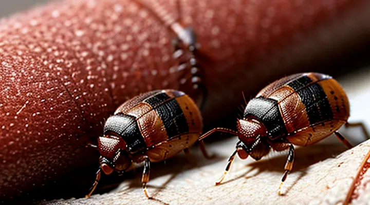
Bedbugs (Cimex species) share a characteristic overall body shape regardless of sex. Both male and female insects are dorsoventrally flattened, which facilitates movement within narrow crevices of bedding and furniture. The abdomen is broad and oval, tapering slightly toward the posterior, while the thorax is narrower and houses the legs and wings (vestigial in this genus). Adults measure approximately 4.5–5.5 mm in length; females may reach the upper end of this range due to the expanded abdomen needed for egg development, but the basic silhouette remains consistent.
Key morphological features of the shared body plan:
- Flattened profile: reduces resistance when crawling under fabric.
- Elongated oval outline: provides a streamlined shape for penetrating host skin.
- Segmented thorax: three pairs of legs attached to the mesothorax and metathorax.
- Reduced wings: tiny, non‑functional elytra covering the dorsal surface.
- Antennae: four‑segmented, positioned near the head, identical in both sexes.
The only noticeable deviation concerns the abdomen’s volume: females exhibit a more pronounced swelling when gravid, while males retain a uniformly tapered abdomen. Aside from this, the overall body architecture is virtually indistinguishable between the sexes.
Sexual Dimorphism in Length
Male bedbugs are consistently shorter than females. Average body length for males ranges from 4.0 mm to 4.8 mm, while females typically measure between 5.0 mm and 5.9 mm. This size gap persists across developmental stages, becoming most pronounced after the final molt.
Key points:
- Females possess a broader abdomen to accommodate egg production, contributing to overall length increase.
- Males exhibit a more streamlined thorax, reflecting their role in seeking mates.
- Length measurements obtained with calibrated microscopy show less than 0.2 mm overlap between the largest males and smallest females, allowing reliable sex determination in field samples.
- Geographic variation can shift average sizes by up to 0.3 mm, but the male‑female disparity remains evident.
Understanding this dimensional dimorphism aids accurate identification, population monitoring, and the assessment of reproductive potential within infestations.
Reproductive Anatomy and Its Visual Cues
Male Aedeagus and Its Location
Male bedbugs possess an aedeagus, a sclerotized tube that functions as the intromittent organ during copulation. The aedeagus is situated within the posterior abdomen, specifically inside the genital capsule that occupies the ventral region of the ninth abdominal segment. It remains concealed beneath the dorsal exoskeleton and is only observable after careful dissection or microscopic examination.
Key characteristics of the male aedeagus include:
- Rigid, needle‑like structure composed of chitin.
- Paired lateral valves that support the central tube.
- Muscular attachments that facilitate extension during sperm transfer.
The female reproductive system contains an ovipositor and a spermatheca, located in the same abdominal segment but positioned ventrally and laterally to the male’s genital capsule. Externally, male and female bedbugs appear nearly identical; differentiation relies on internal anatomical features such as the aedeagus in males and the ovipositor in females.
Female Parietal Organ (Cimex lectularius)
The female parietal organ of Cimex lectularius is a cuticular structure located on the dorsal abdomen near the posterior margin. It appears as a raised, sclerotized plate with a series of fine, evenly spaced pores. Microscopic examination shows the organ’s surface covered by a thin waxy layer that reduces desiccation and may serve as a pheromone release site.
Morphologically, the organ differs from the male counterpart in size and pore density. Females possess a broader plate, typically 0.12 mm wide, with approximately 30 pores per square millimeter, whereas males display a narrower, less porous version. These differences contribute to the distinct visual profiles of the sexes when observed under magnification.
Key characteristics:
- Position: dorsal abdominal segment VII–VIII
- Composition: hardened cuticle with waxy coating
- Pore arrangement: regular, species‑specific pattern
- Dimorphic traits: larger surface area and higher pore count in females
The organ’s function aligns with reproductive behavior; the emitted chemicals attract males and facilitate mating. Its structural resilience also supports the female’s prolonged blood‑feeding cycles, allowing efficient nutrient storage for egg production.
Behavioral Differences Affecting Appearance
Mating Scars on Females
Male and female bedbugs (Cimex lectularius) are often examined together to assess reproductive status. Females that have mated display a distinct scar on the dorsal surface of the abdomen, directly posterior to the ventral shield. The scar is a pale, crescent‑shaped line produced by the male’s traumatic insemination, where the copulatory organ pierces the female’s integument.
Key characteristics of the scar:
- Color: lighter than surrounding cuticle, ranging from whitish to yellowish.
- Shape: concave curve following the contour of the abdomen.
- Length: typically 0.3–0.5 mm, extending across the dorsal midline.
- Texture: smooth, lacking the punctate pattern of normal exoskeletal markings.
The scar appears within 24 hours after copulation and persists for the female’s lifespan. Its presence confirms successful insemination, while its absence in adult females suggests virgin status or recent emergence from the last molt. The scar does not impair locomotion or feeding, but repeated traumatic inseminations can enlarge the wound, causing localized melanization and occasional hemolymph loss.
When male and female specimens are photographed side by side, the contrast between the male’s slender, elongated abdomen and the female’s broader, scarred dorsum becomes a reliable visual cue for field identification. Researchers use this marker to estimate population breeding rates, monitor control interventions, and differentiate between species with similar external morphology.
Post-Mating Body Condition
Male and female bedbugs emerge from copulation with distinct physiological states that affect their external appearance. The female’s abdomen expands rapidly as she stores a blood meal required for egg development; the cuticle becomes more translucent, and the dorsal surface often exhibits a slight darkening due to increased hemolymph volume. The male’s abdomen typically returns to a pre‑mating size, and the genital capsule may show signs of wear or slight discoloration from the transfer of seminal fluids.
When the pair is observed side by side after mating, the size contrast is pronounced: the female appears markedly larger and more rounded, while the male retains a slender profile. The female’s posture often shifts toward a more sedentary stance as she seeks a suitable oviposition site, whereas the male remains mobile, frequently searching for additional mates.
Key indicators of post‑mating body condition include:
- Female abdominal girth increase (30‑50 % larger than pre‑mating measurement)
- Female cuticle translucency and slight darkening
- Male abdominal reduction to baseline dimensions
- Presence of genital wear on the male’s aedeagus
- Behavioral shift: female immobility, male continued locomotion
These characteristics provide a reliable visual cue for researchers assessing reproductive status and for pest‑management personnel identifying recently mated populations in field surveys.
Microscopic and Advanced Identification Techniques
Examining the Genitalia Under Magnification
Male and female bedbugs can be distinguished by the shape of their terminal abdominal segments when viewed under a microscope. The male’s aedeagus is a slender, curved structure ending in a hook-like tip, while the female’s spermatheca appears as a rounded, bulbous cavity attached to a short duct. Both sexes possess a translucent exoskeleton that reveals the underlying musculature, but the genitalia provide the most reliable diagnostic characters.
Key morphological differences observable at 40–100× magnification:
- Aedeagus (male): elongated, slightly curved, sclerotized, with a distal hook; visible ventrally when the abdomen is dissected.
- Spermatheca (female): oval sac, less sclerotized, positioned laterally on the ventral side; connected to a short, flexible duct.
- Parameres (male): paired lateral lobes flanking the aedeagus, each bearing fine setae.
- Ovipositor remnants (female): faint, filamentous structures near the posterior margin, absent in males.
When both specimens are placed side by side on a slide, the contrast between the male’s pointed aedeagus and the female’s rounded spermatheca becomes immediately apparent. The surrounding abdominal tergites are identical in coloration and pattern, confirming that genital morphology is the primary criterion for sex determination in Cimex lectularius.
Using Stains for Detailed Analysis
Researchers apply staining techniques to reveal the combined morphology of male and female bedbugs. By treating mixed‑sex samples with appropriate dyes, investigators obtain clear differentiation of sex‑specific structures while preserving overall body form.
Vital dyes such as methylene blue and Nile red penetrate living specimens, highlighting cuticular patterns and abdominal coloration. Histological stains, including hematoxylin–eosin and Giemsa, enhance internal organ visibility after fixation. Fluorescent markers—e.g., Alexa‑fluor conjugates attached to sex‑linked proteins—provide high‑contrast imaging under epifluorescence or confocal microscopy.
The standard protocol follows these steps:
- Collect a representative cohort containing both sexes.
- Fix specimens in 70 % ethanol or formalin for 30 minutes.
- Rinse and apply chosen stain for a duration calibrated to tissue thickness.
- Mount samples on glass slides with appropriate mounting medium.
- Examine under light, phase‑contrast, or fluorescence microscopy, recording images for comparative analysis.
Benefits of stain‑based examination include:
- Precise separation of male and female external features such as genitalia and abdominal segment width.
- Detection of blood‑meal residues, aiding assessment of feeding status across sexes.
- Identification of pathogen presence within tissues, supporting epidemiological studies.
- Quantitative measurement of morphological parameters using image‑analysis software.
By integrating multiple staining modalities, researchers achieve a comprehensive visual profile that captures the joint appearance of male and female bedbugs and supports detailed taxonomic, physiological, and disease‑vector investigations.
Importance of Accurate Identification
Pest Control Strategies
Male‑female bedbug pairs can be identified by the larger, rounded abdomen of the female and the slimmer, elongated body of the male, often observed in close contact during copulation. Recognizing these pairs aids in targeting active breeding sites, which is essential for effective eradication.
Effective pest‑control measures include:
- Heat treatment: Raise ambient temperature to 50 °C (122 °F) for at least 90 minutes; eliminates all life stages, including concealed mating clusters.
- Steam application: Direct steam at 100 °C (212 °F) on seams, cracks, and bed frames; disrupts mating pairs and disperses eggs.
- Insecticide fogging: Use products containing pyrethroids or neonicotinoids approved for indoor use; apply to voids where paired insects congregate.
- Encasement of mattresses and box springs: Seal with certified covers; prevents re‑infestation from hidden couples.
- Vacuuming: Remove visible pairs and shed skins; dispose of contents in sealed bags to avoid redistribution.
- Monitoring traps: Deploy interceptor devices under legs of furniture; capture both sexes, providing data on population dynamics.
Integration of these tactics into a coordinated program reduces reproductive capacity and accelerates population collapse. Regular inspections for male‑female pairings confirm treatment efficacy and guide follow‑up actions.
Understanding Bed Bug Biology
Bed bugs (Cimex lectularius) are hematophagous insects belonging to the family Cimicidae. Adults measure 4–5 mm in length, exhibit a flattened, oval body, and possess a reddish‑brown exoskeleton that deepens after a blood meal. The species displays minimal sexual dimorphism, yet several morphological markers distinguish males from females when observed side by side.
- Size: females are slightly larger, averaging 5 mm, while males range 4–4.5 mm.
- Abdomen shape: the female abdomen is broader and more rounded, accommodating egg development; the male abdomen tapers toward the posterior.
- Scent glands: males possess a conspicuous dorsal glandular patch used during courtship; females lack this structure.
- Genitalia visibility: male genital capsule is visible at the terminal abdominal segment, whereas the female ovipositor is concealed beneath the ventral cuticle.
During copulation, the male mounts the female’s dorsal surface, aligning the genitalia for sperm transfer. The pair remains coupled for 30–60 minutes, during which the female’s abdomen expands to receive the spermatophore. This interaction does not alter external coloration but may cause temporary swelling of the female’s abdomen.
The life cycle proceeds through five nymphal instars before reaching adulthood. Each stage requires a blood meal to molt, with development time ranging from 5 weeks under optimal temperature (≈27 °C) to several months in cooler conditions. Females lay 1–5 eggs per day, depositing them in crevices near host resting sites. Egg shells are pale, oval, and approximately 0.5 mm in length; they hatch within 6–10 days.
Understanding these anatomical and behavioral characteristics clarifies how male and female bed bugs appear together, aids in accurate identification, and informs control strategies targeting both sexes throughout their reproductive cycle.
