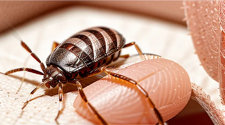
Understanding Bed Bug Morphology
General Bed Bug Anatomy
Head and Thorax Features
The head of a bedbug provides reliable cues for sex determination. Female specimens possess a broader, more rounded clypeus, while males exhibit a narrower, slightly pointed clypeus. Antennal segments are uniformly sized in females; in males, the second and third segments are marginally elongated, giving a subtly tapered appearance. The eyes of females are set closer together, forming a tighter ocular pair, whereas male eyes are spaced slightly wider apart.
The thorax also displays distinguishing characteristics. In females, the pronotum is smooth and lacks conspicuous punctures; males show a lightly pitted pronotum with fine, evenly distributed depressions. The dorsal surface of the mesothorax in females is uniformly matte, while males often have a faint sheen due to a thin layer of cuticular wax. Leg morphology differs as well: female forelegs have a more robust femur, and the tibial spines are shorter; male forelegs are slimmer, and tibial spines are elongated, facilitating mating grasp.
Abdominal Segments
The abdomen provides the most reliable characters for separating female bedbugs from males. Females possess a broader, more rounded terminal segment, while males display a narrower, tapered final segment.
Key abdominal differences:
- Spermatheca presence – a visible, swollen sac located on the ventral side of the fifth abdominal segment in females; absent in males.
- Genital capsule – males have a distinct, sclerotized genital capsule on the eighth abdominal segment; females lack this structure.
- Segment count visibility – the ninth abdominal segment is elongated and concealed by the ovipositor in females; in males it remains exposed and short.
- Size variation – overall abdominal length is typically greater in females, especially the distal two segments, due to egg‑carrying capacity.
- External morphology – females exhibit a smoother tergite surface on the last three segments, whereas males show subtle ridges and setae patterns.
Inspecting these features under magnification yields accurate sex determination without reliance on external coloration or behavior.
Key Differences in Sexual Dimorphism
Abdominal Shape and Structure
Female Abdomen: Rounded and Larger
Female bedbugs can be recognized by the shape and size of their abdomen. The abdomen of a female is noticeably broader and more convex than that of a male, which tends to be flatter and narrower. This enlargement accommodates the ovaries and developing eggs, giving the female a distinctly rounded silhouette when viewed from the side or from above.
Key visual cues include:
- A visibly swollen abdomen that appears dome‑shaped rather than flat.
- A width that exceeds the thorax by a clear margin, often making the overall body outline appear more oval.
- A smoother curvature without the subtle taper seen in males.
When examining a specimen, focus on the posterior third of the body. In females, the abdomen expands uniformly, creating a bulbous appearance that remains consistent across different life stages after the first molt. Males retain a relatively slender profile throughout their development.
Male Abdomen: Pointed and More Elongated
The male bedbug’s abdomen exhibits a distinct shape that aids identification. The terminal segment tapers sharply, forming a pointed tip rather than the rounded end seen in females. This tip is noticeably more elongated, extending farther beyond the preceding abdominal segments.
Key characteristics of the male abdomen:
- Apex converges to a narrow point.
- Overall length exceeds that of the female’s abdomen relative to body size.
- Segment proportions display a stretched appearance, especially in the posterior region.
- Under magnification, the genital capsule is visible at the tip, confirming male sex.
These morphological cues provide reliable criteria for sex determination without reliance on behavioral observation.
External Genitalia Examination
Female Genitalia: Hidden and Internal
Female bedbugs can be sexed only by examining the reproductive structures concealed beneath the abdomen. The female genital apparatus consists of an internal ovipositor tube, a spermatheca for sperm storage, and paired accessory glands. These components remain hidden beneath the dorsal cuticle and become visible only after the terminal abdominal segments are cleared and observed under magnification.
The ventral surface of the abdomen reveals a small, rounded genital opening (the gonopore) in females, whereas males possess a conspicuous, elongated paramere. Behind the gonopore, the spermatheca appears as a compact, opaque sac when the tissue is cleared with potassium hydroxide or clove oil. The ovipositor presents as a thin, translucent tube extending into the posterior abdomen, detectable only after careful dissection.
Accurate identification therefore requires:
- Preparation of the specimen with a clearing agent to render cuticle transparent.
- Placement of the abdomen ventral side up on a microscope slide.
- Observation at 40–100× magnification to locate the gonopore and underlying spermatheca.
- Confirmation of the ovipositor tube by gently probing the posterior segment.
These steps reveal the internal female genitalia that differentiate a female from a male bedbug, providing a reliable method for sex determination when external coloration or size offers no clues.
Male Genitalia: Visible Aedeagus and Parameres
Male bedbugs possess external genital structures that provide reliable visual cues for sex determination. The aedeagus, a hardened tube extending from the abdomen’s posterior margin, is readily observable under magnification. Its tip terminates in a curved, needle‑like apex that contrasts with the smooth ventral surface of females. Lateral to the aedeagus, the parameres appear as paired, flattened lobes projecting outward from the genital capsule. In males, these lobes are distinct, often bearing fine setae and a slight curvature toward the midline; females lack comparable projections.
Key diagnostic features include:
- Aedeagus length: typically 0.2–0.3 mm, exceeding the ventral abdominal width.
- Apex shape: pointed and slightly hooked in males; absent in females.
- Parameres: symmetrical, visible on both sides of the aedeagus; missing or reduced in females.
- Surface texture: males exhibit a glossy, sclerotized cuticle; females display a softer, less reflective cuticle.
Observation protocol:
- Place the specimen on a clean slide with a drop of ethanol to immobilize.
- Use a stereomicroscope at 40–60× magnification.
- Align the abdomen ventrally to expose the genital region.
- Identify the aedeagus and assess paramere morphology.
These characteristics enable precise differentiation of male individuals from females without dissection or molecular analysis.
Practical Identification Methods
Visual Inspection Under Magnification
Using a Magnifying Glass or Microscope
A magnifying glass or a light microscope provides the resolution needed to separate male and female bedbugs without molecular analysis. A hand lens with 10‑30× power reveals overall body proportions, while a compound microscope at 40‑100× displays terminal abdominal structures.
Females typically present a broader, more convex abdomen that occupies a larger portion of the thorax‑abdomen junction. The ventral side often contains a visible ovipositor tip and, in gravid individuals, a cluster of eggs visible through the cuticle. Males exhibit a narrower, tapered abdomen ending in a distinct genital capsule that appears as a small, hardened projection on the dorsal surface of the last abdominal segment.
Observable characteristics:
- Abdomen width: female > male, measured across the dorsal midline.
- Abdomen shape: female rounded, male pointed.
- Terminal segment: male shows a hardened genital capsule; female shows a smoother, less sclerotized tip.
- Egg presence: females may display translucent egg masses within the abdomen.
- Size: adult females average 5.5 mm, males 4.5 mm; magnification clarifies this difference.
To perform the examination, place the specimen on a clean slide, orient the dorsal side upward, and focus on the posterior abdomen. Adjust lighting to enhance contrast between sclerotized and membranous regions. Record measurements if precise identification is required.
Live specimens may contract, obscuring features; gentle immobilization or preservation in ethanol improves visibility. Consistent magnification and careful observation of the listed traits reliably differentiate the sexes.
Best Practices for Specimen Handling
Accurate sex determination of bedbugs requires careful specimen management from capture through analysis. Follow these procedures to preserve diagnostic features and ensure reliable results.
- Capture live insects using an aspirator or fine brush; avoid crushing the abdomen, where female genital structures reside.
- Transfer each bug into a labeled vial containing 70 % ethanol; ethanol prevents decay while maintaining tissue integrity for microscopic examination.
- Record collection data—date, location, trap type, and environmental conditions—on a waterproof label attached to the vial.
- Prior to mounting, rinse specimens in distilled water to remove ethanol residues that can obscure surface details.
- Position the insect on a clean petri dish dorsal side up; gently straighten legs and antennae with fine forceps to expose the abdomen.
- Place a drop of mounting medium (e.g., Hoyer’s) on a glass slide, then transfer the bug using a fine needle. Cover with a coverslip, ensuring no air bubbles remain.
- Examine the abdomen under a compound microscope at 40–100× magnification. Female bedbugs exhibit a broader, rounded abdomen and a visible spermatheca; males display a narrower abdomen and a distinct paramere.
- Capture images of the examined structures; annotate with scale bars and specimen identifiers.
- Store slides in a humidity‑controlled cabinet at 20–25 °C to prevent fungal growth and preserve clarity.
- Dispose of waste ethanol and biological material according to institutional biosafety protocols.
Adhering to these steps minimizes distortion of sexual characters, facilitates reproducible observations, and supports accurate reporting in entomological studies.
Behavioral Indicators (Limited Use)
Mating Scars on Females (Traumatic Insemination)
Female bedbugs can be recognized by the presence of distinctive wounds left during traumatic insemination. The male pierces the female’s dorsal abdominal wall with his intromittent organ, depositing sperm directly into her hemocoel. This process creates a permanent scar that is absent in males.
Key characteristics of the scar:
- Located on the dorsal surface of the abdomen, typically between the third and fourth abdominal segments.
- Appears as a small, pale, crescent‑shaped depression or raised ring.
- Remains unchanged after each mating; repeated copulations may enlarge the scar slightly but never eliminate it.
- Visible without magnification in most adult specimens; a hand lens or low‑power microscope improves detection.
In contrast, male bedbugs lack any dorsal abdominal incision and exhibit a smoother exoskeleton. When sorting specimens, inspecting the abdomen for the traumatic insemination scar provides a reliable, rapid method to separate females from males.
Observed Mating Behavior (Caution: Not Definitive)
Researchers have recorded several mating‑related actions that can hint at a bedbug’s sex, though none provide a reliable identification on their own. During copulation, the female typically remains stationary while the male positions himself beneath her abdomen, using his abdomen to insert the intromittent organ. This posture creates a visible gap between the male’s thorax and the female’s dorsal surface, which can be seen under magnification.
Observed cues include:
- Male’s elongated, curved genital capsule visible at the rear of the abdomen after mating.
- Female’s expanded abdomen, often appearing swollen due to egg development.
- Male’s rapid, repetitive probing motions with his rostrum before successful coupling.
- Female’s tendency to emit a short, high‑frequency vibration immediately after copulation.
These behaviors should be interpreted with caution; environmental stress, incomplete mating cycles, or observer angle can obscure or mimic the described traits. Consequently, morphological examination of genital structures remains the definitive method for sex determination.
