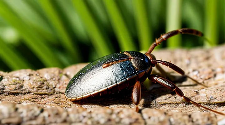
«Immediate Actions After a Tick Bite»
«Tick Removal Techniques»
«Using Tweezers Correctly»
Proper removal of a tick from the skin reduces infection risk and minimizes tissue damage. The following protocol ensures accurate handling of tweezers.
- Choose fine‑point, non‑slip tweezers made of stainless steel.
- Grip the tick as close to the skin surface as possible, grasping the head, not the legs.
- Apply steady, downward pressure to pull straight out without twisting or jerking.
- Avoid squeezing the body; compression can release saliva and pathogens.
- After extraction, inspect the mouthparts; if any remain, repeat the grip and pull.
- Disinfect the bite area with an approved antiseptic solution.
- Dispose of the tick in a sealed container or by incineration; do not crush it.
Post‑removal care includes monitoring the site for redness, swelling, or rash for at least two weeks. If symptoms develop, seek medical evaluation promptly.
«Avoiding Common Mistakes»
When caring for a tick attachment, several preventable errors can compromise recovery and increase infection risk.
- Do not ignore the need for prompt removal; delaying even a few hours allows saliva to enter the wound and heighten pathogen transmission.
- Avoid crushing the tick’s body during extraction; squeezing the abdomen forces internal fluids into the skin, potentially spreading disease.
- Do not use home remedies such as petroleum jelly, heat, or chemicals to detach the parasite; these methods often cause the tick to release more saliva and may leave mouthparts embedded.
- Refrain from applying excessive pressure or bandages that obstruct circulation; tight dressings can cause tissue damage and delay healing.
- Do not neglect post‑removal inspection; failing to verify complete removal can leave mouthparts behind, leading to local inflammation or secondary infection.
- Avoid using unsterilized tools; non‑sterile tweezers or fingers increase the chance of bacterial contamination.
- Do not disregard symptom monitoring; dismissing early signs of rash, fever, or joint pain postpones medical intervention for tick‑borne illnesses.
Proper technique includes grasping the tick close to the skin with fine‑pointed, sterilized tweezers, pulling upward with steady, even pressure, and disinfecting the site immediately after removal. Follow with a clean, breathable dressing and observe the area for at least two weeks, seeking professional care if systemic symptoms develop.
«Cleaning the Bite Area»
«Antiseptic Solutions»
Antiseptic solutions are the primary chemical agents used to reduce microbial contamination at a tick‑bite location. Selection depends on spectrum of activity, tissue tolerance, and availability. Common options include:
- Povidone‑iodine (5‑10 %): Broad‑spectrum, rapid bactericidal effect; apply with a sterile swab, allow to dry, do not rinse.
- Chlorhexidine gluconate (0.5‑4 %): Effective against Gram‑positive and Gram‑negative bacteria; apply thin layer, let stand for 30 seconds before gentle removal of excess.
- Alcohol‑based solutions (70 % isopropanol or ethanol): Fast‑acting, evaporative; apply with a sterile pad, avoid prolonged contact to limit skin irritation.
- Hydrogen peroxide (3 %): Limited antibacterial activity; use only for superficial cleaning, rinse after 1‑2 minutes.
Application protocol:
- Wash hands thoroughly, wear disposable gloves.
- Rinse the bite area with sterile saline to remove debris.
- Apply the chosen antiseptic using a sterile applicator, covering the entire wound margin.
- Allow the solution to remain in contact for the recommended duration (generally 30 seconds to 2 minutes).
- Pat dry with a sterile gauze; avoid rubbing.
- Cover with a non‑adhesive dressing if the site is exposed to friction.
Avoid solutions containing harsh surfactants or high concentrations of hydrogen peroxide, as they can damage epidermal cells and delay healing. Re‑apply antiseptic if the wound becomes contaminated or after dressing changes. Monitoring for signs of infection—redness, swelling, heat, or purulent discharge—remains essential throughout treatment.
«Soap and Water Protocol»
The Soap and Water Protocol addresses the immediate care of a tick‑bite wound. Prompt cleansing reduces bacterial load, removes residual saliva, and prepares the site for further treatment.
- Wet the bite area with clean running water.
- Apply a mild, fragrance‑free soap.
- Lather for at least 20 seconds, ensuring coverage of the bite and surrounding skin.
- Rinse thoroughly with running water to eliminate soap residues.
- Pat the area dry with a disposable, sterile gauze or clean paper towel.
- Apply a topical antiseptic (e.g., povidone‑iodine or chlorhexidine) if available.
- Cover with a sterile, non‑adhesive dressing when the tick has been removed.
- Observe the site for redness, swelling, or increasing pain over the next 48 hours; seek medical evaluation if symptoms develop.
«Post-Removal Care and Monitoring»
«Signs of Infection to Watch For»
«Redness and Swelling»
Redness and swelling are the most common early signs of a tick bite reaction. They indicate localized inflammation caused by the tick’s saliva and, occasionally, an early infection.
Assess the bite site promptly. Measure the diameter of the erythema, note any increase in temperature, and observe whether the swelling expands beyond the immediate area. Record the presence of pain, itching, or a raised border, which may suggest a progressing infection.
Treat the reaction with a three‑step protocol:
- Clean the area with mild soap and water; apply an antiseptic such as povidone‑iodine.
- Reduce inflammation by applying a cold compress for 10–15 minutes, three times daily.
- Administer an oral antihistamine (e.g., cetirizine 10 mg) to control itching and a low‑potency topical corticosteroid (e.g., hydrocortisone 1 %) to diminish erythema.
Monitor the site for 48–72 hours. Seek professional medical evaluation if any of the following occur:
- Erythema enlarges by more than 2 cm or spreads outward.
- Swelling becomes markedly painful or tense.
- Systemic symptoms appear, such as fever, chills, headache, or fatigue.
- A rash develops elsewhere on the body, suggesting a possible tick‑borne disease.
Prompt, systematic care of redness and swelling minimizes tissue damage and reduces the risk of secondary infection following a tick bite.
«Pus or Discharge»
Pus or discharge emerging from a tick bite signals a possible secondary bacterial infection. The presence of thick, yellow‑white fluid indicates that immune cells are responding to invading pathogens, and it warrants immediate attention.
First, cleanse the area with mild soap and sterile saline. Follow with a gentle pat dry to avoid further irritation. If the fluid persists, apply a thin layer of an over‑the‑counter antiseptic ointment, such as bacitracin or mupirocin, then cover with a sterile gauze pad.
Consider systemic therapy when any of the following conditions exist:
- Fever above 38 °C (100.4 °F)
- Expanding redness or swelling extending beyond the bite margin
- Increasing pain despite local care
- Presence of multiple or large pustules
In these cases, a short course of oral antibiotics effective against Staphylococcus aureus and Streptococcus species (e.g., doxycycline, amoxicillin‑clavulanate) should be initiated promptly. Consult a healthcare professional before starting treatment to confirm appropriate drug selection and dosage.
Monitor the wound daily. Discontinue topical agents once discharge ceases and the skin appears clean. Seek medical evaluation if the site deteriorates, develops necrosis, or if systemic symptoms such as chills, headache, or joint pain arise.
«Fever and Rash»
A tick bite that is accompanied by fever and a rash signals possible infection and requires prompt care. Initial measures focus on wound hygiene and early detection of systemic signs.
Clean the bite site with soap and water, then apply an antiseptic. Observe the area for expanding redness, a “bull’s‑eye” pattern, or other changes. Record the date of the bite and the appearance of any fever, chills, or rash, noting the rash’s distribution and characteristics.
If fever exceeds 38 °C (100.4 °F) or a rash develops, seek medical evaluation without delay. Clinicians may order laboratory tests to identify pathogens such as Borrelia burgdorferi, Rickettsia spp., or other tick‑borne agents. Empiric antibiotic therapy, typically doxycycline, is initiated when bacterial infection is suspected, even before test results return.
Supportive care includes:
- Antipyretics for temperature control.
- Hydration to maintain fluid balance.
- Analgesics for localized pain.
Monitoring continues for at least 48 hours after treatment initiation. Persistence or worsening of fever, rash expansion, or new neurological symptoms warrants reassessment and possible adjustment of therapy.
Preventive advice: remove attached ticks promptly with fine‑point tweezers, grasping close to the skin and pulling steadily. Perform regular skin inspections after outdoor activities in endemic areas.
«When to Seek Medical Attention»
«Symptoms of Lyme Disease»
Tick bites that may transmit Borrelia burgdorferi require prompt care to reduce the risk of Lyme disease. Early recognition of infection relies on awareness of characteristic symptoms.
The most common early manifestation is erythema migrans, a expanding red rash usually 5–10 cm in diameter, often with central clearing. The lesion appears 3–30 days after the bite and may be accompanied by mild fever, chills, headache, fatigue, muscle and joint aches, and swollen lymph nodes. Absence of a rash does not exclude infection; systemic signs may develop without visible skin changes.
If the disease progresses, disseminated Lyme disease can present with multiple erythema migrans lesions, neurological involvement such as facial nerve palsy, meningitis, or radiculitis, and cardiac abnormalities like atrioventricular block. Late-stage disease may cause arthritis, most frequently affecting the knees, and chronic musculoskeletal pain.
Key clinical indicators to monitor after a tick bite include:
- Expanding erythematous rash with central clearing
- Fever, chills, or flu‑like symptoms
- Headache, neck stiffness, or facial weakness
- Palpitations, dizziness, or syncope suggesting cardiac involvement
- Joint swelling, especially of large joints
Prompt removal of the tick, thorough cleansing of the bite site, and assessment for these symptoms guide the decision to initiate antibiotic therapy. Early treatment with doxycycline or amoxicillin typically prevents progression to disseminated or late manifestations. Continuous observation for the outlined signs ensures timely intervention and minimizes long‑term complications.
«Allergic Reactions»
Tick bites can trigger immediate hypersensitivity reactions that require swift medical attention. Symptoms may include intense itching, swelling beyond the bite margin, hives, respiratory difficulty, or systemic urticaria. Prompt identification of these signs prevents progression to anaphylaxis.
Initial care focuses on wound cleansing and allergy management. Wash the area with soap and water, apply a cold compress to reduce edema, and avoid scratching. If localized allergic manifestations appear, administer a non‑sedating antihistamine according to dosage guidelines. For extensive swelling or systemic involvement, prescribe a short course of oral corticosteroids.
Severe reactions demand emergency intervention. Administer intramuscular epinephrine immediately, monitor airway and circulation, and arrange transport to a medical facility. Follow‑up evaluation should include allergy testing to identify tick‑specific IgE and counsel on future avoidance strategies.
Key steps for clinicians:
- Inspect bite site for expanding erythema or hives.
- Record onset time of allergic symptoms.
- Provide antihistamine; add corticosteroid if edema persists.
- Deliver epinephrine for anaphylactic signs; activate emergency services.
- Document reaction and advise patient on tick‑avoidance measures.
«Incomplete Tick Removal»
Incomplete removal occurs when part of a tick’s mouthparts or body remain embedded after an attempt to detach the parasite. Residual fragments can continue to secrete saliva, increasing the risk of local inflammation, secondary infection, and transmission of tick‑borne pathogens.
Immediate management includes:
- Use fine‑pointed tweezers to grasp the visible portion of the tick as close to the skin as possible.
- Apply steady, downward pressure to extract the remaining parts without crushing the mouthparts.
- Disinfect the bite area with an antiseptic solution (e.g., povidone‑iodine or chlorhexidine).
- Cover the site with a sterile dressing to prevent bacterial entry.
After removal, observe the wound for signs of erythema, swelling, or fever. Seek medical evaluation if:
- The bite area enlarges or develops a necrotic center.
- Systemic symptoms such as headache, muscle aches, or rash appear.
- The tick was attached for more than 24 hours or the species is known to transmit disease.
Healthcare providers may prescribe antibiotics for bacterial superinfection, administer prophylactic treatment for specific infections (e.g., Lyme disease), and recommend follow‑up examinations to ensure complete healing.
«Preventive Measures and Future Considerations»
«Personal Protection Strategies»
«Appropriate Clothing»
After a tick attaches, the surrounding skin must be kept clean and protected. Appropriate garments prevent further irritation, reduce the risk of secondary infection, and allow the wound to heal without obstruction.
When treating the bite, remove any tight or abrasive clothing that contacts the area. Gently cleanse the site with antiseptic solution, then apply a sterile dressing. Cover the wound with breathable fabric that adheres lightly but does not constrict blood flow.
- Loose‑fitting cotton or linen shirts, allowing air circulation.
- Soft, moisture‑wicking underlayers that keep the area dry.
- Non‑adhesive gauze pads secured with hypoallergenic tape.
- Light, waterproof outerwear if exposure to rain or humidity is expected, but ensure it can be removed easily for wound inspection.
Avoid synthetic fibers that trap sweat, as moisture promotes bacterial growth. Wash all garments in hot water after each use and replace any item that becomes soiled or frayed. Selecting the right clothing facilitates effective care and minimizes complications at the tick bite location.
«Insect Repellents»
Insect repellents form a central element of managing a tick‑bite wound. They reduce the risk of additional ticks attaching to the exposed area and help prevent secondary irritation.
Effective formulations include:
- DEET (N,N‑diethyl‑meta‑toluamide) at 20‑30 % for reliable protection on skin.
- Picaridin (KBR 3023) at 10‑20 % for comparable efficacy with a milder odor.
- IR3535 (ethyl‑butyl‑acetyl‑amino‑propionate) at 10‑20 % for a non‑synthetic alternative.
- Oil of lemon eucalyptus (PMD) at 30 % for plant‑derived protection, suitable for short‑term use.
Application guidelines:
- Apply a thin layer to all uncovered skin, avoiding eyes, mouth and broken skin.
- Re‑apply every 4–6 hours, or sooner after swimming, heavy sweating, or towel drying.
- Use a separate repellent‑treated garment for clothing; spray or soak fabrics with a 0.5 % permethrin solution, allowing it to dry before wear.
Immediate post‑bite care:
- Clean the bite site with soap and water, then apply an antiseptic such as povidone‑iodine.
- Observe the area for expanding redness, fever, or rash; seek medical evaluation if symptoms develop.
- After cleaning, a thin coating of a skin‑safe repellent can discourage nearby ticks from re‑attaching while the wound heals.
Additional preventive steps:
- Wear long sleeves and trousers, tucking pants into socks.
- Perform systematic tick checks at least once daily in endemic areas.
- Remove any attached tick promptly with fine‑point tweezers, grasping close to the skin and pulling straight upward; disinfect the bite site afterward.
By integrating appropriate repellents with proper wound hygiene and regular inspections, the likelihood of complications from a tick bite diminishes significantly.
«Tick-Borne Disease Awareness»
«Common Diseases and Symptoms»
A tick bite requires prompt removal of the attached arthropod, thorough cleansing of the area, and systematic observation for signs of infection. Use fine‑point tweezers to grasp the tick as close to the skin as possible, pull upward with steady pressure, and avoid crushing the mouthparts. After extraction, wash the site with soap and water or an antiseptic solution, then apply a clean dry dressing.
Common illnesses transmitted by ticks present with characteristic symptoms that guide clinical decisions.
- Lyme disease – erythema migrans rash expanding from the bite, fever, headache, fatigue, joint pain.
- Rocky Mountain spotted fever – fever, headache, macular‑palpable rash beginning on wrists and ankles, progressing to trunk.
- Anaplasmosis – fever, chills, muscle aches, leukopenia, thrombocytopenia.
- Babesiosis – hemolytic anemia, fever, chills, jaundice.
- Ehrlichiosis – fever, rash, leukopenia, elevated liver enzymes.
- Tick‑borne relapsing fever – recurrent febrile episodes, headache, myalgia, possible meningismus.
Management of the bite site includes:
- Tick extraction – as described above.
- Local care – cleanse, apply antiseptic, cover with sterile gauze.
- Symptom monitoring – record temperature, rash development, joint swelling, or neurological changes for up to 30 days.
- Prophylactic antibiotics – consider a single dose of doxycycline (200 mg) within 72 hours for high‑risk Ixodes scapularis exposures in endemic areas, provided the tick was attached ≥36 hours.
- Medical evaluation – seek immediate care if fever exceeds 38.5 °C, rash appears, or systemic symptoms develop; laboratory testing for specific pathogens may be required.
Early detection of disease-specific manifestations combined with proper wound care reduces the likelihood of severe complications and supports optimal recovery.
«Geographical Risks»
Tick‑borne disease risk differs sharply across regions, and this variation determines the clinical approach to a bite wound. Knowledge of the local tick species and the pathogens they transmit guides decisions on wound care, prophylactic therapy, and follow‑up.
- North‑East United States, Central Europe, and parts of Asia – Ixodes ricinus or I. scapularis; primary concern is Borrelia burgdorferi (Lyme disease). Recommended prophylaxis: a single dose of doxycycline (200 mg) within 72 hours of removal if the bite occurred in a high‑incidence area and the tick was attached ≥36 hours.
- Southern United States, Central America – Dermacentor variabilis and Amblyomma americanum; risk of Rickettsia rickettsii (Rocky Mountain spotted fever) and Ehrlichia chaffeensis. Treatment of suspected infection: doxycycline 100 mg twice daily for 7–14 days.
- Australia, New Zealand – Ixodes holocyclus; potential for tick‑induced paralysis and bacterial infection. Management includes immediate removal of mouthparts, observation for neurotoxic signs, and empirical amoxicillin if bacterial infection is suspected.
- Sub‑Saharan Africa, parts of the Middle East – Hyalomma spp.; transmit Crimean‑Congo hemorrhagic fever and tick‑borne relapsing fever. Prompt initiation of appropriate antiviral or antibiotic therapy (e.g., tetracycline) is advised after laboratory confirmation.
Effective wound management begins with careful extraction of the tick using fine‑point tweezers, grasping the head as close to the skin as possible, and pulling steadily without crushing the body. The bite site should be cleansed with an antiseptic solution; no chemical agents are applied to the tick itself. After removal, the patient’s location dictates whether a single prophylactic dose of doxycycline is warranted, whether broader‑spectrum antibiotics are indicated, or whether observation alone suffices. Documentation of the bite’s geographic origin, duration of attachment, and any emerging symptoms (fever, rash, arthralgia, neurologic signs) is essential for timely diagnosis and targeted therapy.
