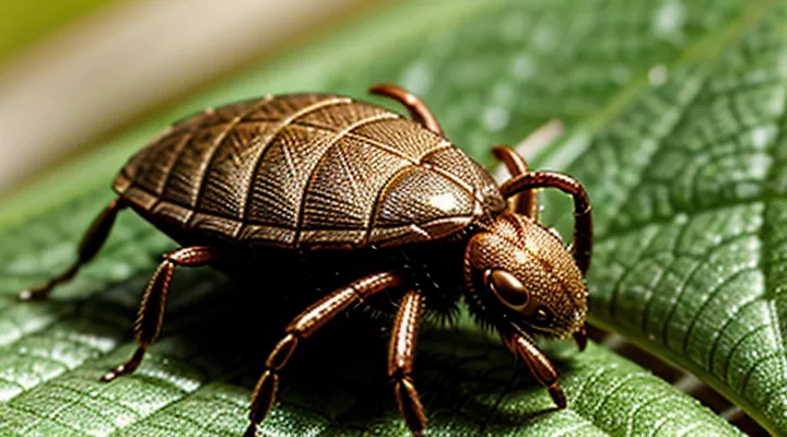
What Ticks Are and Why They Are a Concern
Understanding Tick Bites and Disease Transmission
Common Tick Species and Their Habitats
Ticks vary in appearance and preferred environments, which influences the likelihood of attachment to human skin. Recognizing the most common species and where they are typically found improves early detection.
- Deer tick (Ixodes scapularis) – dense deciduous forests, leaf litter, and shaded grass in the eastern United States. Frequently encountered on hikers and campers during spring and early summer.
- Lone Star tick (Amblyomma americanum) – open fields, woodland edges, and suburban lawns in the southeastern and mid‑Atlantic regions. Active from March through September, often attaching to exposed limbs.
- Western black‑legged tick (Ixodes pacificus) – coastal redwood forests, chaparral, and high‑elevation meadows of the western United States. Peaks in late spring and early fall.
- American dog tick (Dermacentor variabilis) – grassy pastures, gardens, and open woodland in the central and eastern United States. Most active in summer months, commonly found on the lower legs.
- Rocky Mountain wood tick (Dermacentor andersoni) – alpine meadows, pine forests, and shrublands at elevations above 6,000 ft in the western United States. Seasonal activity centers on late spring.
Each species exhibits a characteristic size and coloration that assists identification once attached. Adult females enlarge noticeably after feeding, while nymphs are small and may go unnoticed. Habitat awareness enables targeted skin checks after outdoor exposure: examine scalp, neck, armpits, groin, and any area that contacted vegetation. Prompt removal reduces the risk of disease transmission.
Health Risks Associated with Tick Bites
Ticks transmit pathogens that can cause serious illness. Early identification of a feeding tick reduces exposure time, decreasing the likelihood of infection.
Common diseases transmitted by ticks include:
- Lyme disease, caused by Borrelia burgdorferi, leading to rash, joint pain, and neurological symptoms.
- Rocky Mountain spotted fever, caused by Rickettsia rickettsii, presenting with fever, headache, and a characteristic rash.
- Anaplasmosis, caused by Anaplasma phagocytophilum, resulting in fever, muscle aches, and low blood counts.
- Babesiosis, caused by Babesia microti, producing hemolytic anemia and flu‑like symptoms.
- Ehrlichiosis, caused by Ehrlichia chaffeensis, causing fever, fatigue, and organ dysfunction.
Risk factors increase with prolonged attachment, outdoor exposure in wooded or grassy areas, and lack of prompt removal. Symptoms may appear days to weeks after the bite; some infections progress silently, underscoring the need for vigilance.
If a tick is suspected, inspect the skin thoroughly, focusing on hidden regions such as scalp, armpits, and groin. Remove the arthropod with fine tweezers, grasping close to the skin, and pull steadily without twisting. Clean the site with antiseptic and monitor for rash, fever, or other systemic signs. Seek medical evaluation if any symptoms develop, providing details about the bite and the tick’s appearance when possible.
Initial Steps for Tick Detection
Visual Inspection of the Skin and Clothing
Areas Most Prone to Tick Attachment
Ticks favor body regions where skin is thin, moisture is higher, and clothing provides a protected environment. The most frequently targeted locations include:
- Scalp and hairline
- Behind the ears
- Neck, especially the posterior aspect
- Axillary folds (underarms)
- Groin and inner thigh creases
- Behind the knees
- Around the waistline, including belt loops and underwear seams
- Between fingers and toes, particularly in socked feet
These sites offer easy access to blood vessels and remain concealed from visual inspection. The combination of warmth, humidity, and limited airflow encourages attachment and feeding. Regular examination of the listed areas after outdoor exposure is essential for early detection of engorged or attached specimens. Prompt removal reduces the risk of pathogen transmission.
What to Look For: Size, Color, and Shape of Ticks
Detecting an attached tick requires careful visual inspection of the skin surface. The most reliable indicators are the insect’s dimensions, pigmentation, and outline.
- Size – Engorged ticks expand dramatically, often reaching 5 mm or more in length, while unfed specimens remain under 2 mm. A noticeable increase in size compared to nearby insects suggests a feeding tick.
- Color – Unfed ticks appear brown or reddish‑brown; during feeding they become grayish‑white or darkened as blood accumulates. A shift in hue, especially a mottled or translucent appearance, signals attachment.
- Shape – A flat, oval body indicates an unattached stage, whereas a swollen, balloon‑like abdomen denotes a feeding tick. The mouthparts may be visible as a small, dark, protruding point embedded in the skin.
When these characteristics coincide—enlarged, color‑altered, and bulging morphology—confidence in the presence of a feeding tick is high, prompting immediate removal.
Performing a Thorough Self-Examination
Using a Mirror for Hard-to-Reach Areas
A mirror enables inspection of regions that are difficult to view directly, such as the scalp, neck, behind the ears, and the upper back. Position a full‑length or hand‑held mirror at eye level, adjust lighting to eliminate shadows, and tilt the reflective surface until the target area is clearly visible.
When the area appears in the mirror, focus on the following characteristics:
- Small, rounded body resembling a seed or pinhead
- Darkened abdomen indicating engorgement
- Visible legs or legs clustered at the front
- Mouthparts embedded in the skin, often appearing as a tiny black dot
If any of these signs are present, the parasite has begun feeding and must be removed promptly.
To verify attachment with the mirror:
- Align the mirror so the suspected spot is centered.
- Observe the creature from multiple angles to confirm that the mouthparts are anchored.
- Compare the size to a grain of rice; a fed tick expands rapidly, confirming attachment.
- Document the finding with a photograph if needed for medical reference.
Using a mirror in this systematic manner provides a reliable method for detecting attached ticks in otherwise inaccessible locations.
Importance of Checking Pets and Children
Detecting a tick that has latched onto skin requires a systematic visual inspection. Early identification prevents disease transmission and reduces the need for extensive medical treatment.
Pet owners should examine animals after outdoor activity. Focus on areas where ticks commonly attach: ears, neck, armpits, between toes, and the tail base. Run a fine-toothed comb through the coat, feeling for small, raised bumps. Any attached parasite appears as a dark, rounded object embedded in the skin. Remove it with tweezers, grasping close to the mouthparts, and pull straight upward.
Children should be checked before and after play in grassy or wooded environments. Inspect the scalp, behind ears, under arms, groin, and behind knees. A tick may be visible as a tiny, brown or black speck. If the body is obscured by hair, part the hair and look for a firm, raised spot. Prompt removal follows the same technique used for pets.
Routine checks provide several benefits:
- Immediate detection limits the time a tick can feed.
- Reduces the risk of Lyme disease, Rocky Mountain spotted fever, and other tick-borne illnesses.
- Allows owners to monitor pets for signs of infection, such as fever or lethargy.
- Encourages habit formation in families, reinforcing preventive health practices.
Establish a daily inspection schedule during peak tick season. Carry a pair of fine tweezers and a small container for the removed tick in case identification is required. Document any findings and seek medical advice if the bite site becomes inflamed or if symptoms such as fever, rash, or joint pain develop.
Recognizing Signs and Symptoms of a Tick Bite
Physical Sensations and Localized Reactions
Itching, Irritation, or a Numb Sensation
When a tick secures its mouthparts, the host often experiences localized sensations that differ from ordinary skin irritation. The presence of an itch, a burning irritation, or a transient numbness may indicate that the parasite is attached and feeding.
- Itch: A persistent, localized itch that intensifies after a few minutes suggests the tick’s hypostome has penetrated the epidermis.
- Irritation: Sharp, burning discomfort that does not subside with typical antihistamine use points to mechanical irritation from the tick’s barbed mouthparts.
- Numb sensation: A brief loss of feeling around the bite site can occur when the tick’s saliva anesthetizes the area to facilitate blood intake.
If any of these sensations appear after outdoor exposure, conduct a thorough skin inspection. Part the hair, use a magnifying lens, and look for a small, dark, oval body embedded in the skin. Remove the tick promptly with fine‑tipped tweezers, grasping close to the skin surface, and pull straight upward to avoid leaving mouthparts behind. After removal, clean the area with antiseptic and monitor for lingering symptoms.
Redness or Swelling at the Bite Site
Redness or swelling around a suspected bite is one of the most immediate indicators that a tick may have become embedded. The reaction typically appears within hours of the attachment and can range from a faint pink halo to a pronounced, raised welt. A localized, tender swelling often suggests that the tick’s mouthparts have penetrated the epidermis and are feeding on blood.
Key characteristics to assess:
- Color: pink to deep red, sometimes with a darker central area where the tick’s head is positioned.
- Size: a diameter of 5 mm or more, expanding as the feeding period lengthens.
- Texture: firm to the touch, may feel like a small lump rather than a flat rash.
- Duration: persists beyond 24 hours without fading, indicating ongoing attachment.
Distinguishing this response from a simple insect bite involves examining the site for the tick itself. A visible, engorged arthropod, often partially obscured by skin, confirms attachment. If the tick is not immediately apparent, gently part the skin around the swelling and use a magnifying device to search for a tiny, darkened mouthpart. Persistent redness, especially when accompanied by a palpable bump, should prompt removal of the tick and monitoring for secondary symptoms such as fever or rash.
Distinguishing a Tick from Other Skin Irregularities
Moles, Scabs, or Pimples vs. Ticks
Ticks differ from moles, scabs, and pimples in several observable ways. A tick is a small, oval arachnid that attaches firmly to the skin, often appearing as a raised, engorged nodule. Unlike a mole, which has smooth, uniform pigmentation and does not change size rapidly, a tick’s body expands as it feeds, creating a noticeable bulge that may change color from light brown to dark gray‑black within hours.
Key distinguishing features:
- Mobility: Ticks remain attached and do not shift position on their own; moles, scabs, and pimples are static.
- Surface texture: Ticks have a leathery, slightly rough exoskeleton; moles are smooth, scabs are crusty, and pimples are soft and may contain pus.
- Color variation: Ticks exhibit a gradient from pale abdomen to darker dorsal shield; moles show consistent coloration, while scabs are brown‑red and pimples range from red to white.
- Location: Ticks favor warm, moist areas such as the scalp, groin, or armpits; moles appear anywhere, scabs form over injuries, and pimples develop primarily on facial or torso skin.
- Reaction to pressure: Pressing a tick reveals a firm, attached body; a mole yields no movement, a scab may bleed, and a pimple may release fluid.
If a small, rounded lump enlarges, darkens, and remains anchored despite gentle pressure, the likelihood of a tick attachment is high. Immediate removal with fine‑tipped tweezers, grasping the tick close to the skin and pulling upward with steady force, reduces infection risk. After extraction, clean the area with antiseptic and monitor for rash or fever, which may indicate disease transmission.
The Characteristic Appearance of an Embedded Tick
An embedded tick can be identified by several distinct visual cues. The body appears as a round or oval, slightly flattened structure that adheres tightly to the skin. In early attachment the tick measures 2–5 mm and displays a reddish‑brown hue; as it feeds, the abdomen expands and turns a lighter, bluish‑gray color.
The head region, often called the capitulum, remains visible as a dark, pin‑shaped point protruding from the skin. This point houses the hypostome, the barbed feeding organ, and may be mistaken for a small puncture but is actually the tick’s mouthparts anchored in the epidermis.
Engorgement produces a noticeable bulge. The abdomen swells to a size of 5–10 mm or larger, depending on feeding duration. The surface becomes smooth and glossy, contrasting with the rougher texture of an unattached tick.
Key characteristics to examine:
- Rounded, flat body attached without a clear gap between the tick and skin.
- Visible capitulum or dark central point.
- Color change from reddish‑brown to lighter gray as feeding progresses.
- Abdomen enlargement, creating a raised, smooth bump.
Recognition of these features enables prompt removal and reduces the risk of disease transmission.
What to Do if You Find a Tick
Safe Tick Removal Techniques
Using Fine-Tipped Tweezers
Fine‑tipped tweezers allow a precise assessment of whether a tick is still embedded. First, inspect the lesion for a swollen, rounded abdomen and for any visible legs or mouthparts protruding from the skin. If the tick’s body appears enlarged and the head is not easily lifted, attachment is likely.
To verify attachment, follow these steps:
- Grasp the tick as close to the skin surface as possible with the tweezers’ tips, avoiding compression of the body.
- Apply steady, upward pressure to lift the tick. If resistance persists and the mouthparts remain embedded, the tick is still anchored.
- If the tick detaches cleanly without the mouthparts, attachment has been broken.
After removal, place the tick on a white surface and examine the head. Presence of the capitulum (mouthparts) indicates that the tick was removed before full detachment; a missing capitulum suggests incomplete extraction and possible residual parts in the skin. In such cases, repeat the removal process with fresh tweezers, targeting any remaining fragments.
Document the event, clean the area with antiseptic, and monitor for signs of infection or rash over the next several days.
Proper Disposal of the Tick
After confirming that a tick is firmly attached, safe disposal prevents reinfestation and disease transmission. The removal process should end with immediate and secure elimination of the specimen.
- Place the detached tick into a sealable plastic bag.
- Add a small amount of rubbing alcohol or a few drops of 70 % isopropyl solution to kill the arthropod.
- Expel air, seal the bag tightly, and store it in a freezer for at least 24 hours if immediate disposal is not possible.
- After freezing, dispose of the bag in regular household waste; do not flush or compost.
For environments with high tick activity, maintain a dedicated disposal container containing alcohol or bleach. Replace the solution weekly and clean the container with a disinfectant to avoid cross‑contamination. Documentation of the tick’s removal date and location can aid health professionals in assessing potential disease risk.
Post-Removal Care and Monitoring
Cleaning the Bite Area
When a tick is suspected, the first step after confirming attachment is to clean the bite site. Proper cleaning reduces the risk of secondary infection and prepares the area for further inspection or removal.
- Wash hands thoroughly with soap and water before touching the bite.
- Apply a mild antiseptic solution (e.g., chlorhexidine or povidone‑iodine) to the skin surrounding the tick. Avoid harsh chemicals that could irritate the tissue.
- Gently scrub the immediate area with a clean gauze pad or soft cloth. Do not press directly on the tick, which could cause it to embed deeper.
- Rinse the region with sterile saline or clean water to remove residual antiseptic.
- Pat the skin dry with a disposable towel; do not rub.
After cleaning, re‑examine the site for signs of attachment: a firm, rounded swelling, a dark spot at the mouthparts, or a visible tick body partially embedded. Document the appearance and note any changes during the next 24‑48 hours. If the bite becomes red, swollen, or painful, seek medical evaluation promptly.
Observing for Signs of Infection or Disease
After outdoor activity, examine the exposed skin for any small, dark or light spot that may indicate a tick’s mouthparts embedded in the epidermis. A firm, raised area often appears where the tick’s hypostome has penetrated.
Key indicators of infection or disease following attachment include:
- Local redness extending beyond the bite margin
- Swelling that persists or enlarges over 24‑48 hours
- Warmth or tenderness at the site
- A central puncture wound that remains open or bleeds
- Development of a bullseye‑shaped rash (erythema migrans)
- Fever, chills, or flu‑like symptoms emerging days after the bite
- Joint pain, headache, or muscle aches without an obvious cause
If any of these signs appear, remove the tick promptly with fine‑point tweezers, grasping as close to the skin as possible and pulling straight upward. Clean the area with antiseptic and monitor for progression. Persistent or worsening symptoms warrant immediate medical evaluation, as early treatment reduces the risk of tick‑borne illnesses such as Lyme disease, Rocky Mountain spotted fever, or anaplasmosis.
