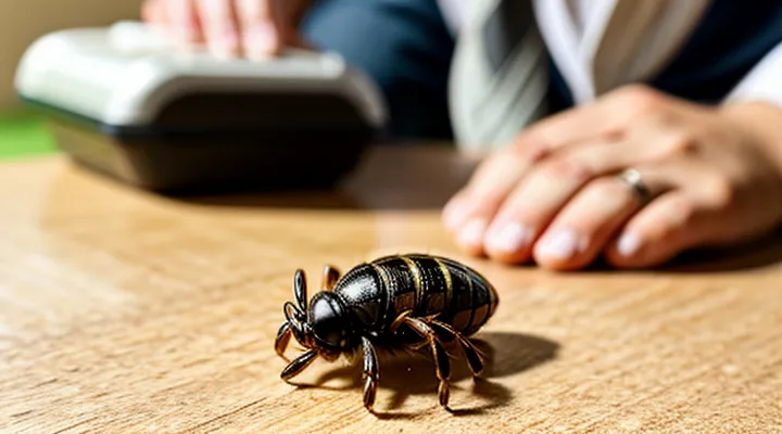
Identifying a Tick Bite
What Ticks Look Like
Ticks are small arachnids, typically measuring 1–5 mm when unengorged. Their bodies consist of two main sections: a forward‑projecting head (capitulum) bearing the mouthparts, and a dorsal shield (scutum) covering the back. The capitulum includes chelicerae for cutting skin and a hypostome that anchors the tick. Six legs emerge from the anterior region; each leg ends in a claw that grips hair or clothing.
Life‑stage differences affect appearance. Larvae, called seed ticks, have six legs and are pale, often translucent. Nymphs possess eight legs, appear darker brown, and are roughly 1 mm in length. Adult females enlarge dramatically after feeding, expanding to 5–10 mm or more and turning reddish‑brown. Adult males remain small, retain the scutum fully covering the back, and stay light brown.
Key visual cues for identification:
- Capitulum shape – elongated, visible from the front.
- Scutum pattern – solid in males, partially covering the abdomen in females.
- Engorgement level – swollen abdomen, rounded silhouette, pale interior.
- Leg count – six in larvae, eight in nymphs and adults.
- Color – varies from yellow‑brown (unfed) to dark brown or reddish after blood intake.
Recognizing these traits assists in confirming the presence of a tick before removal, ensuring the correct technique can be applied safely at home.
Common Symptoms of a Tick Bite
Tick bites often go unnoticed initially, yet early identification of symptoms can prevent complications. Recognizing the body’s response guides timely removal and medical evaluation.
Common manifestations include:
- A small, red bump at the attachment site, sometimes resembling a papule.
- Localized swelling or a raised rash surrounding the bite.
- Itching or mild pain at the puncture point.
- A red, expanding ring known as erythema migrans, typically appearing 3‑30 days after the bite and enlarging up to several centimeters.
- Flu‑like complaints such as fever, headache, fatigue, or muscle aches, especially when the bite is infected with a pathogen.
If any of the following occur, professional assessment is advisable:
- Rapidly enlarging rash or lesions beyond the bite area.
- Persistent fever exceeding 38 °C (100.4 °F) for more than 24 hours.
- Neurological signs, including facial weakness, numbness, or difficulty concentrating.
- Severe joint pain or swelling that does not resolve within a few days.
Prompt removal of the tick, followed by monitoring for these symptoms, reduces the risk of disease transmission.
Essential Supplies for Tick Removal
Tweezers: The Preferred Tool
When a tick attaches to skin, prompt extraction lowers the chance of infection. Fine‑point tweezers provide the most reliable grip without crushing the parasite.
Ideal tweezers have smooth, narrow jaws that fit around the tick’s head. Stainless‑steel models resist corrosion and can be sterilized. Slanted tips improve visibility and allow a shallow angle of approach, reducing pressure on the body.
Removal procedure
- Clean the area with soap and water.
- Grip the tick as close to the skin as possible, holding the mouthparts, not the abdomen.
- Apply steady, gentle pressure and pull straight upward.
- Release the tick once it detaches; avoid twisting or jerking motions.
- Place the specimen in a sealed container for identification if needed.
After extraction, disinfect the bite site with an antiseptic. Observe the area for several weeks; seek medical advice if redness, swelling, or flu‑like symptoms develop.
Avoid using fingers, blunt objects, or methods that compress the tick’s body, as these can force infected fluids into the wound. Regularly inspect clothing and pets after outdoor activity to prevent attachment.
Antiseptic Solutions
When a tick is extracted at home, antiseptic solutions are essential for preventing bacterial contamination and reducing the risk of infection.
- 70 % isopropyl alcohol – rapid bactericidal action, suitable for skin and instrument disinfection.
- Povidone‑iodine (10 % solution) – broad‑spectrum antimicrobial, safe for direct application to the bite area.
- Chlorhexidine gluconate (0.5 %–2 %) – persistent activity, ideal for post‑removal skin care.
- Hydrogen peroxide (3 %) – useful for initial cleaning, but less effective against spores.
Before removal, apply the chosen antiseptic to the skin surrounding the tick and to the tweezers or forceps that will be used. Wipe the area until it is visibly wet, then allow it to dry for a few seconds. This eliminates surface flora that could be introduced during extraction.
After the tick is grasped as close to the skin as possible and pulled straight upward, immediately treat the bite site with the same antiseptic. Apply a thin layer, let it air‑dry, and repeat the process after 10–15 minutes if the area appears damp. Cover the site with a sterile bandage only if bleeding occurs; otherwise, keep it uncovered to allow airflow.
Avoid solutions that cause tissue irritation, such as undiluted bleach or strong acids. Do not apply antiseptics inside the mouth or on mucous membranes. Observe the bite for signs of redness, swelling, or pus; seek medical attention if these symptoms develop despite antiseptic use.
Gloves and Other Protective Gear
When extracting a tick, direct contact with the parasite should be avoided. Disposable nitrile or latex gloves form a barrier that prevents the tick’s saliva from reaching the skin and reduces the risk of secondary infection. Choose gloves that fit snugly, allowing precise manipulation of tweezers without compromising tactile feedback.
Additional protective equipment enhances safety:
- Fine‑point tweezers made of stainless steel, calibrated to grasp the tick’s head without crushing the body.
- A disposable plastic bag or sealable container for immediate disposal of the removed tick.
- A face mask if the individual is prone to coughing or sneezing during the procedure, limiting aerosol exposure.
- A clean, non‑absorbent cloth or gauze to press the site after removal, controlling bleeding.
Procedure steps that incorporate protective gear:
- Don gloves, ensuring they are intact and free of tears.
- Position tweezers close to the skin, grasp the tick as close to the mouthparts as possible.
- Apply steady, upward pressure to extract the tick in one motion; avoid twisting or jerking.
- Transfer the tick into the sealed bag without touching it directly.
- Remove gloves carefully, turning them inside out, and discard them in a waste container.
- Clean the bite area with antiseptic and cover with sterile gauze if needed.
Using these items minimizes contamination, protects the remover’s hands, and supports a clean, effective extraction.
Step-by-Step Tick Removal Process
Preparing for Removal
Before attempting to extract a tick, ensure the environment is clean and well‑lit. Wash your hands thoroughly with soap and water, then dry them. Gather the necessary instruments: fine‑point tweezers or a specialized tick‑removal tool, disposable gloves, antiseptic solution (e.g., povidone‑iodine), and a small sealable container with a label for the tick. Keep a timer nearby to record the duration of attachment, as this information may be useful for medical assessment.
Verify that the person being treated has no known allergy to antiseptics or latex gloves. If any skin condition or open wound exists near the bite site, note it for later evaluation. Position the individual comfortably, exposing the area while maintaining privacy and minimizing movement.
Check the tick’s location carefully. Identify the head and body orientation to plan a straight pull. If the tick is partially embedded in hair, part the hair with a comb to avoid pulling on surrounding strands. Ensure the removal tool is sterile; if necessary, wipe it with antiseptic before use.
Once preparation is complete, proceed with the extraction promptly to reduce the risk of pathogen transmission.
Grasping the Tick Correctly
Proper grasp of a tick is essential for safe extraction and to prevent pathogen transmission. The bite site should be exposed, and the skin cleaned with alcohol or soap before handling the parasite.
Required tools:
- Fine‑pointed, non‑toothed tweezers (or tick‑removal forceps)
- Disposable gloves (optional)
- Antiseptic solution for post‑removal care
Steps for securing the tick:
- Position tweezers as close to the skin as possible, aiming at the tick’s head where it enters the flesh.
- Clamp the mouthparts firmly without crushing the abdomen; a steady, gentle pressure is sufficient.
- Pull upward with a smooth, continuous motion, avoiding twisting or jerking motions that could detach the mouthparts.
- Release the grip once the tick separates, then place the specimen in a sealed container for identification if needed.
After removal, disinfect the bite area, wash hands thoroughly, and monitor the site for signs of infection over the next several days. If the tick’s mouthparts remain embedded, repeat the grasping procedure using fresh tweezers; do not attempt to dig them out with fingers or sharp objects.
Pulling the Tick Out
Removing a tick by hand requires steady pressure, proper tools, and prompt action to reduce the risk of disease transmission.
A suitable set includes fine‑point tweezers or a dedicated tick‑removal device, antiseptic wipes, gloves, and a sealable container for the specimen.
- Disinfect hands and gloves.
- Grasp the tick as close to the skin as possible, holding the mouthparts, not the body.
- Apply steady, downward force; avoid twisting or jerking.
- Pull until the entire tick separates from the skin.
- Place the tick in the container, add a few drops of alcohol if testing is desired, and seal.
After extraction, cleanse the bite area with antiseptic and monitor the site for redness, swelling, or a rash over the next weeks. Seek medical evaluation if any symptoms develop or if the tick could not be removed completely.
Post-Removal Care
After the tick is removed, clean the bite area with mild soap and lukewarm water. Pat the skin dry with a clean towel; avoid rubbing.
Apply an antiseptic solution, such as povidone‑iodine or alcohol, to the wound. Allow it to air‑dry before covering, if needed.
Monitor the site for signs of infection or irritation. Look for redness expanding beyond the bite, swelling, pus, or increasing pain. If any of these develop, seek medical advice promptly.
Record the date of removal and the tick’s appearance, if possible. This information assists healthcare providers in assessing potential disease transmission.
Typical post‑removal care steps:
- Clean the area with soap and water.
- Disinfect with an appropriate antiseptic.
- Keep the wound uncovered unless it contacts dirty surfaces.
- Observe the bite for 2–4 weeks for abnormal changes.
- Consult a professional if fever, rash, or flu‑like symptoms appear.
Maintain regular hygiene and avoid scratching the bite to reduce irritation and prevent secondary infection.
What NOT to Do When Removing a Tick
Avoid Folk Remedies
Ticks attach firmly to skin and can transmit pathogens within hours. Safe extraction at home depends on using appropriate instruments and following evidence‑based steps, not on traditional or unverified tricks.
Folklore methods—such as applying petroleum jelly, burning, freezing with ice, or using nail polish—lack scientific support. They may irritate the tick, causing it to release additional saliva that contains infectious agents, or they can damage surrounding tissue, complicating removal and increasing infection risk.
Recommended removal procedure
- Choose a pair of fine‑pointed, non‑toothed tweezers.
- Grasp the tick as close to the skin surface as possible, holding the head, not the body.
- Apply steady, downward pressure to pull the tick straight out without twisting.
- Avoid squeezing the body, which could expel gut contents.
- Place the tick in a sealed container for identification if needed.
After the tick is removed, cleanse the bite site with soap and water or an antiseptic. Observe the area for several weeks; seek medical evaluation if a rash, fever, or flu‑like symptoms develop. Professional assessment is essential if removal is difficult, the tick is embedded, or the individual belongs to a high‑risk group (e.g., children, immunocompromised patients).
Do Not Crush or Squeeze the Tick
When extracting a tick, avoid any action that compresses the body of the parasite. Crushing the abdomen can force infected fluids into the host’s skin, increasing the chance of disease transmission. Squeezing also damages the mouthparts, making them more likely to break off and remain embedded, which complicates removal and may cause local irritation.
To prevent crushing or squeezing, follow these steps:
- Use fine‑pointed, non‑slip tweezers.
- Grasp the tick as close to the skin’s surface as possible, holding the head or mouthparts, not the abdomen.
- Apply steady, even pressure to lift the tick straight out without twisting.
- Release the tick into a sealed container for proper disposal; do not crush it between fingers.
- Clean the bite area with antiseptic and wash hands thoroughly.
Monitoring the site for redness, swelling, or a rash over the next several days is advisable; any unusual symptoms should prompt medical consultation. This method minimizes pathogen exposure and ensures complete removal.
Prevent Leaving Parts Behind
Removing a tick without leaving mouthparts behind requires steady hands, proper tools, and adherence to a precise sequence. Incomplete extraction can cause inflammation, infection, or transmission of pathogens.
Use fine‑point tweezers or a specialized tick‑removal device. Disinfect the instrument with alcohol before contact. Grasp the tick as close to the skin as possible, securing the head or mouthparts rather than the body to avoid crushing the abdomen.
- Pull upward with steady, even pressure; do not twist, jerk, or squeeze the body.
- Maintain traction until the entire tick releases from the skin.
- Inspect the bite site and the removed specimen; the tick’s head should be fully visible. If any part remains embedded, repeat the grasping step on the residual fragment.
After extraction, clean the wound with antiseptic and monitor for redness, swelling, or fever over the next several days. Preserve the tick in a sealed container for identification if needed. Prompt, complete removal minimizes the risk of complications.
Aftercare and Monitoring
Cleaning the Bite Area
After extracting the tick, the bite site requires immediate attention to reduce the risk of infection. Begin by washing hands thoroughly with soap and water, then apply the same cleaning method to the affected skin. Use mild antiseptic soap, lather the area, and rinse with clean running water. Pat the skin dry with a disposable paper towel; avoid rubbing, which could irritate the wound.
Apply an antiseptic solution—such as 70 % isopropyl alcohol, povidone‑iodine, or chlorhexidine—directly onto the bite. Allow the antiseptic to remain for at least 30 seconds before gently blotting excess fluid. This step eliminates surface bacteria and minimizes inflammation.
If the bite remains irritated, cover it with a sterile, non‑adhesive dressing. Change the dressing daily or whenever it becomes wet or contaminated. Monitor the site for signs of infection: increasing redness, swelling, warmth, pus, or escalating pain. Should any of these symptoms appear, seek medical evaluation promptly.
Observing for Symptoms of Tick-Borne Illnesses
After a tick is taken off at home, watch the bite site and overall health for the next several weeks. Early detection of infection relies on recognizing specific clinical signs.
- Redness or swelling that expands beyond the bite area
- Fever, chills, or night sweats
- Severe headache, neck stiffness, or facial palsy
- Muscle or joint pain, especially in large joints
- Fatigue, nausea, or abdominal pain
- Rash with a “bull’s‑eye” pattern (central clearing surrounded by a red ring)
Symptoms may appear within days or up to two months after the bite. Record the date of removal, the region of the body where the tick attached, and any visible changes. If any listed sign develops, contact a healthcare professional promptly; early antibiotic therapy can prevent severe disease. Even in the absence of symptoms, a follow‑up evaluation is advisable for high‑risk exposures, such as bites from adult ticks, prolonged attachment, or residence in endemic areas.
When to Seek Medical Attention
Most tick bites can be managed at home, but specific circumstances demand professional evaluation.
- Attachment lasting more than 24 hours or uncertainty about removal time.
- Incomplete extraction, visible mouthparts remaining in the skin.
- Development of a red expanding rash, especially a target‑shaped lesion.
- Onset of fever, chills, headache, muscle aches, or joint pain within days of the bite.
- Signs of infection at the bite site: increasing redness, swelling, pus, or warmth.
Children, pregnant individuals, and persons with weakened immune systems face higher risk of complications and should be assessed promptly.
When any of these indicators appear, contact a healthcare provider without delay. Early consultation enables assessment for tick‑borne diseases, consideration of prophylactic antibiotics, and appropriate wound care.
