Assessment of the Situation
Identifying the Tick
When a tick becomes attached, the first step is to confirm its presence and species. Accurate identification guides removal technique and assesses disease risk.
Observe the lesion closely. A tick appears as a small, oval or round body, often resembling a tiny seed or bean. The dorsal surface is typically brown, gray, or reddish, sometimes with a mottled pattern. Look for the characteristic “shield” (scutum) on the back of hard ticks (Ixodidae); soft ticks (Argasidae) lack this hard plate. The size varies with feeding stage: unfed nymphs are 1–3 mm, adults range from 3 mm to over 1 cm when engorged.
Examine the attachment point. Ticks embed their mouthparts (hypostome) into the skin at an angle of 30–45°. The mouthparts form a visible central point or a tiny black dot. If the tick’s head is visible, it indicates a recent attachment; a fully embedded tick may show only the lower body.
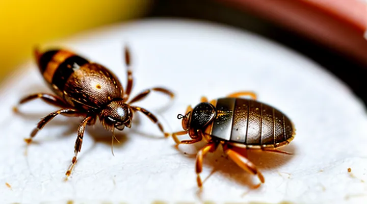
Distinguish ticks from similar arthropods. Fleas are flattened, jump, and lack the elongated mouthparts. Mites are much smaller (under 1 mm) and often hide in skin folds. Lice cling to hair shafts and cannot embed.
Use a magnifying lens or a smartphone camera with zoom to capture details. Photograph the specimen for expert consultation if species identification is uncertain. Recording the following data improves risk assessment:
- Approximate size (mm)
- Color and pattern
- Presence of scutum (hard tick) or absence (soft tick)
- Life stage (larva, nymph, adult)
- Attachment duration estimate (if known)
Having this information enables prompt, appropriate removal and informs whether prophylactic treatment or medical evaluation is required.
Assessing the Bite Location
When a tick attaches, the exact spot of the bite determines the safest removal technique and the risk of infection. Identify the precise region on the skin, noting whether the tick is on a flat surface, a fold, or near a joint. The location influences how much skin can be grasped without damaging surrounding tissue.
- Examine the area under good lighting; use a magnifying lens if needed.
- Confirm that the tick’s head and mouthparts are visible; the mouthparts point away from the body.
- Record whether the bite is on a limb, torso, scalp, or interdigital space.
- Note proximity to sensitive structures such as eyes, mucous membranes, or open wounds.
- Determine if the tick is embedded in hair; part the hair to expose the attachment site.
Accurate assessment guides the choice of removal tool (fine‑point tweezers, tick removal hook) and the angle of traction. It also helps clinicians decide if additional measures—such as prophylactic antibiotics—are warranted based on the bite’s location and the tick’s species.
Safe Tick Removal Techniques
Necessary Tools
When a tick is lodged in the skin, immediate removal requires specific instruments to minimize tissue damage and reduce infection risk.
- Fine‑point tweezers or forceps with a narrow tip
- Tick‑removal device (e.g., a curved hook or specialized plastic tool)
- Disposable nitrile or latex gloves
- Antiseptic solution or wipes (e.g., iodine or alcohol)
- Small, sealable container or puncture‑proof bag for disposal
- Optional magnifying glass for precise visualization
The tweezers must grasp the tick as close to the skin as possible without squeezing the body. The removal device can slide beneath the mouthparts when tweezers are unsuitable. Gloves protect both the extractor and the patient from pathogen exposure. After extraction, apply antiseptic to the bite site and seal the tick in the container before discarding it in household waste. A magnifier assists in confirming complete removal of the mouthparts.
Step-by-Step Removal Process
When a tick penetrates the skin, prompt removal reduces the risk of pathogen transmission. The following procedure outlines each action required to extract the parasite safely and effectively.
- Gather tools: fine‑point tweezers or a specialized tick‑removal device, disposable gloves, antiseptic solution, and a sealed container for disposal.
- Don gloves to avoid direct contact with the tick’s mouthparts.
- Position the tweezers as close to the skin surface as possible, grasping the tick’s head (the part embedded in the skin), not the body.
- Apply steady, gentle upward pressure; avoid twisting, jerking, or squeezing the body, which can cause the mouthparts to break off.
- Continue pulling until the entire tick separates from the skin.
- Inspect the attachment site; if any mouthparts remain, repeat step 3‑5 with a new grip.
- Disinfect the bite area with an appropriate antiseptic.
- Place the removed tick in the sealed container; label with date and location for potential testing.
- Dispose of the container by sealing it in a trash bag or flushing, following local regulations.
After removal, monitor the bite site for signs of infection or rash over the next several weeks. Seek medical evaluation if redness expands, a fever develops, or a characteristic bullseye lesion appears. Documentation of the tick’s species and removal date aids healthcare providers in assessing disease risk.
Post-Removal Care
Cleaning the Bite Area
Immediately after removing a tick, the skin around the attachment site must be disinfected to reduce the risk of infection. First, wash hands thoroughly with soap and water, then apply the same cleaning method to the bite area. Use a mild antiseptic solution—such as a 70 % isopropyl alcohol swab, povidone‑iodine, or chlorhexidine gluconate—applying gentle pressure for at least 30 seconds. Rinse with sterile saline or clean water, pat the skin dry with a sterile gauze pad, and avoid rubbing, which could irritate the tissue.
If an antiseptic is unavailable, a solution of diluted hydrogen peroxide (3 % concentration, mixed 1:1 with water) can be used as a temporary measure, followed by a thorough rinse. After cleaning, cover the site with a sterile adhesive bandage to protect against external contaminants. Monitor the area for signs of redness, swelling, or pus; any such developments warrant medical evaluation.
Finally, document the date and location of the bite, along with the cleaning method employed, for reference in case symptoms of tick‑borne illness appear later. This record assists health professionals in assessing potential exposure and determining appropriate treatment.
Monitoring for Symptoms
After a tick attaches, observe the bite site and overall health for the next several weeks. Early detection of illness relies on recognizing specific signs that may develop within hours to days.
Watch for the following symptoms:
- Fever or chills
- Headache, especially if severe or persistent
- Muscle or joint aches
- Fatigue or malaise
- Rash, particularly a red expanding spot or a bull’s‑eye pattern
- Nausea, vomiting, or abdominal pain
If any of these manifestations appear, seek medical evaluation promptly and mention the recent tick exposure. Continuous self‑monitoring enhances timely diagnosis and treatment of tick‑borne diseases.
When to Seek Medical Attention
Incomplete Tick Removal
If the head or mouthparts of a tick remain lodged after an attempt to pull it out, the situation requires prompt, precise action. Leaving fragments embedded can increase the risk of local infection and facilitate transmission of tick‑borne pathogens.
- Grasp the visible portion of the tick as close to the skin as possible with fine‑pointed tweezers.
- Apply steady, downward pressure to pull the mouthparts straight out; avoid twisting or jerking, which may break the hypostome.
- If resistance is felt or the mouthparts do not detach, stop pulling immediately. Continued force can cause the tick’s mandibles to fragment further.
When removal cannot be completed safely:
- Disinfect the area with an antiseptic solution (e.g., povidone‑iodine or chlorhexidine).
- Cover the site with a sterile dressing.
- Seek professional medical care without delay. Clinicians may use specialized instruments or a minor surgical procedure to extract residual parts.
After professional removal, monitor the bite site for:
- Redness expanding beyond the immediate area.
- Swelling, warmth, or pus formation.
- Flu‑like symptoms, fever, or rash, especially a bull’s‑eye pattern.
Report any of these signs to a healthcare provider promptly, as they may indicate early infection or disease transmission. Documentation of the tick’s attachment time, species (if identifiable), and removal details assists clinicians in risk assessment and treatment decisions.
Signs of Infection
If a tick remains attached, removal should be followed by a careful observation of the bite site for any indication of infection. Early warning signs include:
- Redness that expands beyond the immediate margin of the bite.
- Swelling that increases in size or becomes firm to the touch.
- Warmth localized around the area, noticeably hotter than surrounding skin.
- Increasing pain or tenderness that does not diminish after a few hours.
- Pus or other fluid discharge, suggesting bacterial involvement.
- Fever, chills, or malaise developing within 24–48 hours.
- Enlarged, tender lymph nodes near the bite, especially in the armpit or groin.
The appearance of any of these symptoms warrants prompt medical evaluation. Delayed treatment can lead to cellulitis, abscess formation, or systemic infection, potentially complicating the initial tick exposure. Immediate consultation with a healthcare professional enables appropriate antibiotic therapy and prevents progression to more severe conditions.
Symptoms of Tick-Borne Diseases
A tick attached to the skin can transmit several pathogens, each producing a characteristic set of clinical signs. Recognizing these manifestations promptly guides early treatment and reduces the risk of severe complications.
Common tick‑borne illnesses and their principal symptoms include:
- Lyme disease – expanding erythema migrans rash, fever, chills, headache, fatigue, joint pain, and, in later stages, neurologic deficits such as facial palsy or meningitis.
- Rocky Mountain spotted fever – abrupt fever, severe headache, myalgia, nausea, and a maculopapular rash that often begins on the wrists and ankles before spreading to the trunk.
- Anaplasmosis – high fever, muscle aches, chills, nausea, and a mild rash; laboratory tests frequently reveal low platelet count and elevated liver enzymes.
- Ehrlichiosis – fever, severe headache, malaise, muscle pain, and a rash in a minority of cases; leukopenia and thrombocytopenia are typical laboratory findings.
- Babesiosis – fever, chills, sweats, hemolytic anemia, jaundice, and dark urine; severe cases may cause organ failure, especially in immunocompromised patients.
- Tularemia – ulcerative skin lesion at the bite site, regional lymphadenopathy, fever, and malaise; respiratory involvement can occur with inhalational exposure.
Symptoms often overlap, and early disease may present with nonspecific signs such as fever, fatigue, and headache. Persistent or worsening manifestations after a tick bite warrant immediate medical evaluation to confirm diagnosis and initiate appropriate antimicrobial therapy.
Prevention of Future Bites
Protective Clothing
When a tick has attached to the skin, immediate removal should be performed while minimizing additional exposure. Wearing appropriate protective clothing reduces the risk of contaminating other body areas and prevents accidental contact with the tick’s mouthparts.
A suitable ensemble includes:
- Long‑sleeved, tightly woven shirts; sleeves should be rolled up to expose forearms for safe handling.
- Long trousers tucked into socks or boots; this prevents the tick from crawling onto the legs.
- Disposable gloves, preferably nitrile, to avoid direct hand contact with the parasite.
- A face mask or protective eye shield if the tick is located near the head, limiting accidental splashes of bodily fluids.
- Closed, non‑porous footwear that can be easily removed after the procedure.
The clothing should be removed carefully after the tick is extracted, turning garments inside out to contain any detached parts. Dispose of the outer layer in a sealed bag before washing the remaining garments at a temperature of at least 60 °C. This protocol ensures that the removal process does not introduce secondary contamination while maintaining personal safety.
Tick Repellents
Tick repellents constitute the primary barrier against tick attachment, reducing the likelihood that a tick will embed and necessitate urgent removal. Effective repellents must be applied to exposed skin and clothing before entering tick‑infested areas and reapplied according to label instructions.
Chemical repellents dominate the market. DEET formulations ranging from 20 % to 30 % provide reliable protection for several hours. Picaridin at 20 % concentration offers comparable duration with a milder odor. IR3535 and ethyl butylacetylaminopropionate (EBAPA) deliver shorter protection windows but are suitable for children and sensitive skin. Permethrin, used to treat clothing, socks, and gear, retains activity after multiple washes and kills ticks on contact.
Natural products are available but generally deliver shorter protection periods. Citronella, lemon eucalyptus (oil of lemon eucalyptus, 30 % PMD), and cedar oil require frequent reapplication and may not meet the efficacy standards of synthetic agents. When used, they should be applied at the maximum recommended concentration and combined with other protective measures.
Proper application limits gaps where ticks can attach. Apply repellent evenly, covering all skin surfaces except the eyes and mouth. For clothing, treat only the outer layer; avoid direct skin contact with permethrin. Reapply after swimming, sweating, or after the time interval specified on the product label. Keep a spare bottle for prolonged exposure.
If a tick has already attached, repellents do not facilitate removal. Immediate steps focus on extraction with fine‑tipped tweezers, grasping the tick close to the skin, and pulling upward with steady pressure. After removal, cleanse the bite area with antiseptic and consider applying a topical antibiotic. Applying a repellent to the surrounding skin can deter additional ticks from attaching during the remainder of the activity.
Recommended repellents and usage guidelines
- DEET 20‑30 % – apply to exposed skin; reapply every 6–8 hours.
- Picaridin 20 % – apply to skin and hair; reapply every 8 hours.
- Permethrin 0.5 % – treat clothing, shoes, and gear; retain efficacy for up to 6 washes.
- Oil of lemon eucalyptus (30 % PMD) – apply to skin; reapply every 4 hours.
- Citronella or cedar oil – apply to skin; reapply every 2 hours; use as supplement, not sole protection.
Checking for Ticks After Outdoor Activities
After any outdoor exposure, a systematic skin inspection is the first defense against tick‑borne disease. The examination should start within an hour of returning home and be repeated 24 hours later, because a tick may detach unnoticed.
- Remove clothing and examine the entire body, beginning with the scalp, neck, armpits, behind the ears, and groin.
- Use a fine‑toothed comb or a hand mirror to view hard‑to‑reach areas.
- Run fingers over the skin to feel for small, raised bumps; adult ticks feel like firm, pea‑sized nodules, while nymphs are barely visible.
- If a tick is found, grasp it with fine‑point tweezers as close to the skin as possible, pull upward with steady pressure, and dispose of it in alcohol.
Document the location, size, and time of removal, then wash the bite site with soap and water. If the tick was attached for more than 24 hours, consult a healthcare professional promptly. Regular self‑checks reduce the likelihood of infection and support early intervention.
