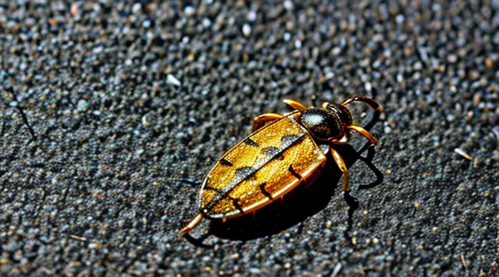
Misconceptions and Dangers of Tick Burning
The Myth of «Sterilization» and «Eradication»
Burning a tick after it has been detached is often presented as a means of sterilization and eradication. In reality, the practice does not guarantee the elimination of pathogens and may give a false sense of safety.
The belief that heat instantly destroys all disease agents rests on several misconceptions:
- Heat kills only the tick’s external structures; internal pathogens can survive if the tick is not fully incinerated.
- Many tick‑borne organisms, such as Borrelia burgdorferi, are resistant to brief exposure to flame.
- The act of burning does not prevent the tick’s saliva or gut contents from entering the bite wound during removal.
Scientific evidence shows that the critical factor in preventing infection is proper removal technique, not post‑removal combustion. The recommended steps are:
- Grasp the tick as close to the skin as possible with fine‑tipped tweezers.
- Pull upward with steady, even pressure, avoiding twisting or crushing.
- Disinfect the bite site with an antiseptic solution.
- Monitor the area for signs of infection or rash over the following weeks.
Burning the specimen may serve a psychological purpose, but it does not replace meticulous extraction and wound care. Relying on flame alone can delay appropriate medical attention if symptoms develop.
Risks to Human Health
Burns and Injuries
Ticks attach with cement‑like saliva that hardens around their mouthparts. Even after the visible body is pulled away, the embedded hypostome can remain embedded in the skin, providing a pathway for bacterial or viral agents. Applying a brief, controlled burn to the bite site denatures proteins, coagulates tissue, and eliminates any remaining tick fragments that might serve as a vector.
A heat‑based post‑removal treatment accomplishes three objectives:
- immediate destruction of residual mouthparts, preventing them from acting as a nidus for infection;
- thermal inactivation of pathogens that survived the mechanical extraction;
- stimulation of local coagulation, which reduces bleeding and seals the puncture wound.
The recommended technique uses a sterile, flame‑producing instrument (e.g., a lighter or electric cautery pen). Hold the flame a few millimeters above the skin, apply heat for 1–2 seconds, and then allow the area to cool naturally. Do not touch the skin with the flame directly; the goal is surface heating, not combustion of tissue. After cooling, clean the site with an antiseptic solution and cover with a sterile dressing if needed. This protocol minimizes the risk of secondary infection and accelerates wound closure.
Inhalation of Pathogens
Ticks carry bacteria, viruses, and protozoa that survive in the exoskeleton after removal. If the carcass is crushed or left intact, mechanical disturbance can release microscopic particles into the air. Inhalation of these particles may lead to respiratory exposure to infectious agents.
Incinerating the tick eliminates viable pathogens and prevents aerosol formation. High temperature denatures nucleic acids and proteins, rendering organisms such as Borrelia burgdorferi, Rickettsia rickettsii, Anaplasma phagocytophilum, and tick‑borne encephalitis virus non‑infectious. Complete combustion ensures that no viable fragments remain to be inhaled.
Key considerations for safe disposal:
- Apply a direct flame until the tick is reduced to ash.
- Verify that the fire is sustained for at least 30 seconds to guarantee thermal inactivation.
- Avoid crushing the tick with fingers or tools; mechanical disruption increases aerosol risk.
- Wash hands thoroughly after handling the tick and after extinguishing the flame.
Following these steps minimizes the chance of respiratory exposure to tick‑borne pathogens.
Environmental Concerns
Release of Toxins
Burning a detached tick eliminates residual salivary proteins that may remain on the skin after extraction. These proteins include anticoagulants, anti‑inflammatory agents, and neurotoxins that can continue to diffuse into surrounding tissue for several minutes. Heat denatures these molecules, preventing further absorption and reducing the risk of localized irritation or systemic reaction.
The thermal process also destroys any viable pathogens that survived the bite. Studies show that temperatures above 60 °C for a few seconds inactivate Borrelia burgdorferi, Anaplasma phagocytophilum, and other common tick‑borne agents. By applying flame directly to the exoskeleton, the temperature at the bite site quickly reaches this threshold, ensuring rapid microbial inactivation.
Key reasons for using flame after removal:
- Immediate denaturation of saliva‑derived toxins.
- Prevention of continued diffusion into host tissue.
- Rapid inactivation of surviving microorganisms.
- Elimination of the tick as a future source of pathogen transmission.
Fire Hazard
Incinerating a tick after it is detached eliminates residual pathogens, but the act introduces a fire risk that must be managed. The heat generated by a small flame can ignite surrounding combustible materials such as dry leaves, paper, or clothing. Open flames placed near flammable surfaces increase the probability of unintended fire spread, especially in indoor environments where ventilation may be limited.
Key fire‑related concerns include:
- Ignition of nearby objects – a spark or ember can contact dry debris, causing rapid combustion.
- Smoke and toxic fumes – burning arthropod tissue releases particulates and chemicals that may irritate respiratory pathways.
- Uncontrolled flame growth – unattended flames can expand beyond the intended area, endangering property and occupants.
Mitigation measures:
- Perform incineration outdoors, away from structures and vegetation.
- Use a dedicated, fire‑proof container (e.g., metal ashtray) to contain the flame.
- Keep a fire extinguisher or water source within reach.
- Extinguish the flame completely before leaving the site.
Adhering to these precautions reduces the likelihood of accidental fire while still achieving effective tick disposal.
Recommended Alternatives for Tick Disposal
Proper Disposal Methods
Alcohol or Antiseptic Solutions
Applying alcohol or an antiseptic solution to a tick bite site after removal serves three primary purposes. First, the liquid disinfects the skin, eliminating microorganisms that may have been introduced by the tick’s mouthparts. Second, the rapid evaporation of alcohol creates a brief cooling effect that can contract tissue, helping to seal the wound and reduce bleeding. Third, the antiseptic can dissolve residual tick saliva, which often contains pathogens, thereby lowering the chance of infection.
Key considerations for selecting an appropriate disinfectant include:
- Efficacy against common tick‑borne organisms – solutions containing isopropyl alcohol (70 % concentration) or povidone‑iodine demonstrate broad antimicrobial activity.
- Skin tolerance – alcohol may cause stinging; iodine can cause irritation in individuals with iodine sensitivity.
- Availability – both agents are widely stocked in first‑aid kits, making immediate application feasible in field situations.
When alcohol or iodine is unavailable, cauterization (burning) the bite site is an alternative method. The heat denatures proteins in residual saliva and seals the wound, but it also carries a risk of additional tissue damage. Consequently, the preferred protocol remains cleaning the area with a proven antiseptic immediately after the tick is extracted.
Sealing in Tape
After a tick is detached from a host, the organism remains alive and may release pathogens if it is crushed or exposed to air. Encasing the specimen in adhesive tape creates a sealed barrier that prevents saliva, feces, or internal fluids from contacting skin, clothing, or surfaces. The method also simplifies disposal by allowing the tape-wrapped tick to be discarded without additional handling steps.
Key advantages of sealing a tick in tape:
- Immediate containment of biological material.
- Elimination of accidental crushing, which can aerosolize infectious agents.
- Compatibility with standard waste protocols; the tape-wrapped tick can be placed in a regular trash bag.
- Minimal equipment required; adhesive tape is readily available in most settings.
The practice supports infection‑control guidelines that recommend rapid inactivation of ectoparasites after removal. By sealing the tick, the risk of secondary exposure to pathogens such as Borrelia, Rickettsia, or Anaplasma is markedly reduced.
Flushing Down the Toilet
Flushing a removed tick down the toilet provides a quick, low‑risk disposal method that eliminates the parasite without exposing household members to a live or partially damaged arthropod. The process requires sealing the tick in a small plastic bag or wrapping it in tissue before flushing, ensuring that the organism cannot escape the plumbing system. This precaution prevents accidental re‑attachment to skin or contamination of surfaces.
Compared with incineration, flushing avoids the need for fire safety measures, specialized equipment, or outdoor burning restrictions. It also reduces the chance of inhaling smoke or creating a fire hazard in confined spaces. However, flushing does not guarantee complete destruction; the tick may survive long enough to exit the sewage system if the waste treatment infrastructure is inadequate.
Key considerations for choosing flushing over burning:
- Containment: Secure the tick to prevent leaks in the toilet bowl.
- Plumbing compatibility: Verify that local sewage systems can handle solid waste without blockage.
- Environmental impact: Recognize that sewage treatment plants are designed to neutralize biological material, reducing ecological risk.
- Regulatory compliance: Some jurisdictions restrict flushing of live organisms; consult local guidelines.
When burning is impractical—due to lack of a safe outdoor area, fire bans, or concerns about smoke inhalation—flushing offers a practical alternative that aligns with public health objectives while maintaining simplicity and speed.
Why Proper Disposal is Crucial
Preventing Disease Transmission
Removing a tick does not guarantee that all infectious material is eliminated. The mouthparts can remain attached to the skin, and residual saliva may contain pathogens that survive brief contact. Immediate destruction of the tick by flame eliminates these sources before they can be transferred to the host.
- Heat denatures proteins and nucleic acids, rendering bacteria, viruses, and protozoa non‑viable.
- Burning ruptures the tick’s exoskeleton, releasing any remaining fluids and exposing them to temperatures that exceed the survival thresholds of most tick‑borne agents.
- The process prevents accidental crushing or squeezing, actions that can force infected material back into the bite wound.
- Visible combustion provides a clear, irreversible indication that the tick has been neutralized, reducing the risk of inadvertent reuse or mishandling.
Alternative disinfection methods, such as alcohol or bleach, may leave viable fragments on the skin or require additional steps to ensure complete inactivation. Flame treatment combines rapid pathogen elimination with a simple, observable outcome, making it an effective measure to curb disease transmission following tick removal.
Protecting the Environment
Burning a tick after it has been removed eliminates a potential vector for disease without introducing chemical contaminants into soil or water. The heat destroys the parasite’s DNA, preventing accidental re‑attachment or spread through waste streams.
Environmental considerations include:
- Minimal emissions when using a low‑temperature, fuel‑efficient incinerator.
- Reduced landfill volume compared with sealing ticks in plastic bags.
- Avoidance of chemical disinfectants that can leach into ecosystems.
When a portable incinerator is unavailable, placing the tick in a sealed, biodegradable container and exposing it to a controlled flame achieves the same result while limiting particulate release. Proper disposal aligns with broader goals of preserving biodiversity and maintaining clean habitats, as it removes a biological hazard without adding synthetic waste.
What to Do After Tick Removal
Cleaning the Bite Area
After a tick is detached, the skin at the attachment point contains saliva, potential pathogens, and residual tissue debris. Prompt decontamination removes these elements, lowering the risk of bacterial infection and reducing the chance that any transmitted agents establish a foothold.
The bite site should be treated as follows:
- Rinse with lukewarm water and a mild, unscented soap; friction eliminates surface contaminants.
- Pat dry with a clean disposable towel; avoid rubbing, which may reopen the wound.
- Apply a broad‑spectrum antiseptic (e.g., povidone‑iodine or chlorhexidine) for at least 30 seconds; this kills remaining microorganisms.
- Cover with a sterile, non‑adhesive dressing only if the area bleeds; otherwise, leave exposed to air for natural drying.
- Observe daily for redness, swelling, or pus; seek medical attention if any signs develop within 72 hours.
Cleaning the bite area also prepares the skin for any additional interventions, such as cauterization, that may be employed to destroy residual tick tissue. Proper hygiene therefore complements other post‑removal measures and enhances overall safety.
Monitoring for Symptoms
After a tick is removed and the bite site is cauterized, close observation for emerging health indicators is essential. The skin around the attachment point should be inspected daily for redness, swelling, or ulceration. Any expansion of the lesion beyond the immediate area warrants prompt medical evaluation.
Key symptoms to monitor include:
- Fever, chills, or unexplained temperature rise
- Severe headache or neck stiffness
- Muscle or joint pain, especially if it appears suddenly
- Nausea, vomiting, or abdominal discomfort
- Rash characterized by concentric circles (bullseye) or other atypical patterns
- Fatigue or malaise persisting beyond a few days
The observation period extends for at least four weeks, as most tick‑borne infections manifest within this timeframe. If any listed symptom arises, seek professional care without delay; early treatment reduces the risk of complications.
Documenting the onset, duration, and progression of each symptom improves diagnostic accuracy. Record the date of tick removal, the method of cauterization, and any subsequent changes in health status. This systematic approach supports timely intervention and optimal outcomes.
When to Seek Medical Attention
After removing a tick, monitor the bite site and overall health for any abnormal signs. Immediate medical evaluation is warranted if any of the following occur:
- Redness or swelling expands beyond the immediate area of the bite, especially if it forms a circular rash.
- A bull’s‑eye rash (a red ring with a clear center) appears, indicating possible Lyme disease.
- Fever, chills, headache, muscle aches, or joint pain develop within two weeks of the bite.
- The tick was attached for more than 24 hours, or its removal was incomplete, leaving mouthparts embedded.
- Signs of infection such as pus, increasing pain, or warmth around the wound.
- The individual is pregnant, immunocompromised, or has a chronic illness that could exacerbate tick‑borne infections.
If none of these symptoms are present, clean the area with soap and water, apply an antiseptic, and observe for at least four weeks. Any delayed reaction, even mild, should still be reported to a healthcare professional to rule out emerging infection. Prompt consultation reduces the risk of complications and ensures appropriate treatment if a pathogen is transmitted.
