Characteristics and Identification
Physical Attributes
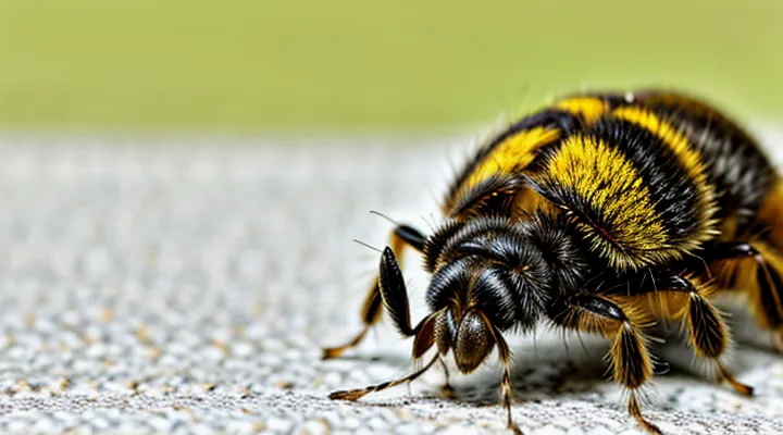
Dermatophagoides farinae is a microscopic arachnid belonging to the family Pyroglyphidae. Adult individuals measure approximately 0.2–0.3 mm in length, with a rounded, oval body that tapers toward the posterior. The cuticle is semi‑transparent, revealing internal structures, and exhibits a pale yellow to light brown coloration under light microscopy.
The mite’s anatomy includes:
- Four pairs of legs, each ending in claw‑like structures that facilitate movement across fabric and dust particles.
- Two fused body regions: the gnathosoma (mouthparts) and the idiosoma (main body), the latter housing the digestive and reproductive systems.
- A set of sensory organs called trichobothria, located primarily on the forelegs, which detect air currents and vibrations.
- Sclerotized plates (tergites) that provide structural support while allowing flexibility necessary for locomotion.
Eggs are ellipsoidal, about 0.1 mm in diameter, and are deposited on surfaces rich in organic debris. Nymphal stages resemble miniature adults, undergoing three molts before reaching maturity. The compact size and lightweight exoskeleton enable the mite to become airborne when disturbed, contributing to its widespread distribution in indoor environments.
Habitat and Environment
Dermatophagoides farinae thrives in indoor environments where human activity provides a steady supply of organic debris. The mite populates household dust, bedding, upholstered furniture, and carpet fibers, exploiting shed skin cells, hair, and textile fibers as food sources. Optimal growth occurs at relative humidity levels between 70 % and 80 % and temperatures ranging from 20 °C to 25 °C (68 °F–77 °F). Low humidity or temperatures below 15 °C (59 °F) markedly reduce population density.
Typical habitats include:
- Mattress and pillow covers, especially those lacking impermeable encasements.
- Sofa cushions and fabric-covered chairs where dust accumulates.
- Carpets and area rugs in rooms with limited ventilation.
- Curtains and draperies that retain moisture and dust particles.
Geographically, the species is widespread in temperate and subtropical regions, reflecting its adaptation to modern indoor climate control. Urban dwellings, office spaces, and schools provide the consistent microclimate necessary for sustained colonization. Regular cleaning, humidity control, and barrier textiles are the primary measures that limit the mite’s presence in these environments.
Life Cycle
Dermatophagoides farinae completes its development within a confined environment of household dust, textiles, and upholstered surfaces. The life cycle progresses through five distinct stages: egg, larva, protonymph, deutonymph, and adult.
- Egg – Laid in clusters on the surface of dust particles; incubation lasts 2–3 days under optimal humidity (70–80 %) and temperature (22–25 °C).
- Larva – Emerges with six legs; feeds on organic detritus and mite feces for 3–4 days before molting.
- Protonymph – Possesses eight legs; undergoes a 2–3 day feeding period, then molts into the deutonymph.
- Deutonymph – Similar to the adult in leg count; development time ranges from 2 to 5 days, depending on environmental conditions.
- Adult – Female mites live 2–4 weeks, producing 20–40 eggs each; males survive slightly shorter, primarily for mating.
A complete cycle, from egg to reproducing adult, can be finished in 10–14 days when temperature and humidity are favorable. Under less ideal conditions, development extends to several weeks, reducing population growth. Rapid reproduction and the accumulation of allergenic proteins in fecal pellets and body fragments make the mite a persistent source of indoor allergens throughout its life stages.
Role in Allergies
Dermatophagoides farinae, commonly called the American house dust mite, produces a spectrum of airborne allergens that trigger immune responses in sensitized individuals. The mite’s fecal particles, shed skin, and body fragments contain proteins such as Der f 1, Der f 2, Der f 5, and Der f 23. These proteins act as antigens, binding to IgE antibodies on mast cells and basophils, leading to degranulation and release of histamine, leukotrienes, and cytokines. The resulting inflammation manifests as rhinitis, asthma, or atopic dermatitis, depending on the site of exposure.
Key aspects of the allergenic activity include:
- Proteolytic enzymes (e.g., Der f 1) that disrupt epithelial barriers, facilitating allergen entry.
- IgE-binding epitopes that provoke immediate hypersensitivity reactions upon inhalation.
- Chronic exposure that sustains Th2‑biased immune polarization, promoting persistent airway hyperresponsiveness.
Epidemiological data show that up to 80 % of individuals with allergic rhinitis and a similar proportion of asthmatics exhibit sensitization to D. farinae. Indoor environments with high humidity and abundant textiles provide optimal conditions for mite proliferation, increasing airborne allergen concentrations. Control measures—such as reducing indoor humidity below 50 %, regular laundering of bedding at ≥60 °C, and use of allergen-impermeable covers—lower exposure levels and correlate with measurable reductions in symptom scores and medication use.
In clinical practice, specific IgE testing for D. farinae allergens guides diagnosis and informs immunotherapy selection. Sublingual or subcutaneous allergen immunotherapy targeting Der f components has demonstrated long‑term efficacy in decreasing symptom severity and medication dependence for patients with mite‑induced respiratory allergy.
Allergenic Components
Major Allergens
Dermatophagoides farinae is a house dust mite whose secreted proteins constitute a primary source of indoor allergens. The species produces several well‑characterized allergenic molecules, notably Der f 1, Der f 2, Der f 4, and Der f 6. These proteins trigger IgE‑mediated responses in sensitized individuals, leading to symptoms such as rhinoconjunctivitis, asthma, and atopic dermatitis.
Major allergen groups that dominate clinical practice include:
- Dust‑mite allergens – Der f 1 and Der f 2 from D. farinae, alongside homologous proteins from Dermatophagoides pteronyssinus.
- Pollen allergens – Bet v 1 from birch, Amb a 1 from ragweed, and related seasonal proteins.
- Animal‑dander allergens – Fel d 1 from cats, Can f 1 from dogs, and associated lipocalins.
- Mold spores – Alt a 1 from Alternaria, Asp f 1 from Aspergillus.
- Cockroach allergens – Bla g 1 and Bla g 2 from Blattella germanica.
Exposure to D. farinae occurs through settled dust in bedding, upholstered furniture, and carpets. Concentrations exceeding 1 mg g⁻¹ of dust routinely elicit sensitization. Diagnostic panels incorporate recombinant Der f allergens to improve specificity, while allergen‑specific immunotherapy employs standardized extracts to induce tolerance.
In summary, D. farinae contributes a substantial portion of the indoor allergen burden, with its major proteins forming a core component of the broader spectrum of clinically relevant allergens.
Mechanisms of Allergic Reaction
Dermatophagoides farinae is a common house dust mite whose fecal particles and body fragments contain proteins that act as potent allergens. When inhaled, these particles encounter the respiratory epithelium, where they are captured by dendritic cells. The cells process the proteins and present peptide fragments to naïve T lymphocytes in regional lymph nodes, prompting differentiation toward a Th2 phenotype. Th2 cells secrete interleukins IL‑4, IL‑5, and IL‑13, which drive class switching in B cells to produce IgE antibodies specific to mite allergens.
IgE molecules bind to high‑affinity FcεRI receptors on mast cells and basophils. Subsequent exposure to the same mite allergens cross‑links surface‑bound IgE, triggering rapid degranulation of these effector cells. The released mediators—histamine, prostaglandins, leukotrienes—induce immediate symptoms such as bronchoconstriction, increased vascular permeability, and mucus secretion. A secondary wave of inflammation follows, characterized by recruitment of eosinophils, further cytokine release, and tissue remodeling.
Key steps in the allergic cascade caused by Dermatophagoides farinae:
- Antigen uptake by dendritic cells and presentation to Th2 cells
- Th2 cytokine production (IL‑4, IL‑5, IL‑13)
- B‑cell class switching to IgE synthesis
- IgE binding to mast cell and basophil FcεRI receptors
- Allergen‑induced IgE cross‑linking and cell degranulation
- Early‑phase mediator release (histamine, leukotrienes)
- Late‑phase cellular infiltration and chronic inflammation
These mechanisms explain how exposure to the mite translates into the clinical manifestations of allergic disease.
Impact on Health
Allergic Rhinitis
Dermatophagoides farinae, commonly known as the American house dust mite, is a microscopic arthropod that thrives in warm, humid indoor environments. It feeds on human skin scales and fabric fibers, proliferating in bedding, upholstered furniture, and carpets. The mite’s fecal pellets and fragmented bodies contain a mixture of protein allergens that become airborne and readily inhaled.
When these allergens reach the nasal mucosa, they trigger an IgE‑mediated immune response in sensitized individuals. The resulting inflammation of the nasal lining produces the characteristic signs of allergic rhinitis: sneezing, rhinorrhea, nasal congestion, and itchy or watery eyes. Persistent exposure can exacerbate symptoms and increase the risk of secondary complications such as sinusitis or asthma.
Diagnostic confirmation relies on skin prick testing or serum-specific IgE assays that identify reactivity to Dermatophagoides farinae allergens. Positive results, combined with a clinical history of nasal symptoms, establish the link between the mite and allergic rhinitis.
Effective control combines environmental measures with medical therapy:
- Reduce indoor humidity to below 50 % using dehumidifiers or air conditioning.
- Wash bedding weekly in water ≥ 60 °C; dry on high heat.
- Encase mattresses and pillows in allergen‑impermeable covers.
- Vacuum with HEPA‑filtered equipment and clean floors with damp mops.
- Apply intranasal corticosteroids or antihistamine sprays to alleviate inflammation.
- Consider allergen‑specific immunotherapy for long‑term tolerance.
By limiting exposure to Dermatophagoides farinae and implementing targeted pharmacologic treatment, most patients achieve substantial symptom relief and improved quality of life.
Asthma
Dermatophagoides farinae is a microscopic arthropod that thrives in indoor environments containing human skin scales, textile fibers, and low‑level humidity. Adults measure 0.2–0.3 mm, lack eyes, and reproduce rapidly, producing large numbers of fecal pellets that contain potent protein allergens. These allergens persist on bedding, upholstered furniture, and carpet, becoming airborne during normal household activity.
In susceptible individuals, inhalation of D. farinae allergens initiates an IgE‑mediated immune response that targets the airways. The reaction induces mast‑cell degranulation, release of histamine and leukotrienes, and recruitment of eosinophils, leading to airway hyper‑responsiveness, mucus overproduction, and bronchoconstriction. These pathophysiological changes underlie the development and exacerbation of asthma symptoms such as wheezing, coughing, and shortness of breath.
Clinical management of mite‑related asthma includes:
- Environmental control: regular washing of bedding at ≥60 °C, use of allergen‑impermeable mattress covers, reduction of indoor humidity below 50 %, and removal of carpets or heavy drapes.
- Pharmacotherapy: inhaled corticosteroids and bronchodilators to mitigate airway inflammation and obstruction.
- Immunotherapy: sublingual or subcutaneous administration of mite extracts to induce tolerance in selected patients.
Recognition of D. farinae as a prominent asthma trigger guides both diagnostic testing (skin‑prick or serum IgE assays) and targeted interventions aimed at reducing exposure and modulating immune reactivity.
Atopic Dermatitis
Dermatophagoides farinae is a microscopic acarid that thrives in indoor dust, feeding on shed human skin cells and textile fibers. The organism measures 0.2–0.3 mm, reproduces rapidly in warm, humid environments, and is a principal component of household dust across temperate regions.
The mite produces a suite of proteins that bind immunoglobulin E, initiating immediate‑type hypersensitivity reactions. Upon inhalation or skin contact, these allergens trigger mast‑cell degranulation, leading to pruritus, erythema, and the release of cytokines that amplify inflammatory pathways.
In patients with atopic dermatitis, exposure to D. farinae intensifies epidermal barrier disruption and Th2‑dominant inflammation. Studies report higher specific IgE levels to mite antigens in individuals whose eczema flares correlate with increased indoor dust mite counts. The allergen‑induced cytokine milieu aggravates skin lesions, prolongs healing time, and elevates the risk of secondary infection.
Control strategies focus on reducing mite burden and mitigating immune activation:
- Maintain indoor relative humidity below 50 %.
- Wash bedding weekly in water ≥ 60 °C; dry thoroughly.
- Use allergen‑impermeable covers for mattresses and pillows.
- Remove carpets, upholstered furniture, and heavy curtains where dust accumulates.
- Apply topical corticosteroids or calcineurin inhibitors during flare‑ups; consider systemic antihistamines or allergen‑specific immunotherapy for persistent cases.
Management and Prevention
Environmental Control
Dermatophagoides farinae, a common house dust mite, thrives in warm, humid environments and is a frequent trigger of allergic reactions such as rhinitis, asthma, and eczema. Reducing indoor exposure requires systematic environmental control.
- Maintain indoor relative humidity below 50 % by using dehumidifiers or air‑conditioning; low moisture limits mite reproduction.
- Wash bedding, curtains, and removable upholstery weekly in water heated to at least 60 °C (140 °F) or freeze items for 24 hours to eradicate mites and allergens.
- Encase mattresses, pillows, and box springs in allergen‑impermeable covers; replace covers annually to preserve barrier integrity.
- Remove carpets, especially in bedrooms; replace with hard‑surface flooring that can be vacuumed with a HEPA‑rated device.
- Vacuum floors, upholstered furniture, and curtains regularly using a vacuum equipped with a HEPA filter; discard collected debris promptly.
- Reduce clutter that accumulates dust; store rarely used items in sealed plastic containers.
- Employ air purifiers with HEPA filtration in high‑use areas to capture airborne mite particles.
Implementing these measures consistently lowers mite populations, thereby diminishing allergen load and mitigating symptom severity for sensitized individuals.
Medical Treatments
Dermatophagoides farinae is a house dust mite whose fecal particles contain potent allergens that trigger IgE‑mediated responses in sensitized individuals. Exposure to these particles can provoke rhinitis, asthma exacerbations, and atopic dermatitis.
Accurate diagnosis relies on skin‑prick testing or specific IgE assays that include D. farinae extracts. Positive results confirm sensitization and guide therapeutic decisions.
Pharmacologic management focuses on reducing inflammation and relieving symptoms:
- Inhaled corticosteroids for persistent asthma or allergic rhinitis.
- Short‑acting β2‑agonists for acute bronchospasm.
- Intranasal antihistamines or corticosteroid sprays for nasal congestion.
- Topical corticosteroids and calcineurin inhibitors for eczema flare‑ups.
- Leukotriene receptor antagonists as adjuncts in asthma control.
Allergen‑specific immunotherapy (AIT) offers disease‑modifying potential. Subcutaneous or sublingual formulations containing D. farinae extracts are administered according to standardized protocols, aiming to induce tolerance and decrease medication requirements. Successful AIT reduces symptom scores and improves quality of life in many patients.
Adjunct measures include environmental control: regular vacuuming with HEPA filters, washing bedding at ≥60 °C, and maintaining indoor humidity below 50 % to limit mite proliferation. Combining pharmacotherapy, immunotherapy, and exposure reduction provides a comprehensive approach to managing D. farinae‑related allergic disease.
Immunotherapy
Dermatophagoides farinae is a prevalent house dust mite whose proteins trigger IgE‑mediated responses in sensitized individuals, leading to rhinitis, asthma, and atopic dermatitis. Repeated exposure in indoor environments maintains the allergic cascade, making control of mite allergens a central component of disease management.
Immunotherapy targets the underlying immune dysregulation by introducing controlled doses of mite allergens to induce tolerance. The approach modifies T‑cell subsets, reduces IgE production, and promotes IgG4 antibodies that block allergen‑IgE interaction.
Key elements of mite‑specific immunotherapy include:
- Subcutaneous immunotherapy (SCIT) with standardized Dermatophagoides farinae extracts administered in a build‑up phase followed by maintenance injections.
- Sublingual immunotherapy (SLIT) using tablet or liquid formulations placed under the tongue, offering a self‑administered alternative with a comparable safety profile.
- Component‑resolved diagnostics to identify dominant allergen molecules (e.g., Der f 1, Der f 2) and tailor extract composition.
- Adjunctive strategies such as anti‑IgE monoclonal antibodies to accelerate desensitization and reduce adverse reactions.
Clinical trials demonstrate significant reductions in symptom scores, medication use, and airway hyperresponsiveness after 3–5 years of treatment. Long‑term follow‑up indicates sustained benefit after cessation, supporting immunotherapy as a disease‑modifying option for mite‑induced allergy.
