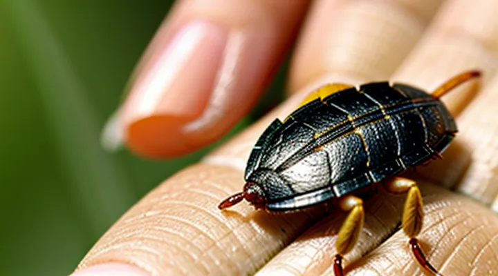
The Basics of Tick Appearance
Size and Shape Variations
Ticks visible on human skin range from a few millimeters to several centimeters, depending on feeding status and species. An unfed nymph typically measures 1–3 mm in length, appearing as a flat, oval body. Adult females of common species such as Ixodes scapularis or Dermacentor variabilis start at 2–5 mm before blood intake. After engorgement, the same females expand to 6–12 mm or more, adopting a rounded, balloon‑like profile that may exceed the width of a pea.
Key dimensions and shape changes:
- Unfed stage – flat, elongated, dorsoventrally compressed; length rarely exceeds 3 mm.
- Partially fed – body elongates slightly, edges become more convex; size increases proportionally to blood volume.
- Fully engorged – body swells uniformly, surface becomes smooth and glossy; length can reach 10–15 mm, width approaches 8 mm.
Species differences affect overall size limits. Amblyomma americanum females may attain 15 mm when fully engorged, while Rhipicephalus sanguineus rarely exceeds 8 mm. Shape remains generally oval, but engorgement produces a markedly rounded silhouette that distinguishes a feeding tick from a detached or dead specimen.
Color and Texture
Ticks attached to human skin appear as small, rounded or oval bodies measuring 2–5 mm in length. Their coloration ranges from light brown to deep reddish‑brown, often matching the surrounding skin tone, which can make early detection difficult. The surface is covered with a smooth, waxy cuticle that feels slightly glossy when dry but becomes moist and slightly sticky after feeding. Key visual cues include:
- A uniform dark‑brown or reddish hue, occasionally with a lighter abdomen.
- A smooth, almost velvety texture that lacks obvious scales or hairs.
- A subtle sheen that increases as the tick engorges with blood.
- Slight flattening of the dorsal side, giving a pancake‑like profile against the skin.
These characteristics distinguish a tick from other skin lesions or insects, aiding accurate identification.
Legs and Mouthparts
A tick attached to a person’s skin presents a rounded, engorged body often dark brown or reddish. The six pairs of legs are short, stout, and positioned around the perimeter of the body. They are not easily seen because they lie flat against the skin, but a careful inspection may reveal tiny, hair‑like segments extending a few millimetres from the tick’s edges.
The mouthparts consist of three primary structures that penetrate the epidermis:
- Hypostome – a barbed, tube‑like organ that anchors the tick and draws blood; it appears as a tiny, dark point at the centre of the body.
- Chelicerae – paired cutting blades that slice the skin during insertion; they are hidden beneath the hypostome and not visible externally.
- Palps – sensory appendages flanking the hypostome; they are short and inconspicuous, aiding the tick in locating blood vessels.
Together, the legs secure the tick’s position while the mouthparts form a concealed feeding apparatus that is rarely visible beyond the central puncture point.
Where Ticks Prefer to Attach
Common Hiding Spots
Ticks embed in skin where they can remain unnoticed for several days. Their small size and tendency to blend with surrounding tissue make detection difficult, especially in areas that are less visible or regularly covered.
- Scalp, particularly near the hairline and behind the ears
- Neck and behind the jawline
- Armpits and under the bra strap
- Groin and inner thigh folds
- Waistline, especially around belts or waistbands
- Between the toes and on the foot arches
- Behind the knees and in the popliteal fossa
- Under the breast tissue and around the nipple
- Abdomen, notably in the region of a diaper or tight clothing
Regular inspection of these locations after outdoor activities, using a magnifying lens if necessary, increases the likelihood of early removal before the tick expands and becomes more visible.
Stages of Engorgement
A feeding tick undergoes distinct visual changes that allow identification on the skin. Early attachment appears as a tiny, flat, gray‑brown speck, roughly the size of a pinhead. As blood intake begins, the body expands, the abdomen swells, and the color shifts to a lighter, more translucent hue. Mid‑engorgement shows a markedly enlarged, balloon‑like abdomen, often reaching several times the original size; the surface may appear glossy and the legs become less visible. Full engorgement presents a rounded, almost spherical shape, with the abdomen dominating the body, skin‑colored or pinkish, and the tick may measure up to 10 mm or more. After detachment, the empty exoskeleton remains as a small, flattened shell that may stay attached for a short period before falling off.
- Flat, unattached stage: tiny, flat, gray‑brown.
- Early engorgement: abdomen begins to swell, color lightens.
- Mid‑engorgement: abdomen markedly enlarged, glossy appearance.
- Full engorgement: round, large, pinkish, dominates body.
- Post‑detachment: empty shell, flat, often still attached briefly.
Differentiating Ticks from Other Blemishes
Moles and Freckles
Moles are benign proliferations of melanocytes that appear as well‑defined, often raised spots. Their color ranges from light brown to black, and they may be flat or slightly elevated. Freckles are clusters of pigment produced by normal melanocytes, typically flat, small (1–2 mm), and uniformly tan or light brown. Both are stable skin features that develop mainly through genetic factors and sun exposure.
Ticks, when attached to the skin, present as elongated, engorged bodies that may resemble a small, dark, oval or round plaque. Unlike moles and freckles, ticks have a visible head and legs, a rough texture, and often a central punctum from which blood is drawn. They can change size rapidly as they feed, whereas moles and freckles remain relatively constant over time.
Key differences:
- Shape: moles – round, sometimes irregular; freckles – round, uniformly small; ticks – oval, often elongated.
- Surface: moles – smooth or slightly raised; freckles – flat, matte; ticks – rough, may feel like a tiny bump.
- Borders: moles – well‑defined; freckles – diffuse edges; ticks – distinct outline with a visible attachment point.
- Mobility: moles and freckles – static; ticks – can shift position as they embed deeper.
- Change over time: moles and freckles – gradual, if any; ticks – rapid enlargement within hours.
Recognizing these characteristics enables accurate identification and appropriate response when a small dark spot is observed on the skin.
Scabs and Insect Bites
Ticks attach to the skin as small, rounded bodies that expand after feeding. The engorged stage appears as a dark brown to grayish sphere, often resembling a tiny bump or a bead. The surrounding skin may show a slight redness, which can be confused with a typical insect bite, but the central lesion remains firm and well‑defined.
Unlike mosquito or flea bites that produce a raised, itchy papule, tick attachment frequently results in a flat or slightly raised area with a central puncture point. When the tick is removed, the puncture may close, leaving a smooth surface that can develop a thin scab within several hours. The scab is usually pale, thin, and adheres tightly to the underlying skin, differing from the thicker crusts seen after allergic reactions to other insects.
Key visual indicators of a tick on human skin:
- Size: 2–5 mm when unfed; up to 10 mm after engorgement.
- Color: reddish‑brown before feeding, darkening to gray‑black after blood intake.
- Shape: oval, slightly flattened dorsally, with a clear anterior mouthpart (hypostome) sometimes visible.
- Surrounding reaction: minimal swelling, occasional mild erythema, no central punctum after removal.
Identification should focus on the combination of a firm, rounded body and the described color change. If a scab forms without significant itching or spreading redness, the lesion likely originated from a tick rather than from a typical allergic insect bite. Persistent redness, expanding rash, or systemic symptoms warrant medical evaluation.
Other Skin Conditions
Ticks attach to the skin as a small, rounded, engorged object, often resembling a dark speck or a tiny, flesh-colored bump. Several unrelated dermatological conditions can mimic this appearance, making accurate identification essential.
- Flea bite: Red papule with a central punctum, surrounded by a halo of erythema; typically appears in clusters on the lower legs.
- Mosquito bite: Raised, itchy wheal that swells rapidly, often with a clear center; resolves within 24‑48 hours.
- Allergic contact dermatitis: Diffuse erythema and scaling, sometimes with vesicles; distribution follows exposure to the irritant.
- Scabies: Linear or serpentine burrows, 2‑5 mm in length, often found between fingers, on wrists, or in the genital area.
- Ringworm (tinea corporis): Annular lesion with a raised, scaly border and a clear central area; may expand outward over weeks.
- Erythema migrans (early Lyme disease): Expanding erythematous rash, often oval, with a central clearing; typically larger than a tick’s size and may develop days after attachment.
Distinguishing factors include lesion shape, size, distribution, presence of itching, and progression over time. Ticks remain firmly attached; removal with fine‑point tweezers reveals a mouthpart embedded in the skin. In contrast, the conditions listed above detach or resolve without a hard, attached body. Accurate visual assessment, combined with patient history of exposure, guides appropriate management.
What to Do if You Find a Tick
Safe Removal Techniques
Ticks appear as small, dark, oval or spherical bodies embedded in the skin, often surrounded by a slight reddening. The head and mouthparts may be visible as a tiny black dot at the center of the lesion. Prompt and proper removal reduces the risk of disease transmission.
Safe removal requires the following steps:
- Grasp the tick as close to the skin surface as possible with fine‑pointed, non‑toothed tweezers.
- Apply steady, downward pressure to pull the tick straight out without twisting or crushing.
- Disinfect the bite area with an antiseptic solution after removal.
- Place the tick in a sealed container with alcohol or a zip‑lock bag for identification, if needed.
- Monitor the site for several weeks; seek medical advice if rash, fever, or expanding redness develop.
Avoid using hot water, petroleum jelly, or sharp objects to detach the tick, as these methods can cause the mouthparts to remain embedded and increase infection risk. Proper technique ensures complete extraction and minimizes complications.
When to Seek Medical Attention
A tick attached to skin appears as a small, rounded, dark‑brown or gray lump, often resembling a tiny bump or a speck of dirt. The body may be slightly enlarged after engorgement, and the head (capitulum) can be seen as a darker point at the surface. The surrounding area may be smooth or slightly raised, without immediate pain.
Medical evaluation is required when any of the following conditions occur:
- The tick remains attached for more than 24 hours, indicating possible engorgement.
- A rash develops, especially a circular red lesion with central clearing (often called “bull’s‑eye” rash).
- Fever, chills, headache, muscle aches, or joint pain appear within days to weeks after the bite.
- Swelling, redness, or pus forms at the bite site, suggesting secondary infection.
- Neurological symptoms arise, such as facial weakness, tingling, or difficulty concentrating.
- The individual is immunocompromised, pregnant, or a young child, increasing risk of complications.
If any of these signs are present, seek professional care promptly. Early treatment reduces the likelihood of severe disease and facilitates appropriate removal techniques.
Monitoring for Symptoms after a Bite
A fed tick attached to the skin appears as a dark, engorged oval that may be partially hidden beneath a thin, translucent cuticle. The body swells as it fills with blood, often measuring several millimeters in length, while the legs remain visible and move slowly.
After removal, cleanse the bite site with antiseptic and store the specimen for identification if possible. Record the date of exposure, the location on the body, and any visible changes in the lesion.
Monitor the following signs:
- Redness expanding beyond the bite margin
- A bull’s‑eye rash (central clearing surrounded by a red ring)
- Fever, chills, or night sweats
- Muscle or joint aches, especially in large joints
- Headache, nausea, or dizziness
- Unexplained fatigue lasting more than 24 hours
Observe the area for at least three weeks. Seek medical evaluation promptly if any listed symptom appears, if the rash enlarges rapidly, or if systemic signs develop, as early treatment reduces the risk of severe infection.
Preventing Tick Bites
Protective Clothing and Repellents
Ticks attach to the skin as small, dark, oval bodies that may swell after feeding. Their size and color often blend with the surrounding tissue, making early detection difficult. Protective measures aim to prevent attachment and reduce the risk of disease transmission.
Effective protective clothing includes:
- Long‑sleeved shirts and full‑length trousers made of tightly woven fabric.
- Pants tucked into socks or boots to seal the gap at the ankle.
- Light‑colored garments that increase visibility of any attached arthropod.
- Insect‑shielded gloves for tasks involving close contact with vegetation.
Topical and spatial repellents complement clothing. DEET concentrations of 20‑30 % provide reliable protection for several hours; picaridin and IR3535 offer comparable efficacy with reduced odor. Permethrin‑treated clothing retains insecticidal activity after multiple washes, creating a chemical barrier that deters ticks upon contact. Application guidelines recommend re‑treating clothing after laundering and re‑applying skin repellents according to product specifications.
Checking Yourself and Pets
Ticks appear as small, oval arachnids that attach firmly to the skin. Unfed specimens are typically 2–5 mm long, pale brown or gray, and may be difficult to see against light-colored skin. After feeding for several days, they expand to 5–15 mm, become dark brown or reddish, and develop a rounded, balloon‑like abdomen. The mouthparts, called the capitulum, protrude forward and may be visible as a tiny black point.
To detect ticks on yourself:
- Conduct a full‑body scan after outdoor activity, using a hand mirror for hard‑to‑see areas.
- Examine scalp, behind ears, neck, armpits, groin, behind knees, and between fingers.
- Pull hair away from the skin to expose the surface.
- Look for a firm, raised bump that may feel like a small bead.
- If a tick is found, use fine‑point tweezers to grasp the head as close to the skin as possible and pull upward with steady pressure.
To check pets:
- Part the fur and visually inspect the head, ears, neck, shoulders, armpits, belly, and tail base.
- Run fingertips along the coat; a tick may feel like a hard lump under the hair.
- Use a tick‑removal tool or tweezers to extract any attached specimens, avoiding crushing the body.
- Dispose of removed ticks in alcohol or sealed container; clean the bite site with antiseptic.
Regular self‑examination and routine pet checks reduce the risk of tick‑borne disease transmission.
Yard Maintenance Tips
Ticks are small, flat arachnids, typically dark brown to reddish, ranging from the size of a grain of rice to a pea when engorged. Their bodies flatten against the skin, often resembling a tiny, smooth bump that may be difficult to see without close inspection. Maintaining a yard reduces the likelihood of these parasites attaching to a person’s skin.
- Mow grass weekly to a height of 2–3 inches; short grass limits humidity and hampers tick movement.
- Trim shrubs and low branches; dense foliage creates a microenvironment favorable to ticks.
- Remove leaf litter, pine needles, and tall weeds; these layers retain moisture and shelter ticks.
- Create a gravel or wood‑chip barrier at the edge of lawns and garden beds; the barrier deters ticks from migrating from wooded areas.
- Keep bird feeders and pet food stations away from high‑traffic zones; food sources attract wildlife that carries ticks.
- Apply an appropriate acaricide to perimeter zones; follow label instructions and reapply according to recommended intervals.
- Inspect pets regularly and use veterinarian‑approved tick preventatives; treated animals reduce tick populations in the yard.
- Conduct a thorough skin check after outdoor activities; prompt removal of attached ticks minimizes disease transmission.
Consistent implementation of these practices maintains a low‑tick environment, decreasing the chance of encountering the small, concealed parasites on human skin.
