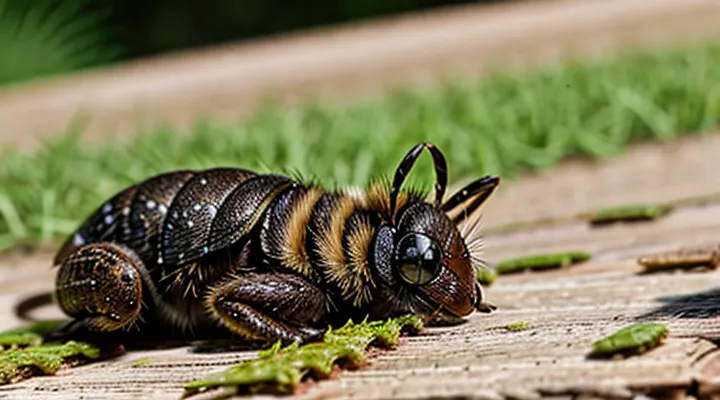
Physical Characteristics
Size and Shape
After a dog has been fed upon, a tick expands dramatically. An unfed adult ixodid measures 2–5 mm in length and 1.5–2 mm in width. Post‑feeding, length typically reaches 8–12 mm, sometimes exceeding 15 mm, while width swells to 5–7 mm. The increase results from the engorgement of the abdomen with blood, often producing a volume rise of 100‑fold.
Shape changes accompany the size growth. The body becomes markedly rounded, with the dorsal surface appearing balloon‑like. The anterior mouthparts remain visible as a small, dark protrusion, but the overall silhouette shifts from an elongated oval to a more spherical profile. The ventral side flattens against the host’s skin, facilitating attachment during the feeding period.
Color Changes
After a canine blood meal, a tick’s external appearance changes markedly. The cuticle, which is initially light‑brown or tan, becomes filled with blood, causing a rapid shift in hue and opacity.
Typical color progression includes:
- Pre‑feeding stage: light brown or tan, semi‑transparent, visible scutum.
- Early engorgement: pinkish‑red tint as hemolymph fills the body; cuticle appears semi‑transparent.
- Full engorgement: deep reddish‑brown or gray‑blue coloration; cuticle becomes opaque and stretched.
- Post‑detachment (drying): darkening to black or charcoal tones as the tick desiccates.
The intensity of the color change correlates with the volume of blood ingested; larger engorged specimens display a more pronounced reddish or grayish hue, while partially fed individuals retain a lighter shade. Species differences affect the exact palette, but the described sequence is consistent across common dog‑infesting ixodid ticks.
Texture and Appearance
After a tick has completed a blood meal on a dog, its body expands dramatically, changing both texture and visual characteristics. The cuticle becomes markedly stretched, giving the abdomen a balloon‑like silhouette that can increase in size three to five times the unfed dimensions.
- Surface texture shifts from smooth and firm to soft and pliable; the engorged cuticle feels moist to the touch.
- Color deepens from light brown or tan to a darker, glossy hue, often resembling a small, semi‑transparent bead.
- The dorsal shield (scutum) remains relatively unchanged in size, creating a noticeable contrast between the rigid, lighter‑colored shield and the swollen, darker posterior.
- Legs appear thinner relative to the enlarged body, sometimes appearing retracted or less prominent.
- The overall shape transforms from a compact, oval form to an elongated, rounded profile with a pronounced, bulging posterior.
These alterations are consistent across common tick species that feed on dogs, providing a reliable visual cue for identification after engorgement.
Comparison: Before and After Feeding
Unfed Tick Appearance
Unfed ticks are small, flat arthropods that resemble tiny brown specks. Their bodies measure 1–3 mm in length and 0.5–1 mm in width, depending on species. The dorsal surface is uniformly brown to reddish‑brown, sometimes with faint patterns, while the ventral side is lighter. Eight legs extend from the anterior region, each ending in a small claw for gripping host fur.
Key morphological features include:
- A hard, shield‑like scutum covering the dorsal surface of adult females; males possess a scutum that does not cover the entire back.
- A pointed capitulum (mouthparts) positioned forward, equipped with chelicerae and a barbed hypostome for anchoring to the host.
- Eyes located on the lateral edges of the scutum in many species, providing limited vision.
When a tick attaches to a dog and engorges, its size expands dramatically, the body becomes rounded and grayish, and the scutum may appear stretched. The contrast between the flat, brown unfed form and the swollen, pale fed form clarifies the visual transformation that occurs after a blood meal.
Visual Differences Post-Feeding
After a dog’s blood meal, a tick undergoes distinct visual changes that facilitate identification of a fed specimen.
The abdomen expands dramatically, becoming spherical or oval and often filling the entire body length. The cuticle stretches, giving the tick a glossy, swollen appearance. Color shifts from light brown or tan to a darker, sometimes reddish hue due to the ingested blood. Legs may appear shorter relative to the enlarged body, and the overall silhouette loses the typical “flat” profile of an unfed tick.
Key visual cues include:
- Abdominal enlargement: markedly distended, dome‑shaped.
- Color alteration: darker, sometimes reddish‑brown.
- Surface texture: smoother, glossier cuticle.
- Proportional changes: legs seem proportionally smaller.
These characteristics reliably differentiate a post‑feeding tick from its unfed counterpart on a canine host.
Why Do Ticks Change So Much?
Blood Meal Absorption
A tick that has completed a blood meal on a dog undergoes rapid morphological transformation driven by the absorption of the host’s plasma and cellular components. The intake of several times its unfed body weight forces the cuticle to expand, the abdomen to swell, and the overall silhouette to become markedly convex.
Visible changes include:
- Size: length may increase from 2–3 mm to 10–12 mm; width expands proportionally, creating a balloon‑like profile.
- Color: cuticle darkens from reddish‑brown to a deep, glossy brown or black, reflecting the concentration of hemoglobin and other pigments.
- Surface texture: the dorsal shield (scutum) remains rigid, while the surrounding integument stretches, producing a smoother, less segmented appearance.
Internally, the midgut epithelium secretes enzymes that break down blood proteins, allowing rapid assimilation of nutrients. Hemolymph pressure rises, stretching the exoskeleton and causing the legs to retract partially, which contributes to the rounded outline. The tick’s metabolic rate spikes, supporting digestion and the synthesis of eggs in engorged females.
Within 24 hours after detachment, the tick reaches maximum engorgement. At this stage, the abdomen occupies the majority of the body mass, and the tick appears as a markedly enlarged, dark, and glossy organism, distinctly different from its flat, pale, unfed counterpart.
Digestive Process
After a canine host supplies a blood meal, the tick’s abdomen expands dramatically as the ingested blood is stored in the midgut. The midgut epithelium secretes digestive enzymes that break down proteins, lipids, and carbohydrates into amino acids, fatty acids, and simple sugars. These nutrients are absorbed directly into the hemolymph, providing energy for egg development and molting.
Key stages of the digestive process:
- Blood intake: The tick draws up to several times its unfed weight in blood, which fills the anterior part of the gut.
- Enzymatic breakdown: Proteases, lipases, and carbohydrases act within the gut lumen, reducing macromolecules to absorbable units.
- Nutrient absorption: Simple molecules cross the gut wall into the hemolymph, raising internal pressure and causing the visible swelling.
- Waste elimination: Undigested remnants are excreted via the anal opening, often leaving a small, darkened patch near the rear.
The resulting appearance is a markedly distended, rounded body with a smooth, glossy surface. The coloration may shift from light brown to a darker, engorged hue due to the blood’s hemoglobin. This physical change directly reflects the internal digestive activity that converts the host’s blood into the resources required for the tick’s reproductive cycle.
Potential Dangers of Engorged Ticks
Disease Transmission
After a dog’s blood meal, a tick swells dramatically, its body often turning pale or reddish‑brown and increasing up to several times its unfed size. The abdomen expands to dominate the organism, while the legs and mouthparts remain proportionally unchanged. This engorged state facilitates pathogen transmission because the tick’s salivary glands are highly active.
Pathogens commonly transferred by an engorged canine tick include:
- Borrelia burgdorferi – agent of Lyme disease; transmitted within 24–48 hours of attachment.
- Ehrlichia canis – causes canine ehrlichiosis; transmission can occur after 6 hours of feeding.
- Anaplasma phagocytophilum – responsible for granulocytic anaplasmosis; infection risk rises after 24 hours.
- Rickettsia rickettsii – Rocky Mountain spotted fever; transmitted rapidly, sometimes within a few hours.
- Babesia spp. – protozoan parasites causing babesiosis; transferred during prolonged feeding.
The enlarged tick’s cuticle becomes thin, allowing easier release of saliva that contains the pathogens. The longer the tick remains attached, the higher the probability of disease transfer. Immediate removal of the engorged tick reduces the chance of infection; careful extraction with fine‑pointed tweezers, grasping the mouthparts close to the skin, prevents mouthpart breakage and subsequent bacterial entry.
Veterinary monitoring after tick removal should include:
- Physical examination for localized inflammation or secondary infection.
- Blood tests targeting the specific agents listed above, especially if the dog shows fever, lethargy, joint pain, or anemia.
- Preventive measures such as regular tick checks, environmental control, and prophylactic medications.
Understanding the visual transformation of a fed tick clarifies why prompt identification and removal are critical components of disease prevention in dogs.
Local Reactions on the Dog
A engorged tick attached to a dog produces a distinct set of cutaneous changes. The skin around the attachment site typically expands as the tick swells, creating a raised, dome‑shaped nodule. The surrounding tissue often turns reddish or pink, indicating localized inflammation. In many cases, the area becomes warm to the touch and may exhibit a thin, clear fluid exudate if the tick’s mouthparts irritate the dermis.
Common local responses include:
- Mild to moderate erythema extending 1–2 cm from the bite point
- Edema that can persist for several days after removal
- Pruritus that intensifies when the dog licks or scratches the area
- Small ulceration or crust formation if the tick’s saliva triggers a hypersensitivity reaction
- Secondary bacterial infection signaled by purulent discharge or increasing pain
Veterinary assessment should focus on the size of the lesion, the presence of necrotic tissue, and any signs of systemic involvement such as fever. Prompt removal of the tick, cleaning of the site with an antiseptic solution, and monitoring for worsening inflammation are essential steps to prevent complications.
What to Do if You Find an Engorged Tick
Safe Removal Techniques
After a tick has drawn blood from a dog, its body expands dramatically. The abdomen becomes rounded, often resembling a small grape, and the cuticle may appear stretched and glossy. The legs may be less visible as the engorged body fills the space between them. Color shifts from brown or reddish‑brown to a darker, sometimes bluish hue, depending on the species and the amount of blood ingested.
Safe removal requires precision to prevent disease transmission and to avoid leaving mouthparts embedded. Follow these steps:
- Use fine‑point tweezers or a specialized tick‑removal tool. Grip the tick as close to the skin as possible, targeting the head where it enters the flesh.
- Apply steady, downward pressure. Pull straight upward with even force; avoid twisting, jerking, or squeezing the body, which can cause the tick’s mouthparts to break off.
- After extraction, inspect the site. If any part remains, repeat the grip and pull technique until the entire tick is removed.
- Disinfect the bite area with an antiseptic solution such as chlorhexidine or povidone‑iodine.
- Place the tick in a sealed container with alcohol or a zip‑lock bag for identification if needed, then discard safely.
- Wash hands thoroughly with soap and water.
If the mouthparts stay embedded despite repeated attempts, consult a veterinarian for professional removal. Monitoring the bite site for redness, swelling, or signs of infection over the next 48 hours is advisable.
Aftercare for Your Dog
After a tick has attached to a dog and finished feeding, it expands dramatically, turning a soft, oval shape into a swollen, balloon‑like body. The abdomen becomes noticeably larger, often reaching the size of a pea or larger, and the color shifts from light brown to a darker, almost black hue. The legs may appear splayed, and the tick’s mouthparts remain embedded in the skin.
Prompt removal is essential. Use fine‑pointed tweezers or a tick‑removal tool to grasp the tick as close to the skin as possible. Pull upward with steady pressure, avoiding twisting or crushing the body. Dispose of the tick in alcohol or a sealed container.
After removal, inspect the bite site. Clean the area with mild antiseptic solution and dry gently. Monitor the skin for redness, swelling, or discharge over the next 48 hours. If inflammation develops or the dog shows signs of lethargy, fever, or loss of appetite, consult a veterinarian.
Regular grooming helps detect ticks early. During walks in grassy or wooded areas, run fingers through the coat and check the ears, neck, and between toes. Apply a veterinarian‑approved tick preventive monthly to reduce future infestations.
If a tick is found attached for more than 24 hours, consider a veterinary examination. Prolonged attachment increases the risk of disease transmission, such as Lyme disease or ehrlichiosis, which may require diagnostic testing and specific treatment.
When to Consult a Veterinarian
After a tick has taken a blood meal from a dog, its body expands dramatically, turning a dull gray‑brown to a swollen, bright reddish‑orange. The abdomen may appear balloon‑like, and the tick often becomes easier to see against the fur because of its size and color change. In some cases the tick’s legs may be obscured as the body fills, making it look like a smooth, fleshy lump.
Veterinary assessment is necessary when any of the following conditions are observed:
- The engorged tick remains attached for more than 24 hours despite attempts to remove it.
- The attachment site shows excessive redness, swelling, or pus.
- The dog exhibits fever, lethargy, loss of appetite, or unexplained weight loss.
- Signs of anemia appear, such as pale gums or persistent weakness.
- Neurological symptoms develop, including tremors, unsteady gait, or facial paralysis.
- The dog has a known history of tick‑borne diseases in the region (e.g., Lyme disease, ehrlichiosis, babesiosis).
Prompt consultation reduces the risk of disease transmission and allows for appropriate treatment, including tick‑preventive medication, wound care, and diagnostic testing for potential infections.
Preventing Tick Infestations
Tick Prevention Products
After a canine has been bitten, the tick expands dramatically. The abdomen swells to a balloon‑like shape, the body darkens to a deep brown or reddish hue, and the legs appear shorter as the engorged organism stretches across the skin. This visual change signals that the parasite has completed a blood meal and may soon detach, potentially transmitting pathogens.
Preventing this stage relies on products that stop attachment or interrupt feeding. Available options include:
- Topical spot‑on treatments – liquid formulations applied along the spine; contain acaricides that repel or kill ticks on contact.
- Oral chewable tablets – systemic medications absorbed into the bloodstream; kill ticks when they bite, preventing engorgement.
- Collars – slow‑release devices that emit repellent vapors; protect the animal for several months without daily application.
- Environmental sprays and powders – chemicals applied to bedding, kennels, or outdoor areas; reduce ambient tick populations, lowering the chance of initial contact.
Each category functions by either deterring attachment, causing rapid paralysis after the tick latches, or destroying the parasite before it can ingest a full meal. Effective control therefore eliminates the visual progression to a swollen tick and the associated health risks.
When selecting a preventive, consider active ingredient, duration of efficacy, species safety, and veterinarian recommendations. Products containing fipronil, permethrin, afoxolaner, or fluralaner have demonstrated consistent results in clinical trials. Regular administration according to label instructions ensures continuous protection, preventing the tick from reaching the engorged stage that follows a dog’s bite.
Environmental Controls
After a canine feed, a tick expands dramatically, reaching up to five times its unfed size. The body becomes round, glossy, and dark brown to reddish, while legs remain visible but shorter relative to the swollen abdomen. This engorged stage persists for several days before the tick detaches.
Detached, engorged ticks fall into the surrounding environment. If conditions remain favorable, they can survive long enough to lay eggs, leading to a new generation of parasites. Consequently, managing the environment where a dog lives directly limits the risk of reinfestation.
- Remove attached ticks promptly; use fine‑point tweezers to grasp the mouthparts and pull straight upward.
- Dispose of removed ticks by submerging them in alcohol or sealing them in a disposable container.
- Wash the dog’s bedding, toys, and grooming tools in hot water (≥60 °C) weekly.
- Vacuum carpets, upholstery, and cracks in flooring; discard vacuum bags or clean canisters immediately.
- Reduce indoor humidity to below 50 % with dehumidifiers; low moisture shortens tick survival time.
- Apply acaricidal sprays or powders to cracks, baseboards, and pet sleeping areas according to label instructions.
- Maintain a cleared perimeter around the home: trim grass, remove leaf litter, and keep shrubs trimmed to below ground‑level.
- Install physical barriers such as fine mesh screens on windows and doors to prevent tick entry.
Regular inspection of the dog’s coat, combined with the above environmental measures, sustains a low‑tick habitat and prevents the establishment of a breeding population after a feeding event.
