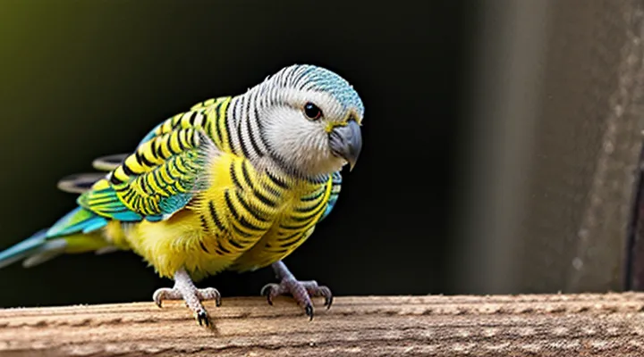
What are Mites?
Types of Mites Affecting Budgerigars
Mite infestations are a common health concern for budgerigars, often visible as tiny, moving specks on skin, feathers, or around the legs. Identification relies on size, coloration, and preferred attachment site, which differ among species.
- Scaly leg mite (Knemidocoptes spp.) – microscopic, elongated, pale‑white bodies; inhabit the scale margins of the legs and feet, causing crusty debris and irritation.
- Feather mite (e.g., Prostigmata spp.) – oval, translucent to light brown; reside within feather barbs, especially on the wings and tail, producing a dull, ragged appearance.
- Northern fowl mite (Ornithonyssus sylviarum) – reddish‑brown, 0.5 mm long; cling to the skin of the vent and abdomen, feeding on blood and leaving small puncture marks.
- Tropical fowl mite (Lipeurus caponis) – similar in size to the northern fowl mite but darker; found on the vent region and surrounding plumage, often causing intense scratching.
- Red mite (Dermanyssus gallinae) – vivid red, 0.4–0.5 mm; temporarily attach to the bird’s skin at night, detach during daylight, leaving tiny hemorrhagic spots.
Recognition of these characteristics enables prompt treatment and reduces the risk of secondary infections. Regular inspection of the legs, vent, and feather clusters is essential for early detection.
How Mites Spread
Mites that infest budgerigars are microscopic, oval‑shaped arthropods measuring 0.2–0.4 mm. Their bodies are semi‑transparent, often appearing as tiny specks moving across the bird’s skin, feather shafts, or around the eyes. Under magnification, the legs are clearly visible, and the abdomen may show a faint reddish hue when engorged with blood.
Mite transmission relies on direct contact and environmental factors. Primary pathways include:
- Physical contact: Mites crawl from one bird to another during mating, feeding, or communal roosting.
- Shared equipment: Cages, perches, feeding bowls, and grooming tools harbor mites that transfer when reused without disinfection.
- Human handling: Hands or clothing that have touched an infested bird can carry mites to a healthy companion.
- Fomites: Dust, litter, and debris in the aviary provide a reservoir where mites survive for several days, facilitating indirect spread.
- Infested wild birds: Contact with wild parakeets or other avian species introduces external mite populations into captive flocks.
Preventive measures focus on breaking these routes. Regular cage cleaning, sterilizing accessories, limiting bird-to-bird contact during outbreaks, and wearing disposable gloves when handling affected individuals reduce infestation risk. Monitoring for the characteristic specks and behavioral signs—such as feather loss or increased preening—allows early detection before widespread transmission occurs.
Visual Identification of Mites on Budgerigars
Common Mite Species and Their Appearance
Mites that infest budgerigars belong to a limited set of species, each with distinctive morphology. Recognizing these features assists in rapid diagnosis and treatment.
-
Proctophyllodes spp. (feather mites) – elongated, oval bodies measuring 0.2–0.4 mm. Color ranges from pale yellow to reddish brown. Legs are short, positioned close to the body, giving a compact appearance. Usually observed on feather shafts and barbs.
-
Dermanyssus gallinae (red poultry mite) – round, soft-bodied, 0.3–0.5 mm in length. Reddish‑brown exoskeleton becomes paler after feeding. Legs are long and slender, ending in hooked claws that enable brief attachment to the bird’s skin.
-
Ornithonyssus sylviarum (northern fowl mite) – oval, 0.25–0.35 mm, dark brown to black. Leg segments are robust, with noticeable spurs on the tarsus. The body surface appears glossy and slightly raised.
-
Knemidokoptes pilae (scaly leg mite) – cylindrical, 0.1–0.2 mm, white to creamy. The cuticle is smooth, and the anterior end is tapered. Infestations produce crusty scales on legs and feet, making the mites visible within the skin layers.
All species are microscopic but can be detected with a hand lens or under a microscope. Their bodies are segmented into gnathosoma (mouthparts) and idiosoma (main body). Coloration often reflects blood ingestion; freshly unfed mites appear lighter, while engorged individuals darken. Size differences are subtle, yet the leg length and body shape provide reliable identification markers for each mite type affecting budgerigars.
Signs and Symptoms of Mite Infestation
Mite infestations on budgerigars produce observable changes that can be identified with careful inspection. The first indicator is a fine, powdery residue on the feathers, especially near the vent, tail, and wing edges. This debris, often mistaken for dandruff, consists of mite excrement and dead skin cells.
Feather damage appears as broken or frayed barbs, giving the plumage a ragged or uneven outline. In severe cases, entire sections of feather may be missing, exposing the skin underneath.
Skin discoloration accompanies the infestation. The affected areas may turn pale, pink, or develop small, darkened spots where mites have burrowed. These lesions can be itchy, prompting the bird to scratch or preen excessively, leading to self‑inflicted wounds.
Behavioral changes provide additional clues. A mite‑infested budgerigar may exhibit reduced activity, loss of appetite, or weight loss due to chronic irritation and stress.
Typical signs and symptoms can be summarized as:
- Fine, white or gray powder on feathers and skin
- Broken, frayed, or missing feather barbs
- Pale or reddened skin with tiny dark spots
- Excessive preening, scratching, or feather plucking
- Lethargy, decreased feed intake, and weight loss
Early detection of these manifestations enables prompt treatment, preventing further health decline and minimizing the risk of spread to other birds.
On the Budgerigar's Body
Mites that infest budgerigars are typically microscopic, measuring 0.2–0.5 mm in length. Their bodies are oval, flattened laterally, and covered with fine, translucent cuticle that often appears pale gray or amber under magnification. Eight legs emerge from the anterior region, giving a spider‑like silhouette. When viewed with a hand lens, the mites may be seen as tiny specks moving among the feathers or crawling on the skin.
On the bird’s body, mites concentrate in areas where skin is thin and feathers are dense:
- Around the cere and beak, where skin folds create a protected micro‑environment.
- Along the vent, under the tail feathers, and between the wing and body junction, where humidity is higher.
- On the legs and toes, especially around the scales and claw bases.
Visible indicators of infestation include:
- Fine, white to yellowish particles resembling dust scattered on plumage.
- Small, irregularly shaped lesions or scaly patches on the skin.
- Feather loss or thinning, particularly in the regions listed above.
- Persistent scratching or rubbing against cage bars.
The combination of microscopic size, oval flattened shape, and pale coloration distinguishes these ectoparasites from other insects that may be present on a budgerigar’s plumage.
On the Budgerigar's Environment
Mites that infest budgerigars are microscopic arachnids, typically 0.1–0.3 mm in length, translucent to pale yellow, and often visible as moving specks on the feather shafts or skin folds. Their presence correlates strongly with specific environmental conditions within the bird’s habitat.
The cage environment influences mite proliferation:
- Temperature: Warm ranges (22‑28 °C) accelerate mite life cycles; cooler temperatures slow reproduction.
- Humidity: Relative humidity above 60 % creates favorable microclimates in perches and nesting material.
- Substrate cleanliness: Accumulated droppings, feather debris, and dust provide shelter and food sources.
- Ventilation: Stagnant air promotes humidity buildup, enhancing mite survival.
- Perch material: Rough or porous surfaces retain organic matter, facilitating colonization.
Management of the habitat reduces infestation risk:
- Maintain temperature within the species’ comfort zone, avoiding prolonged heat spikes.
- Regulate humidity with dehumidifiers or proper ventilation to stay near 50 % relative humidity.
- Perform daily cage cleaning, removing waste and replacing litter weekly.
- Disinfect perches and accessories with a dilute bleach solution (1 % sodium hypochlorite) monthly.
- Rotate and replace nesting material regularly, using low‑dust, washable options.
Observing the bird’s plumage for tiny, mobile specks, especially near the vent, wings, and head, assists early detection. Prompt environmental adjustments combined with targeted acaricidal treatment limit mite populations and protect the bird’s health.
Differentiating Mites from Other Conditions
Mites on a budgerigar are typically visible as tiny, moving specks on the skin and feathers. Adult mites measure 0.2–0.3 mm, appear translucent to reddish‑brown, and may be seen crawling along the vent, around the eyes, or between feather barbs. Infestations often cause intense itching, leading to frequent head shaking, feather loss, and crusty or scaly skin patches.
When evaluating a bird, distinguish mite infestations from other conditions by observing the following characteristics:
- Location of lesions – Mites concentrate around the vent, face, and wing joints; nutritional deficiencies produce generalized feather thinning, while fungal infections create dry, flaky patches on the legs and beak.
- Behavior of the bird – Persistent scratching and head tilting are typical of mite irritation; bacterial infections cause swelling, pus, or discharge without the same level of agitation.
- Skin texture – Mite activity creates fine, gritty debris (often called “mite dirt”) and small puncture points; lice leave larger, moving insects and cause a “caked” appearance on the feathers.
- Feather condition – Mite damage results in broken barbs and feather shafts near the affected area; viral feather‑dystrophy yields malformed feathers throughout the plumage, not limited to specific sites.
- Response to treatment – Topical acaricides rapidly reduce mite movement and skin irritation; antifungal or antibiotic therapies show improvement only when the appropriate pathogen is present.
Accurate identification relies on close visual inspection, possibly aided by a magnifying lens, and may be confirmed by microscopic examination of skin scrapings. Prompt acaricidal treatment prevents secondary infections and restores normal feather growth.
Diagnosing Mite Infestations
Veterinary Examination Techniques
Mites are frequent ectoparasites of budgerigars; accurate detection relies on systematic veterinary examination.
During a routine health check, the examiner observes the bird’s plumage and skin. Adult mites measure 0.2–0.4 mm, appear dark brown to reddish, and move rapidly across feather shafts. Infestation often concentrates around the vent, under the wings, and at the base of the tail, where feather loss, erythema, or crusted debris may be present.
Effective diagnostic steps include:
- Visual inspection: Use a bright, magnified light source to scan ventral and dorsal surfaces, noting any motile organisms or feather damage.
- Feather plucking: Gently remove a few feathers from suspect areas; examine the shafts and follicles for attached mites or eggs.
- Skin scraping: Collect a small sample from irritated skin with a sterile scalpel; place the material on a glass slide for microscopic evaluation.
- Microscopy: Apply a drop of mineral oil or saline to the slide; identify mites by their rounded bodies, short legs, and characteristic dorsal shields.
- Dermatoscopy (optional): Employ a handheld dermatoscope to visualize mites in situ without removing feathers, useful for minimally invasive assessment.
Microscopic morphology differentiates common species: Knemidokoptes spp. display a rounded opisthosomal shield, while Lorryia spp. possess elongated bodies and prominent setae. Recognizing these features guides targeted treatment.
Routine examinations at monthly intervals, especially during breeding season, reduce parasite load and prevent secondary infections. Early identification through the outlined techniques ensures prompt therapeutic intervention and maintains avian health.
At-Home Observation Methods
Mite infestation on a budgerigar can be detected through direct visual inspection of the bird’s skin, feathers, and vent area. The parasites are microscopic to the naked eye, but adult individuals appear as tiny, oval-shaped bodies, usually 0.2–0.4 mm long, with a pale or translucent coloration that may be faintly visible against the feather shafts. In heavy infestations, clusters of mites create a dusty or powdery coating, especially around the wings, tail, and under the beak.
Effective observation at home relies on a systematic approach:
- Secure the bird in a calm, restrained position using a towel or a specialized handling cage.
- Illuminate the plumage with a bright, focused light source; a handheld LED flashlight or a desk lamp with a white filter works well.
- Examine the feather base and skin folds for moving specks or small white dots; use a low‑magnification loupe (10×–15×) to enhance visibility.
- Gently part the feathers along the wing edge, tail, and vent, looking for mite activity or a fine, powdery residue.
- Record any observations with a smartphone camera set to macro mode for later comparison.
Additional tools improve detection accuracy:
- A portable digital microscope provides 20×–40× magnification, revealing the mite’s legs and body segmentation.
- A fine‑toothed comb can separate feathers without damaging the skin, exposing hidden parasites.
- A transparent sheet placed under the bird catches falling debris, allowing inspection of dropped mite bodies.
Handling procedures must minimize stress: keep the environment quiet, maintain a stable temperature, and limit examination time to a few minutes per session. Repeating the inspection weekly during breeding season or after introducing new birds helps catch infestations early, preventing secondary skin irritation and feather damage.
Treatment and Prevention
Effective Treatment Options
Mites on budgerigars appear as tiny, moving specks on the skin, often visible as fine white or yellowish dots near the beak, eyes, and feather bases. Infestations cause itching, feather loss, and skin irritation, requiring prompt intervention.
Effective treatment options include:
- Topical ivermectin or selamectin applied according to the label dosage; repeated after 7‑10 days to eliminate emerging larvae.
- Moxidectin oral solution, administered under veterinary guidance; dose schedule mirrors that of ivermectin.
- Pyrethrin‑based dusts (e.g., permethrin) applied to the bird’s plumage; thorough coverage of the head, neck, and vent is essential, followed by a second application after 5 days.
- Diatomaceous earth sprinkled in the cage; replace daily and ensure the bird does not ingest large amounts.
- Environmental sanitation: wash all perches, toys, and nesting material in hot water; vacuum cages and surrounding areas; treat the room with an approved acaricide spray.
Monitoring after treatment involves daily inspection for live mites and assessment of skin condition. If symptoms persist beyond two weeks, a veterinarian should reassess the case and consider alternative systemic medications or combination therapy.
Preventing Future Infestations
Mites on budgerigars appear as tiny, elongated bodies about 0.2 mm long, translucent to pale yellow, often visible as moving specks near the beak, eyes, and feather bases. Their legs are short, and they may leave fine, white debris (fecal pellets) on the bird’s plumage.
Preventing future infestations requires consistent environmental and bird‑care practices:
- Isolate new or returning birds for at least 14 days; conduct a full visual inspection before integration.
- Clean cages, perches, and accessories with hot, soapy water weekly; follow with a disinfectant approved for avian use.
- Replace bedding and substrate regularly; avoid reusable fabric liners that retain moisture.
- Maintain low humidity (40‑50 %) and good ventilation to discourage mite reproduction.
- Apply a dust‑free, veterinary‑approved miticide to the bird’s environment after any confirmed case; repeat according to product guidelines.
- Schedule routine health checks, including microscopic feather examinations, at least quarterly.
- Store feed in sealed containers; discard any food that has been exposed to pests or moisture.
Implementing these steps creates a hostile environment for mites, reducing the likelihood of re‑colonization and protecting the health of budgerigars.
Cage Hygiene
Mites on a budgerigar are most often visible as tiny, moving specks on the skin or feathers, sometimes resembling dust or small black dots. Their presence indicates a compromised environment, making regular cage sanitation essential for early detection and prevention.
Effective cage hygiene includes:
- Daily removal of uneaten seed, fresh vegetable scraps, and droppings.
- Weekly full‑cage cleaning: disassemble perches and toys, scrub with a mild, bird‑safe detergent, rinse thoroughly, and allow to dry before reassembly.
- Monthly deep sanitation: soak all accessories in a diluted bleach solution (1 part bleach to 32 parts water), rinse repeatedly, and disinfect the cage frame with an approved avian sanitizer.
- Routine replacement of substrate or liner material to eliminate residual eggs and larvae.
- Periodic inspection of the bird’s plumage and skin, focusing on the vent, wing folds, and neck for the characteristic specks associated with mite activity.
Maintaining these practices reduces the likelihood of infestation, supports the bird’s health, and facilitates prompt identification of any mite‑related symptoms.
Quarantining New Birds
Quarantining newly acquired birds prevents the introduction of parasites, including feather mites, into established flocks. A budgerigar that has not undergone isolation may carry microscopic arthropods that are difficult to detect without close examination.
Mite identification relies on visual cues. Adult mites appear as tiny, elongated bodies measuring 0.2–0.5 mm, often translucent or pale yellow. Under magnification, their legs are fine and jointed, and the abdomen may show a faint oval shape. On a budgerigar, mites cluster around the vent, under the wings, and at the base of tail feathers. Affected birds may exhibit feather loss, scaly skin, or a dusty residue known as “mite debris.”
Effective quarantine follows a structured protocol:
- Separate the new bird in a dedicated cage away from resident birds.
- Maintain temperature and humidity within species‑appropriate ranges.
- Conduct daily visual inspections of skin and feathers, using a magnifying lens.
- Clean the cage, perches, and accessories with a disinfectant safe for avian use.
- Perform a 30‑day observation period; extend if any signs of infestation appear.
- If mites are detected, apply a veterinarian‑approved acaricide and repeat examinations for two weeks after treatment.
Documentation of each step ensures traceability and supports timely intervention. Consistent quarantine reduces the risk of mite transmission and safeguards the health of the entire aviary.
Regular Health Checks
Regular health examinations are essential for early detection of ectoparasites on budgerigars. During each check, the veterinarian or experienced keeper inspects the bird’s plumage, skin, and beak for the characteristic signs of mite infestation.
Key observations include:
- Small, translucent or reddish specks moving across the feathers, especially near the vent and wing joints.
- Fine, thread‑like lines or webbing that indicate mite tunnels in the skin.
- Crusted or scaly patches on the face, neck, or tail area.
- Excessive preening, feather loss, or a ragged appearance of the plumage.
A systematic approach—visual inspection, feather brushing, and, when necessary, microscopic slide preparation—provides reliable identification. Recording findings at each visit creates a health history that simplifies treatment decisions and prevents severe infestations.
