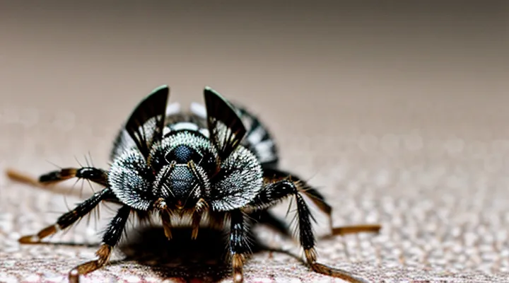
What is a Clothes Louse?
Distinguishing Clothes Lice from Other Parasites
Clothing lice are small, wingless insects that live in fabrics and feed on human blood. Adults measure 2–4 mm, have a flattened, elongated body, and display a uniform gray‑brown coloration. Their heads are concealed beneath a short, forward‑projecting “headplate,” and each segment bears six legs with clawed tarsi. Nymphs resemble miniature adults, lacking fully developed wings and genitalia, and are semi‑transparent, allowing internal structures to be faintly visible in close‑up photographs.
Key visual differences from other human parasites include:
- Head lice (Pediculus humanus capitis): larger body (3–4 mm), prominent eyes, and a visible “crab‑like” head with a broader shape; typically found on the scalp, not in clothing.
- Pubic lice (Pthirus pubis): broader, crab‑shaped body, larger claws adapted for coarse hair; coloration is darker, often reddish‑brown.
- Bed bugs (Cimex lectularius): oval, flattened, reddish‑brown after feeding; visible antennae and distinct abdominal segments; usually located in mattress seams, not fibers.
- Fleas (Siphonaptera): laterally compressed, strong jumping legs, dark brown to black coloration; often seen on pets rather than in garments.
- Mites (e.g., Dermanyssus gallinae): microscopic (0.3–0.5 mm), translucent, with only two pairs of legs visible in photos; lack the robust body of lice.
Photographic guidelines for accurate identification:
- Use macro or close‑up lenses to capture details of the headplate, leg claws, and body segmentation.
- Illuminate specimens with diffuse light to reduce shadows that obscure translucent nymphal structures.
- Include a scale reference (e.g., a ruler millimeter mark) to convey size.
- Capture multiple angles—dorsal, lateral, and ventral—to display the full morphology.
By focusing on size, body shape, leg morphology, and typical habitat, photographs can reliably separate clothing lice from other ectoparasites.
Life Cycle of the Clothes Louse
The clothing louse (Liposcelis spp.) progresses through a simple, incomplete metamorphosis that can be identified in photographic records. Eggs appear as minute, oval, translucent capsules measuring 0.3–0.5 mm; they cling to fabric fibers and are often invisible without magnification. First‑instar nymphs emerge as pale, legged larvae, slightly larger than the eggs, with a soft, whitish body and visible antennae. Subsequent molts produce second‑ and third‑instar nymphs, each stage showing incremental darkening of the cuticle and development of the characteristic flattened, oval shape. The final molt yields the adult, a wingless, brownish‑gray insect about 1–2 mm long, with a distinct head capsule, three long cerci, and reduced eyes. Adults are the most frequently captured in photographs because their size and coloration contrast with textiles.
Key visual cues across the cycle:
- Eggs: transparent, clustered in seams; often require focus stacking to resolve.
- Early nymphs: light coloration, visible setae; appear as tiny specks on high‑resolution macro images.
- Later nymphs: darker hue, more defined body segmentation; distinguishable from debris by consistent shape.
- Adults: uniform brownish tone, flattened dorsoventral profile; recognizable by three posterior cerci and lack of wings.
Understanding these visual markers enables accurate identification of each developmental stage in photographic evidence, facilitating monitoring and control efforts.
Physical Characteristics of the Clothes Louse
Size and Shape
In photographic documentation, a clothing louse measures approximately 2 mm to 5 mm in total length. The body is flattened laterally, giving a low profile that aids concealment among fabrics. Width ranges from 0.5 mm at the thoracic region to 1 mm near the abdomen, creating an oval silhouette when viewed from above. The head is reduced, forming a blunt, rounded cap that merges smoothly with the thorax. Legs are short, positioned near the anterior third, and do not protrude beyond the body outline. Antennae are slender, typically 0.3 mm long, and lie flat against the dorsal surface.
Key dimensional characteristics:
- Length: 2–5 mm
- Maximum width: 0.8–1 mm
- Thoracic width: ~0.5 mm
- Antenna length: ~0.3 mm
- Leg length: <0.4 mm
Overall shape: dorsoventrally flattened, oval to slightly elongated, with a tapered posterior that rounds into a smooth edge. This morphology is consistently observable across macro‑photographs taken with standard magnification.
Coloration and Transparency
Clothing lice (Pediculus humanus corporis) appear as small, wingless insects measuring 1–4 mm. In photographic documentation, their coloration ranges from pale yellow‑brown to reddish‑brown, depending on the age of the specimen and the degree of engorgement after feeding. Newly emerged nymphs exhibit a uniform, translucent hue that allows internal structures to be faintly visible, while mature adults display a more opaque, darker exoskeleton.
Key visual cues related to color and transparency include:
- Translucent cuticle in early stages: Light passes through the thin body wall, revealing the digestive tract and hemolymph as faint pink or greenish tones.
- Progressive darkening after blood meals: The abdomen becomes markedly opaque, often taking on a deep reddish shade due to ingested hemoglobin.
- Glossy surface on adult exoskeleton: Microscopic reflections create a slight sheen, distinguishing the insect from surrounding fabric fibers.
- Contrast with textile background: Lice typically stand out against light-colored fabrics because their natural pigments absorb more light, whereas on dark fabrics they may blend, requiring close‑up focus or angled lighting to capture detail.
Accurate identification in images relies on recognizing these coloration patterns and the shift from translucency to opacity as the insect matures and feeds.
Body Segments and Appendages
In photographs, a clothing‑louse exhibits a distinct three‑part body plan. The anterior region, or head, is small, rounded, and bears a pair of short, filiform antennae that extend forward and are often visible as thin, dark lines. Adjacent to the head, the thorax consists of three fused segments, each bearing a pair of legs. The legs are slender, jointed, and end in tiny claws adapted for gripping fabric fibers; they appear as pale, hair‑like structures in close‑up images.
The posterior region, the abdomen, is elongated and segmented, typically displaying six visible divisions. The abdominal segments are separated by faint transverse sutures, giving the body a slightly striped appearance. Each segment may show a subtle increase in width toward the middle, creating a gentle hourglass silhouette. The ventral side bears tiny, hair‑like setae that can catch light and become discernible in high‑resolution photos.
Key appendages observable in clear images include:
- Antennae: two, slender, dark, extending from the head.
- Legs: six, thin, jointed, ending in microscopic claws.
- Mouthparts: concealed beneath the head capsule, appearing as a small, rounded aperture when the louse is viewed from the side.
- Setal brushes: short, fine hairs covering the thorax and abdomen, providing texture contrast against the louse’s reddish‑brown cuticle.
Overall, photographic depictions reveal a compact, segmented insect with a brownish‑gray exoskeleton, clearly defined head, thorax, abdomen, and the characteristic appendages that enable it to navigate and remain attached to clothing fibers.
Head Structure
The head of a clothing‑infesting louse is a compact, hardened capsule measuring 0.3–0.5 mm in length. The dorsal surface bears a pair of small, oval compound eyes positioned laterally, providing limited visual perception. Two short, segmented antennae arise from the anterior margin; each antenna consists of three flagellomeres ending in sensory cones that detect chemical cues from fabric fibers.
Mouthparts are of the piercing‑sucking type. A slender, stylet‑like labrum penetrates the epidermis of the host material, while the mandibles and maxillae form a tube through which digestive enzymes are injected. The labium, folded beneath the head, guides the stylet during feeding.
The head is reinforced by a sclerotized exoskeleton, which includes a well‑defined occipital plate protecting the posterior region. Musculature attached to the head capsule controls antennae movement and mouthpart operation.
Key components of the head structure:
- Compound eyes (lateral, oval)
- Antennae (three‑segmented, sensory cones)
- Sclerotized exoskeleton with occipital plate
- Piercing‑sucking mouthparts (labrum, mandibles, maxillae, labium)
- Associated musculature for feeding and sensory functions
These anatomical features enable the louse to locate, attach to, and extract nutrients from textile fibers, facilitating its survival in indoor environments.
Thorax and Legs
A clothing louse displays a compact thorax that bridges the head and abdomen. The thorax consists of three fused segments, each bearing a pair of legs. Dorsally, the surface appears smooth, often covered with fine scales that give a slightly iridescent sheen in photographs. Lateral margins may show faint ridges where the legs attach, creating a clear demarcation between thoracic plates.
The six legs are uniformly short and robust, adapted for clinging to fabric fibers. Each leg has three main segments—coxa, femur, and tibia—ending in a pair of curved tarsal claws. The claws are visible as tiny, hook‑shaped extensions that grasp fibers, a characteristic feature in close‑up images. Leg joints are clearly articulated, allowing the louse to maneuver through clothing seams.
- Three pairs of legs, one per thoracic segment
- Jointed segments: coxa, femur, tibia, tarsus
- Curved tarsal claws for fiber attachment
- Fine scales on thorax enhance reflectivity in photos
These morphological details enable reliable identification of the thorax and legs when examining photographic records of clothing lice.
Abdomen and Spiracles
The abdomen of a clothing louse appears as a tapered, cylindrical segment extending behind the thorax. It is typically segmented into 7–9 visible rings, each delineated by subtle ridges. In photographs the cuticle exhibits a pale, translucent to light brown hue, allowing internal contents to be faintly discernible. The posterior end often narrows to a pointed tip, occasionally ending in a pair of short, bristle‑like setae.
Spiracles are the only external respiratory openings and are positioned laterally on the abdomen. Most images show a pair of small, oval openings on each of the middle abdominal segments (usually segments 3–5). The openings are surrounded by a thin, sclerotized rim that contrasts slightly with the surrounding cuticle. In high‑resolution macro photos the spiracles may appear as tiny, darkened pores, sometimes highlighted by a faint halo caused by light reflection.
Key visual identifiers:
- Cylindrical abdomen with 7–9 faintly ridged segments.
- Light brown to translucent coloration.
- Lateral, oval spiracles on mid‑abdominal segments, bordered by a sclerotized ring.
- Posterior taper ending in a pointed tip, occasionally with short setae.
Clothes Louse Eggs: Nits
Appearance of Nits on Clothing
Nits attached to garments appear as tiny, oval or elongated bodies measuring 0.5–1 mm in length. Their color ranges from translucent white to light brown, becoming darker after feeding. The shells are smooth, slightly glossy, and often cling to the fabric’s fibers at a shallow angle, creating a “pearl‑like” line along seams, cuffs, or the inner collar.
Key visual identifiers include:
- Size comparable to a pinhead, clearly smaller than lint or fabric fibers.
- Uniform shape: consistently oval, without the irregular edges of fabric fuzz.
- Attachment pattern: clusters clustered near seams or hair‑rich areas, rarely scattered randomly.
- Slight opacity: semi‑transparent when viewed against a contrasting background, revealing a faint interior.
Photographic evidence typically shows nits as a faint, linear series of tiny beads embedded in the weave, distinguishable from dust by their regular spacing and smooth surface. Proper lighting—diffuse, side‑illumination—highlights their three‑dimensional form, allowing accurate identification.
Location of Nits
Nits are the eggs of the clothing‑dwelling louse, firmly cemented to fabric fibers. In photographic evidence they appear as tiny, oval, translucent or brownish specks that remain attached even after the garment is washed.
Typical attachment sites include:
- seams and stitching lines, where fibers overlap
- cuffs, collars, and waistbands, areas that experience repeated friction
- folds and creases, especially in trousers and shirts
- under armpit panels and behind the neck, where clothing is snug
- pockets and interior linings, where moisture may accumulate
Photographs reveal nits clustered along these zones, often in rows or scattered patterns. The eggs are slightly raised from the surface, casting a faint shadow that distinguishes them from lint. Close‑up images show the cemented base of the nit attached to a single fiber, confirming its location on the garment rather than on human hair.
Visual Differences: Nymphs vs. Adults
In photographic identification, nymphs and adults of the clothing louse can be separated by size, body shape, and anatomical details.
- Size: Nymphs measure 1–2 mm, roughly half the length of mature specimens that reach 2–4 mm.
- Body shape: Nymphs exhibit a more rounded abdomen and lack the pronounced elongation seen in adults.
- Antennae: Nymphal antennae are shorter, with fewer visible segments; adult antennae display three clearly defined segments.
- Legs: Nymph legs are proportionally shorter and lack the robust setae that characterize adult legs.
- Coloration: Nymphs often appear lighter, ranging from pale yellow to light brown, while adults tend toward a darker brown or reddish hue.
- Wing pads: Adult males may show rudimentary wing pads; nymphs never develop these structures.
These visual cues enable reliable differentiation when reviewing photographs of clothing lice.
How to Confirm a Clothes Louse Infestation Visually
Where to Look for Clothes Lice
To locate clothing lice in photographic material, focus on areas where the insects naturally reside and where visual cues are most distinct.
- Seams and stitching lines, especially along side panels and back panels, often trap small insects.
- Cuffs, hem edges, and collar bands provide dark, confined spaces that enhance contrast against the fabric.
- Pocket interiors and zipper pulls create shadowed zones that can conceal the lice’s bodies.
- Under folds, pleats, and layered garments, where fabric overlaps, the insects may be hidden but visible when the layer is lifted.
- Buttons, snaps, and belt loops present rigid structures that can hold the lice in place, making them easier to spot.
In photographs, the lice appear as tiny, elongated bodies ranging from 2 mm to 4 mm, typically gray‑brown to reddish in color. Their heads are slightly narrower than the thorax, and legs are visible as short, jointed extensions. When captured in high‑resolution images, the insects often display a glossy exoskeleton that reflects light, creating a subtle sheen distinct from the matte texture of most fabrics. Close‑up shots with adequate depth of field reveal the characteristic segmentation, allowing reliable identification without magnification tools.
Common Misidentifications
Photos of clothing lice often lead to confusion because the insects are small, translucent, and frequently captured against patterned fabrics. The most frequent mistakes involve mixing these parasites with harmless or unrelated objects.
- Dust particles or lint: Both appear as light‑colored specks, but lice have distinct, segmented bodies and jointed legs, while dust lacks any anatomical structure.
- Moth larvae or caterpillars: Larvae are larger, cylindrical, and possess a smooth, often fuzzy surface; lice are flattened, oval, and display clearly defined legs and antennae.
- Flesh flies or other dipterans: Flies have wings and a robust thorax; lice are wingless and their bodies are compressed to fit between fabric fibers.
- Dead skin cells (dandruff) or hair fragments: These are irregularly shaped and lack the repetitive segmentation seen in lice.
- Fabric fibers or loose threads: Fibers are uniform in thickness and texture, whereas lice show a segmented abdomen and visible head capsule.
Accurate identification relies on recognizing three key visual cues: a flattened, oval body; six short, clawed legs positioned near the rear; and a head with short antennae. When these features are visible in a photograph, the organism can be confidently classified as a clothing louse rather than a non‑parasitic element.
Impact of Clothes Lice: Visual Evidence
Skin Reactions and Bite Marks
Clothing‑dwelling lice produce localized skin irritation that appears as small, red papules. Bites are usually 1–2 mm in diameter, surrounded by a faint halo of erythema. Lesions often cluster near seams, cuffs, or waistbands where the insects feed, creating linear or staggered patterns that follow the garment’s contours.
Typical manifestations include:
- Pruritic welts that develop within hours of exposure
- Slight swelling that may persist for 24–48 hours
- Secondary excoriation from scratching, leading to crusted lesions
- Rarely, brief urticarial patches extending beyond the bite site
The reaction timeline is predictable: initial redness appears within 30 minutes, peak itching occurs after 2–4 hours, and resolution begins after 3–5 days if re‑infestation is prevented. Persistent or spreading lesions suggest secondary infection and require medical assessment.
Distinguishing clothing‑lice bites from other arthropod feeds relies on distribution and context. Flea bites are typically scattered on lower extremities; bed‑bug marks show a “breakfast‑lunch‑dinner” line of three lesions. In contrast, clothing‑lice bites align with garment seams and lack the grouped “cannonball” arrangement seen with other pests. Recognizing these patterns aids accurate identification and appropriate treatment.
Fecal Matter and Shed Exoskeletons
Fecal deposits and molted skins provide the most reliable visual cues for confirming the presence of clothing lice in photographic evidence.
Fecal matter appears as tiny, dark specks or smears on fabric fibers, seams, and the interior of pockets. In close‑up images the particles are usually black or dark brown, often irregularly shaped, and may accumulate in folds or along stitching lines where the insects rest and feed. The deposits are typically glossy under direct lighting, contrasting sharply with the surrounding textile texture.
Shed exoskeletons, or nymphal skins, are translucent to pale yellow‑brown and retain the characteristic segmented outline of the louse. In photographs they manifest as thin, paper‑like shells that cling to fibers, especially near seams, cuffs, and collar edges. The exuviae often display the distinct head capsule and thoracic plates, allowing size estimation; adult specimens range from 2 to 4 mm, while nymphal skins are proportionally smaller.
Key visual identifiers:
- Dark, glossy specks on fabric surfaces.
- Accumulation of specks in creases, seams, and pockets.
- Thin, translucent shells with segmented outlines.
- Exuviae adhering to fibers near garment edges and seams.
