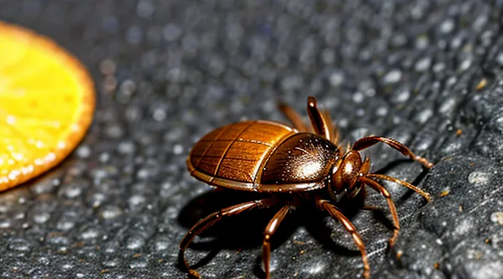
Understanding the Tick's Anatomy
The Hypostome's Role
The hypostome is the central feeding apparatus of a tick, located on the ventral side of the mouthparts. It consists of a hardened plate bearing backward‑pointing barbs that embed into the host’s epidermis. Cement glands secrete a proteinaceous substance that hardens around the hypostome, creating a firm attachment.
During extraction, the orientation of the hypostome determines the mechanical advantage of rotating the tick. The barbs are aligned so that a clockwise twist (when the tick’s head points toward the host) tends to disengage them, whereas a counter‑clockwise motion pushes the barbs deeper into the tissue. Consequently, rotating the tick in the direction that follows the natural curvature of the barbs reduces the risk of tearing the mouthparts.
- Grasp the tick as close to the skin as possible with fine‑pointed tweezers.
- Apply steady, gentle pressure while turning the tick clockwise (if the mouthparts point upward) or in the direction that aligns with the hypostome’s curvature.
- Avoid jerking motions; maintain a smooth rotation until the tick releases.
Correct rotation minimizes hypostome fragmentation, limits the chance of pathogen entry, and prevents residual mouthparts from remaining embedded in the skin.
Barbs and Attachment
Ticks attach by inserting their mouthparts into the host’s skin. The hypostome, a barbed structure, secures the parasite while the surrounding cement glands produce a proteinaceous glue that hardens around the feeding site. Barbs face outward in a spiral pattern, allowing the tick to maintain a firm grip as it expands during engorgement.
Because the barbs are arranged radially, a rotational force applied in either direction can engage the same set of projections. The cement layer, however, adheres uniformly to the epidermis, making the direction of twist irrelevant to the detachment process. The primary risk of rotation is that the mouthparts may break off, leaving portions embedded in the tissue and increasing the likelihood of infection.
Effective removal therefore relies on a steady, upward traction that separates the cement and disengages the barbs without shear. If a slight twist is necessary to loosen the cement, the motion should be gentle and brief; the direction—clockwise or counter‑clockwise—does not affect the outcome.
Key points:
- Barbs are radially symmetric; direction of rotation does not alter their engagement.
- Cement glue bonds uniformly; rotational force offers no advantage in breaking this bond.
- Gentle, vertical pull is the recommended method to avoid mouthpart fragmentation.
- Minimal twisting, if used, should be smooth and brief; orientation is inconsequential.
Recommended Tick Removal Techniques
Straight Pull Method
When a tick is attached, the decision about rotating it clockwise or counter‑clockwise becomes irrelevant if the Straight Pull Method is applied. This technique removes the parasite without any twisting motion, thereby minimizing the chance of mouthpart fragmentation.
The method consists of three precise actions:
- Grip the tick as close to the skin as possible with fine‑point tweezers.
- Apply steady, upward traction directly away from the host.
- Release the tick once it separates; do not rotate, jerk, or squeeze the body.
Avoiding rotation prevents the mandibles from breaking off inside the skin, which can introduce pathogens. The constant, linear force also reduces tissue trauma compared with a twisting maneuver.
For optimal results, disinfect the tweezer tips before use, clean the bite site after removal, and store the tick in a sealed container if laboratory identification is required. The Straight Pull Method thus resolves the directional dilemma by eliminating rotation entirely.
Using Fine-Tipped Tweezers
Fine‑tipped tweezers provide the most reliable grip for tick extraction. Grasp the tick as close to the skin as possible, avoiding compression of the body. A secure hold prevents the mouthparts from snapping off and remaining embedded.
Position the tweezers so the jaws align with the tick’s head. Apply steady, gentle pressure to lift the parasite from the skin surface. Once the tick is elevated, rotate the instrument in a smooth motion. A clockwise turn or a counter‑clockwise turn is effective; the critical factor is maintaining continuous pressure without jerking.
Key steps:
- Pinch the tick’s head with fine‑tipped tweezers, close to the skin.
- Pull upward with constant force; do not squeeze the body.
- Rotate the tweezers smoothly (either direction) while maintaining traction.
- Release the tick once the mouthparts disengage, then disinfect the bite area.
Avoid twisting the tick after it has been lifted, as this increases the risk of mouthpart breakage. Complete removal in a single, controlled motion to minimize tissue irritation and infection risk.
Tools for Tick Removal
Effective tick extraction depends on using instruments that grip the parasite close to the skin without compressing its abdomen. Fine‑point tweezers, preferably stainless‑steel, provide the necessary precision. Their narrow tips allow the operator to hook the tick’s head or mouthparts and pull straight upward. Some models feature serrated edges that improve grip on slippery exoskeletons.
Tick‑removal devices, often marketed as “tick key” or “tick remover,” consist of a shallow, U‑shaped notch. The tick is positioned in the notch and a gentle downward pressure releases the mouthparts. These tools minimize the need for twisting, reducing the risk of mouthpart retention.
Specialized forceps, such as curved‑tip or angled forceps, enable access to ticks lodged in hard‑to‑reach areas like the scalp or interdigital spaces. Their design permits a controlled, vertical pull while maintaining a secure hold.
When employing any of these tools, the following steps ensure consistent results:
- Disinfect the instrument with isopropyl alcohol before use.
- Grasp the tick as close to the skin as possible.
- Apply steady, upward traction; avoid squeezing the body.
- If rotation is required, turn the tick in the direction that aligns with the natural curvature of its mouthparts, typically counter‑clockwise for most species.
- Inspect the bite site for retained fragments; remove any visible remnants with the same tool.
- Clean the area with antiseptic and monitor for signs of infection.
Choosing the appropriate instrument eliminates the need for excessive twisting, thereby decreasing the likelihood of incomplete removal and subsequent disease transmission.
Tick Removal Cards
Tick removal cards are compact reference tools that convey evidence‑based guidance for extracting attached ticks safely. They combine visual cues with concise instructions, allowing laypersons and clinicians to act quickly without consulting lengthy manuals.
Research indicates that rotating a tick counter‑clockwise aligns with the natural orientation of the mandibles, minimizing the risk of mouthpart breakage. Clockwise rotation tends to compress the mouthparts against the skin, increasing the chance of incomplete removal and subsequent infection. The cards therefore emphasize a gentle, steady counter‑clockwise twist as the preferred technique.
Typical card layout includes:
- A clear illustration of a tick’s anatomy, highlighting the mouthparts.
- A step‑by‑step sequence:
- A brief warning about avoiding squeezing the body, which can force pathogens into the host.
By presenting the direction recommendation and procedural steps on a single sheet, tick removal cards reduce uncertainty, promote consistent practice, and help prevent complications associated with improper tick extraction.
Tick Removal Hooks
Tick removal hooks are specialized instruments designed to grasp the tick’s mouthparts without compressing the abdomen. The tip is typically a narrow, curved metal or plastic prong that slides beneath the tick’s capitulum. By pulling straight upward, the hook disengages the hypostome from the skin, minimizing the risk of leaving mouthparts embedded.
When using a hook, the direction of rotation is irrelevant because the device does not rely on twisting. Instead, the recommended technique involves:
- Positioning the hook as close to the skin as possible, directly under the mouthparts.
- Applying steady, vertical traction until the tick releases.
- Inspecting the attachment site for any retained fragments and cleaning with antiseptic.
Hooks are preferred over tweezers when the tick is attached in narrow or delicate areas, such as the eyelid or scalp, where squeezing could force regurgitated fluids into the wound. Their design also reduces the likelihood of the tick’s body rupturing, which can increase the chance of pathogen transmission.
For optimal results, select a hook with a fine, smooth tip, sterilize before each use, and store in a sealed container to maintain cleanliness. Regularly replace worn or damaged hooks to ensure precise engagement with the tick’s mouthparts.
Why Twisting is Not Recommended
Risk of Leaving Mouthparts
Leaving portions of a tick’s mouthparts embedded in the skin can trigger local inflammation, infection, and prolonged irritation. When the body attempts to expel retained fragments, a small ulcer may develop, increasing the chance of secondary bacterial entry. In some cases, the remaining hypostome can act as a nidus for tick‑borne pathogens, allowing transmission of disease agents even after the visible part of the tick has been removed.
The likelihood of fragment retention rises when excessive force is applied to the tick’s body rather than to its head. Gripping the tick’s abdomen and pulling or twisting sharply can crush the mouthparts, causing them to break off. A controlled, steady motion that focuses on the mouthparts reduces this risk.
Key points to minimize retained fragments:
- Use fine‑point tweezers to grasp the tick as close to the skin as possible.
- Apply steady, gentle traction without squeezing the body.
- Rotate the tick minimally, just enough to disengage the hypostome if resistance is felt.
- Inspect the bite site after removal; any visible fragments warrant immediate medical attention.
Prompt removal with proper technique lowers the probability of lingering mouthparts, thereby decreasing the potential for local tissue damage and pathogen transmission.
Potential for Infection
Removing a tick with any technique that squeezes or twists the mouthparts can increase the likelihood of pathogen transmission. Pressure on the engorged foregut may force bacteria, viruses, or protozoa into the host’s bloodstream, and ruptured parts left in the skin become a focus for secondary bacterial infection.
- Bacterial superinfection at the bite site occurs when tick remnants or damaged tissue harbor skin flora such as Staphylococcus aureus or Streptococcus pyogenes.
- Transmission of tick‑borne pathogens (e.g., Borrelia burgdorferi, Anaplasma phagocytophilum, Rickettsia spp.) is facilitated by prolonged attachment and by mechanical disruption of the tick’s salivary glands.
- Local inflammation and necrosis can develop if the tick’s hypostome is broken, creating a portal for opportunistic microbes.
The safest removal method employs fine‑pointed tweezers to grasp the tick as close to the skin as possible and to exert steady, upward traction. Rotational movement—whether clockwise or counter‑clockwise—should be avoided because it increases the chance of mouthpart breakage and subsequent infection. Immediate cleaning of the bite area with antiseptic and monitoring for signs of infection further reduce risk.
Stressing the Tick
Applying force to a tick before removal does not improve outcomes. The tick’s mouthparts embed in skin tissue; additional compression increases the risk of breaking the anchoring barbs. The preferred method relies on a steady, vertical lift without rotation.
- Grasp the tick as close to the skin as possible with fine‑point tweezers.
- Maintain a firm, straight line of pull.
- Avoid any twisting motion, whether to the right or to the left.
- Continue pulling until the entire organism separates from the host.
If a twist is introduced, the direction—clockwise or counter‑clockwise—has no advantage and may cause the hypostome to fracture, leaving remnants in the wound. Consistent upward traction eliminates the need for rotational stress and reduces the likelihood of infection.
Post-Removal Care
Cleaning the Bite Area
After a tick is extracted, the skin surrounding the attachment point requires immediate cleaning to lower the chance of bacterial invasion. The surface should be washed with plain soap and lukewarm water, scrubbing gently to remove any residual saliva or debris. Rinse thoroughly, then apply a broad‑spectrum antiseptic such as povidone‑iodine or chlorhexidine; allow it to dry before covering the site.
- Use disposable gloves to avoid contaminating the wound.
- Do not apply petroleum‑based products, as they can trap bacteria.
- Re‑clean the area if it becomes soiled or if drainage appears.
- Observe the bite for redness, swelling, or discharge over the next 48 hours; seek medical attention if these signs develop.
Proper decontamination complements the removal technique and contributes to a lower incidence of secondary infection.
Monitoring for Symptoms
When a tick is detached, observe the bite site and the individual for any emerging signs. Immediate inspection should confirm that the mouthparts are fully removed; residual fragments can provoke localized inflammation.
Key symptoms to watch for include:
- Redness expanding beyond the attachment area
- Swelling or tenderness at the bite spot
- Fever, chills, or malaise within 24–72 hours
- Headache, muscle aches, or joint pain
- Rash resembling a target or “bull’s‑eye” pattern
If any of these manifestations appear, seek medical evaluation promptly. Documentation of the removal date, tick size, and observed symptoms assists healthcare providers in diagnosing tick‑borne illnesses such as Lyme disease, Rocky Mountain spotted fever, or anaplasmosis. Continuous monitoring for at least four weeks after removal is advisable, as some infections have delayed onset.
When to Seek Medical Attention
After extracting a tick, monitor the bite site and the person’s overall condition. Seek professional medical care if any of the following occur:
- The tick was attached for more than 24 hours or could not be removed completely.
- The bite area becomes increasingly red, swollen, or develops a rash that expands beyond the immediate site.
- Flu‑like symptoms appear within two weeks, such as fever, headache, muscle aches, or fatigue.
- A bullseye‑shaped erythema emerges around the bite, indicating possible Lyme disease transmission.
- The individual experiences joint pain, neurological signs (numbness, tingling, facial weakness), or cardiac irregularities.
- The person is pregnant, immunocompromised, or has a chronic condition that could exacerbate tick‑borne infections.
Prompt evaluation enables appropriate testing, antibiotic therapy, and prevention of complications associated with tick‑borne pathogens. If uncertainty remains about the removal technique or the tick’s identification, contact a healthcare provider without delay.
Preventing Tick Bites
Protective Clothing
Protective clothing reduces the risk of secondary infection and limits skin exposure while a tick is being extracted. Wearing appropriate barriers allows the practitioner to apply steady force without compromising personal safety.
- Disposable nitrile gloves: prevent direct contact with tick saliva and avoid contaminating surrounding skin.
- Long‑sleeved, tightly woven shirts: shield arms from accidental bites and keep the removal site visible.
- Elastic or closed‑leg trousers: cover lower limbs and reduce the chance of ticks crawling onto the practitioner.
- Eye protection: guards against splashes of bodily fluids that may occur during the pull.
When the tick is grasped with fine‑pointed tweezers, a consistent rotational motion releases the mouthparts from the host’s skin. Studies show that rotating the instrument counter‑clockwise, matching the natural curvature of the tick’s hypostome, minimizes resistance and lowers the probability of mouthpart breakage. Counterclockwise torque, combined with steady upward traction, yields a clean extraction while the protective layers keep the surrounding area free of contamination.
Insect Repellents
Insect repellents contain compounds such as DEET, picaridin, IR3535, or oil of lemon eucalyptus. These agents create a volatile barrier that deters ticks from questing and attaching to skin. Laboratory tests show that formulations with at least 20 % DEET reduce tick attachment rates by more than 90 % on treated surfaces.
When a tick becomes attached despite repellent use, the removal technique should avoid twisting motions. The recommended procedure is:
- Pinch the tick’s head with fine‑pointed tweezers as close to the skin as possible.
- Apply steady upward pressure until the mouthparts disengage.
- Disinfect the bite site and the tweezers after extraction.
Research comparing clockwise and counter‑clockwise rotation finds no advantage for either direction. Rotational force can increase mouthpart breakage, leaving fragments in the skin and raising infection risk. A straight pull minimizes tissue damage and ensures complete removal.
Effective tick bite prevention combines regular application of a proven repellent with prompt, non‑rotational extraction if attachment occurs. This approach lowers the incidence of tick‑borne diseases and reduces the need for corrective measures after a bite.
Checking for Ticks
When a bite is suspected, the first step is to verify the presence of a tick before attempting any removal technique. Accurate identification prevents unnecessary manipulation and reduces the risk of secondary infection.
Inspect exposed skin areas, especially around the scalp, neck, armpits, groin, and behind the knees. Use a magnifying glass if necessary. Run fingertips over the surface to feel for any attached arthropod; a live tick may be partially embedded, appearing as a small, rounded bump. Examine clothing seams and undergarments where a tick could cling unnoticed.
If a pet is present, check its fur and ears thoroughly. Part the hair with a comb, looking for engorged or flat specimens. Record the attachment site and note the duration of attachment, as longer feeding periods increase pathogen transmission probability.
After detection, document the tick’s size, color, and stage (larva, nymph, adult). Photograph the specimen if possible; this aids later identification and medical consultation.
Key points for effective checking:
- Conduct a full-body survey within minutes of outdoor exposure.
- Use bright lighting and a magnifier for small stages.
- Include clothing, footwear, and personal items in the examination.
- Extend the search to pets and any nearby animals.
- Record details of the tick and the bite location before removal.
A systematic check ensures that the subsequent decision—whether to rotate the tick clockwise, counterclockwise, or employ another removal method—rests on verified presence rather than speculation.
