Challenges of Crushed Specimens
Structural Integrity and Identification
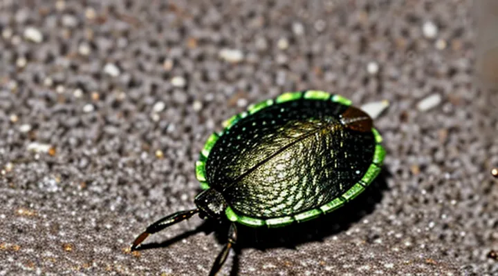
Analyzing a tick that has been physically compressed is feasible because the exoskeleton retains sufficient structural information to permit identification. The chitinous cuticle, despite deformation, preserves characteristic patterns of festoons, spiracular plates, and genital aperture that remain discernible under magnification. These morphological markers enable taxonomic classification to the species or genus level.
Microscopic examination, often combined with scanning electron microscopy, reveals surface topography and internal remnants. DNA extraction remains viable; the hemolymph and internal tissues, protected by the cuticle, survive compression long enough for polymerase chain reaction amplification. Successful genetic analysis confirms species identity and can detect pathogen presence.
Key factors influencing analytical outcome:
- Degree of compression: moderate flattening preserves cuticular landmarks; extreme crush can obscure diagnostic features.
- Preservation method: ethanol fixation or rapid drying prevents further degradation.
- Instrumentation: high‑resolution optics or electron microscopy enhances visibility of minute structures.
- Sample handling: avoiding additional mechanical stress maintains integrity for both morphological and molecular assays.
When structural integrity is sufficiently retained, both morphological assessment and molecular techniques provide reliable identification, demonstrating that a crushed tick can indeed be analyzed with accurate results.
Degradation of Genetic Material
Crushing a tick ruptures cellular membranes, exposing nucleic acids to endogenous nucleases and environmental agents that accelerate degradation. Immediate mechanical disruption releases DNases that cleave phosphodiester bonds, reducing fragment length within minutes. Heat generated by friction further destabilizes double‑helix structure, promoting hydrolysis of the sugar‑phosphate backbone.
External factors compound the loss of intact genetic material. Atmospheric oxygen drives oxidative damage, converting guanine to 8‑oxoguanine and inducing strand breaks. Moisture facilitates hydrolytic cleavage of adenine and cytosine residues, while ultraviolet radiation causes pyrimidine dimers. Together, these processes diminish the quantity of recoverable DNA and lower the average fragment size.
Successful molecular analysis depends on preserving sufficient length and integrity for polymerase chain reaction (PCR) or sequencing. The following conditions improve outcomes:
- Rapid cooling of the specimen after crushing
- Addition of nuclease inhibitors or EDTA to chelate divalent cations
- Storage in a dry, low‑temperature environment
- Use of extraction kits optimized for fragmented DNA
When these measures are applied, detection of tick species, pathogen presence, or blood‑meal sources remains feasible despite the initial physical trauma. Conversely, failure to limit degradation results in insufficient template for amplification, rendering analysis unreliable.
Contamination Risks
Analyzing a damaged tick can be performed, but the process creates immediate contamination hazards that must be managed. When a tick is crushed, internal fluids containing bacteria, viruses, or protozoa are released into the surrounding environment, increasing the likelihood of exposure for laboratory personnel and compromising the safety of adjacent workspaces.
Key contamination risks include:
- Aerosol generation that can carry pathogens to respiratory tracts.
- Surface contamination of benches, equipment, and protective gear.
- Cross‑contamination of other specimens or cultures.
- Degradation of nucleic acids and proteins, reducing analytical accuracy.
Effective control measures require strict biosafety practices: use of a certified biological safety cabinet, double gloves, disposable lab coats, and face protection; implementation of decontamination protocols for all contact surfaces; and disposal of crushed material in sealed biohazard containers. Adhering to these procedures minimizes exposure and preserves the integrity of the analytical results.
Methods for Analysis
Microscopic Examination
Microscopic examination can be performed on a tick that has been physically disrupted. The specimen is collected, placed on a slide, and covered with a suitable mounting medium that preserves cellular detail. Clearing agents such as lactophenol or potassium hydroxide render the cuticle transparent, allowing internal structures to be visualized without additional dissection. Staining with dyes like Giemsa or hematoxylin enhances contrast for nuclei, gut contents, and pathogen organisms.
Through light microscopy, the following features become accessible:
- Fragments of the exoskeleton that retain species‑specific sculpturing.
- Salivary gland remnants that may contain spirochetes, rickettsiae, or protozoa.
- Mid‑gut contents where blood meals and associated microorganisms are detectable.
- Egg or larval remnants that provide reproductive status information.
The analysis yields reliable data despite the loss of overall body shape. Morphological markers on cuticle fragments enable species identification when reference collections are consulted. Molecular techniques, such as PCR, can be applied to the same slide preparation because DNA remains intact within crushed tissues. This dual approach allows confirmation of pathogen presence and tick taxonomy.
Limitations arise when fragmentation eliminates key diagnostic structures, such as the capitulum or spiracular plates, which are essential for certain identification keys. In such cases, reliance on molecular markers increases. Nevertheless, the combination of microscopic observation and DNA analysis provides sufficient evidence to answer whether a damaged tick can be examined and to explain the underlying mechanisms that make the process viable.
Polymerase Chain Reaction (PCR)
Polymerase chain reaction (PCR) provides a rapid, sensitive method for detecting tick‑borne pathogens after the arthropod has been physically disrupted. When a tick is crushed, its internal tissues release nucleic acids that can be captured in a simple extraction buffer. PCR amplifies specific DNA fragments of target organisms, allowing identification even when only trace amounts remain.
Key factors enabling successful analysis of a pulverized tick:
- Efficient lysis of tick cells to release genomic material.
- Removal of PCR inhibitors commonly present in arthropod hemolymph, such as heme and polysaccharides, through purification kits or silica‑based columns.
- Selection of primers that target conserved regions of pathogen genomes (e.g., 16S rRNA for bacteria, 18S rRNA for protozoa, or specific viral genes).
- Use of a thermocycler with precise temperature control to ensure reliable denaturation, annealing, and extension cycles.
- Inclusion of appropriate positive and negative controls to validate assay performance.
The rationale for employing PCR in this context lies in its ability to amplify minute quantities of DNA, making it possible to detect pathogens that would otherwise be undetectable after mechanical disruption. Moreover, the technique yields results within a few hours, facilitating timely diagnosis and epidemiological investigations.
Immunofluorescence Assays
Immunofluorescence assays provide a direct method for examining a homogenized tick. Crushing the arthropod releases cellular and extracellular antigens, allowing antibodies to access target molecules without the need for intact morphology.
The assay relies on antibodies that bind specific pathogen proteins. A primary antibody recognizes the antigen; a secondary antibody conjugated to a fluorophore binds the primary antibody. When illuminated with the appropriate wavelength, the fluorophore emits light that can be captured by a fluorescence microscope, producing a visible signal indicative of the presence of the target.
Typical preparation of a crushed tick includes:
- Homogenization in a physiological buffer to preserve protein structure.
- Fixation with paraformaldehyde or acetone to immobilize antigens.
- Permeabilization with detergent to facilitate antibody penetration.
- Blocking with serum or protein solution to reduce nonspecific binding.
- Incubation with primary and fluorescent secondary antibodies under controlled temperature and time.
Immunofluorescence is suitable for this application because:
- It detects intracellular and extracellular pathogens simultaneously.
- Fluorescent signals appear within minutes after incubation, enabling rapid diagnosis.
- The technique distinguishes multiple agents by using antibodies labeled with different fluorophores.
- It preserves spatial information, allowing observation of pathogen distribution within tick tissues.
Constraints include the necessity for highly specific antibodies, potential fluorescence background from tick debris, and the requirement for a calibrated fluorescence microscope. When these factors are managed, immunofluorescence assays reliably reveal the presence of disease‑causing organisms in a crushed tick specimen.
Why Analyze a Crushed Tick?
Disease Transmission Risk Assessment
Analyzing a flattened tick provides direct evidence of pathogen presence, which refines disease transmission risk assessment. Physical disruption does not destroy nucleic acids; DNA and RNA remain recoverable for molecular testing.
Molecular techniques applicable to crushed specimens include:
- Polymerase chain reaction (PCR) targeting species‑specific gene fragments.
- Reverse transcription PCR (RT‑PCR) for RNA viruses.
- Quantitative PCR (qPCR) to estimate pathogen load.
- Sequencing of amplified products for strain identification.
Immunological assays remain viable:
- Enzyme‑linked immunosorbent assay (ELISA) detecting antigens released during crushing.
- Lateral flow immunoassays for rapid field screening.
Microscopic examination can reveal residual spirochetes or protozoan forms, especially when combined with fluorescent labeling.
The results inform risk models by:
- Confirming infection status of ticks that could not be examined intact.
- Adjusting prevalence estimates in vector populations.
- Guiding targeted control measures in areas where crushed ticks are encountered (e.g., after removal attempts).
Consequently, crushed tick analysis is technically feasible and enhances the precision of disease transmission risk assessments.
Pathogen Identification
Analyzing a tick that has been physically disrupted is feasible because crushing breaks the arthropod’s cuticle, exposing internal tissues where pathogens reside. The released material can be collected directly for nucleic‑acid extraction, allowing molecular assays to detect bacterial, viral, or protozoan agents.
Molecular detection methods applicable to crushed specimens include:
- Polymerase chain reaction (PCR) targeting species‑specific genes.
- Real‑time PCR for quantification of pathogen load.
- Metagenomic sequencing to identify mixed infections.
- Reverse transcription PCR for RNA viruses, provided RNA integrity is preserved.
Culture techniques remain viable for bacteria and protozoa when the homogenate is inoculated onto selective media under appropriate conditions. Immunoassays such as ELISA can be performed on the lysate to detect antigens or antibodies produced by the tick‑borne organism.
Advantages of crushing the vector:
- Immediate access to pathogen DNA/RNA without dissection.
- Homogenate preparation simplifies sample handling and standardizes input for downstream assays.
- Increased yield of nucleic acids improves sensitivity of detection.
Potential drawbacks:
- Mechanical shearing may fragment nucleic acids, reducing assay efficiency for long‑read sequencing.
- Release of host proteins can inhibit enzymatic reactions if not removed during purification.
- Loss of anatomical context prevents correlation of pathogen location within the tick.
Overall, the practice enables reliable identification of tick‑borne pathogens when combined with rigorous extraction protocols and appropriate controls to mitigate contamination and degradation.
Epidemiological Surveillance
Epidemiological surveillance monitors the distribution and dynamics of pathogens transmitted by arthropods, including ticks that carry bacteria, viruses, and protozoa. Accurate identification of infected vectors is essential for assessing disease risk and directing public‑health actions.
When a tick is physically damaged, such as being crushed, morphological characteristics become unusable, but molecular analysis remains viable. DNA can be extracted from the remnants of the cuticle and internal tissues, allowing polymerase‑chain‑reaction (PCR) or sequencing assays to detect pathogen genetic material. The main constraints are reduced DNA yield and potential contamination from environmental sources.
Key points for surveillance programs:
- Sample preparation: homogenize the crushed specimen, apply a lysis protocol optimized for low‑biomass material.
- Molecular detection: use broad‑range primers for Borrelia, Rickettsia, Anaplasma, and other common tick‑borne agents; confirm positives with species‑specific assays.
- Quality control: include negative controls to identify contamination; quantify DNA to assess extraction efficiency.
Analyzing crushed ticks expands the pool of usable specimens, especially when intact specimens are unavailable, thereby improving the sensitivity of pathogen detection and enhancing the overall quality of surveillance data.
Limitations and Considerations
Specimen Condition and Age
A crushed tick can still be examined, but the reliability of results depends heavily on the specimen’s physical state and its temporal characteristics. Physical damage disrupts external morphology, making species identification through visual keys difficult. However, internal structures such as the gut and salivary glands may remain intact enough for molecular extraction. Preservation of nucleic acids and proteins is contingent upon the degree of crushing; excessive compression can shear DNA, reduce yield, and introduce contaminants from the crushing surface.
Age influences analytical outcomes through developmental stage, engorgement level, and post‑mortem interval. Young nymphs contain less tissue, limiting material for extraction, while adult ticks provide larger sample volumes. The time elapsed since the tick detached from a host determines DNA integrity: prolonged exposure to heat, humidity, or UV radiation accelerates degradation. Additionally, pathogen viability declines over time; some bacteria and viruses persist longer in desiccated ticks, whereas others become undetectable after days.
Key considerations for successful analysis of a damaged tick:
- Assess remaining tissue mass; prioritize samples with visible internal sections.
- Perform rapid DNA extraction to mitigate degradation.
- Use quantitative PCR or next‑generation sequencing, which tolerate fragmented DNA.
- Record the tick’s life stage and estimated time since detachment to interpret pathogen load accurately.
- Store the specimen at low temperature (−20 °C or lower) if analysis will be delayed.
By evaluating both condition and age, researchers can determine whether a crushed tick yields sufficient data for species confirmation and pathogen detection.
Expertise and Equipment Requirements
Analyzing a tick that has been physically damaged is feasible because its internal structures and genetic material remain recoverable with proper laboratory techniques.
Expertise required includes:
- Medical entomology knowledge to recognize tick anatomy and disease relevance.
- Molecular biology proficiency for DNA extraction and polymerase chain reaction (PCR) amplification.
- Histopathology skills to prepare and interpret microscopic slides of fragmented tissues.
- Bioinformatics capability to compare genetic sequences against pathogen databases.
- Laboratory safety training to handle potentially infectious material.
Equipment necessary comprises:
- Dissection microscope with high‑magnification optics for precise tissue isolation.
- Sterile micro‑centrifuge tubes, pipettes, and vortex mixers for sample processing.
- Commercial DNA extraction kits optimized for small, compromised specimens.
- Thermal cycler for PCR, along with gel electrophoresis apparatus for product verification.
- Sequencing platform (e.g., Sanger or next‑generation) or access to external sequencing services.
- Biosafety cabinet to protect personnel and prevent cross‑contamination.
Interpretation of Results
Analyzing a flattened tick can generate morphological, molecular, and pathogen‑specific data that must be evaluated in a structured manner. Results from microscopic examination reveal the tick’s developmental stage, sex, and species, which are essential for assessing vector competence and regional disease risk.
Key interpretation points include:
- Species identification confirms whether the tick belongs to a group known to transmit particular pathogens.
- Detection of DNA from bacteria, viruses, or protozoa indicates active infection, but positive findings require verification through controls to rule out contamination.
- Quantitative PCR values provide an estimate of pathogen load; higher copy numbers suggest a greater likelihood of transmission to a host.
- Absence of pathogen DNA does not guarantee freedom from infection, as low‑level or early‑stage infections may fall below detection thresholds.
The final assessment should integrate these data with epidemiological context. If the tick is identified as a competent vector and pathogen DNA is present at significant levels, the finding supports a genuine infection risk in the area. Conversely, inconclusive or negative results, especially when coupled with limited sample quality, demand cautious interpretation and may warrant repeat testing or alternative sampling methods.
