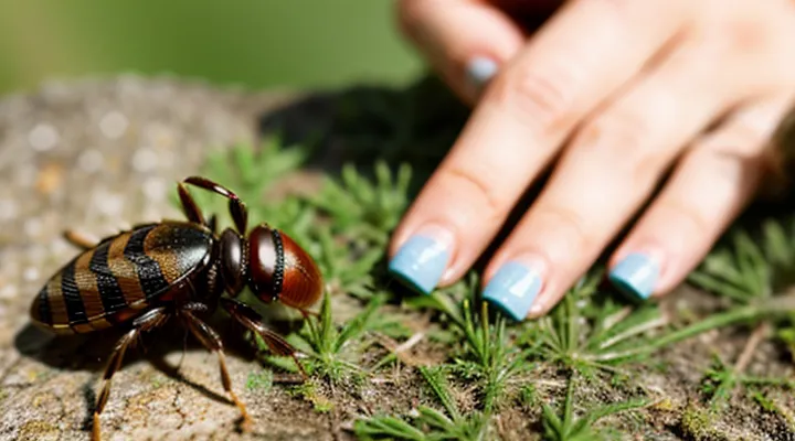
Identifying a Tick
Appearance of a small tick
The small tick measures approximately 1 – 3 mm in length when unfed, expanding up to 5 mm after a blood meal. Its body consists of a rounded anterior segment (capitulum) housing the mouthparts, and a dorsal shield (scutum) that covers the anterior half of the abdomen in males and the entire dorsum in females.
Color ranges from pale beige to reddish-brown, becoming darker and more swollen as the tick feeds. The scutum remains a distinct, often lighter, oval patch on the dorsal surface, while the posterior half of the abdomen may appear translucent when engorged. Legs are short, six‑segmented, and bear tiny claws that enable attachment to hair or skin.
Key visual cues for identification:
- Length: 1 – 3 mm (unfed), up to 5 mm (engorged)
- Shape: oval, dome‑shaped body with a clear scutum
- Color: light beige to reddish‑brown; darker when fed
- Legs: six short pairs, each ending in a claw
- Mouthparts: visible capitulum projecting forward
Recognizing these characteristics allows rapid assessment of the tick’s stage and readiness for safe removal.
Common hiding spots on the body
Ticks often attach in areas where skin is thin, warm, and protected from direct exposure. The most frequent locations include:
- Scalp, especially near the hairline and behind the ears
- Neck, particularly the back of the neck and the area under the jawline
- Armpits, where moisture and warmth create an ideal environment
- Groin and inner thighs, shielded by clothing and body folds
- Behind the knees, especially in the crease where skin is less exposed
- Around the waist, including the belt line and lower back
- Between the fingers and toes, where socks or gloves may conceal the parasite
These regions are prone to harbor small ticks because they are less likely to be inspected during routine self‑examination. Regularly checking each of these spots after outdoor activities reduces the risk of an unnoticed attachment. If a tick is found, use fine‑point tweezers to grasp the mouthparts close to the skin and pull upward with steady pressure, avoiding crushing the body. After removal, cleanse the area with antiseptic and monitor for signs of irritation.
Preparing for Tick Removal
Necessary tools and materials
When extracting a tiny tick, using the right instruments reduces the risk of breaking the mouthparts and minimizes infection. The following items constitute a complete, low‑risk kit.
- Fine‑pointed, flat‑narrow tweezers (metal or stainless steel) – grip close to the skin without crushing the tick.
- Dedicated tick removal device (e.g., a curved hook or plastic “tick key”) – alternative to tweezers for a smooth pull.
- Disposable nitrile gloves – protect hands from pathogen exposure and keep the procedure sterile.
- Antiseptic solution or wipes (70 % isopropyl alcohol, iodine, or chlorhexidine) – cleanse the bite site before and after removal.
- Sealable biohazard bag or puncture‑proof container – immediate disposal of the extracted tick to prevent accidental release.
- Small magnifying glass (optional) – improves visibility of the tick’s attachment point, especially on children’s skin.
- Clean cotton swab or gauze – apply antiseptic and gently press the area after extraction.
Having these tools readily available ensures a controlled, single‑step removal and reduces the chance of secondary complications.
Hand hygiene before removal
Before touching a tick, clean your hands thoroughly. Wash with warm water and antibacterial soap for at least 20 seconds, then rinse completely. Dry with a disposable towel or let air‑dry. If soap and water are unavailable, apply an alcohol‑based hand rub containing a minimum of 60 % ethanol or isopropanol; cover all surfaces of the fingers, palms, and nails, and allow the sanitizer to evaporate fully.
- Use disposable gloves after hand cleaning if you prefer an extra barrier.
- Replace gloves and repeat hand sanitation if you handle multiple ticks or if contamination is suspected.
- Dispose of used gloves and towels in a sealed bag before washing hands again.
Proper hand hygiene reduces the risk of pathogen transfer from the tick’s mouthparts to your skin, ensuring that the removal process remains safe and sterile.
Step-by-Step Tick Removal
Grasping the tick correctly
Grasp the tick as close to the skin as possible, using fine‑point tweezers or a tick‑removal tool. Secure the mouthparts without squeezing the body, which can force pathogens into the host.
- Position the tips of the tweezers around the tick’s head, just above the skin.
- Apply steady, gentle pressure to lift the tick straight upward.
- Avoid twisting or jerking; maintain a smooth motion until the whole organism separates.
- After removal, disinfect the bite area with an antiseptic and store the tick in a sealed container if testing is required.
Prompt, correct handling minimizes tissue damage and reduces the risk of disease transmission.
The pulling motion
The pulling motion is the decisive action for extracting a small tick without crushing its body. Grasp the tick as close to the skin as possible with fine‑point tweezers or a specialized tick‑removal tool. Apply steady, straight traction; avoid twisting, jerking, or squeezing the abdomen, which can release infectious fluids into the host.
- Position tweezers parallel to the skin surface.
- Maintain a firm grip on the tick’s head or mouthparts.
- Pull upward with constant pressure until the tick separates completely.
- Inspect the bite site; if any mouthparts remain, repeat the motion with clean tweezers.
After removal, disinfect the area with an antiseptic, store the tick in a sealed container for identification if needed, and wash hands thoroughly. This method minimizes the risk of pathogen transmission and ensures the tick is fully detached.
Avoiding common mistakes
Removing a small tick without error requires strict adherence to proper technique. Errors such as squeezing the body, using inappropriate tools, or delaying removal increase the risk of pathogen transmission and skin damage.
- Do not crush the tick’s abdomen; use fine‑point tweezers or a tick‑removal hook to grasp the mouthparts close to the skin.
- Avoid pulling at an angle; pull upward with steady, even pressure to prevent mouthpart breakage.
- Do not apply petroleum jelly, heat, or chemicals; these methods stimulate saliva release and heighten infection risk.
- Do not wait for the tick to detach on its own; immediate removal limits attachment time and pathogen exposure.
- Do not reuse tools without disinfecting; clean tweezers with alcohol or bleach solution after each use.
Select a pair of stainless‑steel tweezers with a narrow tip, sterilize before contact, and maintain a firm grip on the tick’s head. Apply continuous upward force until the tick releases completely. After extraction, place the specimen in a sealed container for identification if needed, then cleanse the bite area with antiseptic and monitor for signs of rash or fever over the next several days.
After Tick Removal Care
Cleaning the bite area
After detaching the tick, cleanse the bite site promptly to reduce infection risk. Use a mild antiseptic solution—such as diluted iodine, chlorhexidine, or alcohol swab—and apply it with a clean gauze pad. Gently rub the area for several seconds, ensuring the skin around the puncture is covered. Rinse with lukewarm water to remove residual antiseptic, then pat dry with a sterile cloth.
Maintain cleanliness for the following 24‑48 hours:
- Inspect the wound twice daily for redness, swelling, or discharge.
- Reapply antiseptic if any signs of contamination appear.
- Keep the area uncovered unless it is exposed to dirt; a breathable, non‑adhesive dressing may be used if irritation occurs.
If symptoms such as increasing pain, fever, or a rash develop, seek medical evaluation promptly.
Monitoring for symptoms
After removing a tick, observe the bite site and the person’s overall condition for at least two weeks. Early detection of illness depends on consistent monitoring.
Watch for these signs:
- Redness or swelling that expands beyond the immediate bite area.
- A rash resembling a bull’s‑eye pattern (a central red spot surrounded by a clear ring).
- Fever, chills, or unexplained temperature rise.
- Headache, muscle aches, or joint pain that appear suddenly.
- Nausea, vomiting, or abdominal discomfort.
- Fatigue that worsens rather than improves with rest.
If any symptom emerges, record the date of appearance and contact a healthcare professional promptly. Mention the recent tick exposure and provide details about the tick’s size and the removal method used. Early treatment reduces the risk of complications from tick‑borne diseases.
When to seek medical attention
If a tick has been removed and any of the following signs appear, immediate medical evaluation is required.
- Fever, chills, or flu‑like symptoms within two weeks of the bite.
- Rash that expands, forms a bullseye, or appears on the torso, limbs, or face.
- Persistent headache, muscle aches, or joint pain.
- Swelling or redness at the bite site that worsens after 24 hours.
- Neurological signs such as facial weakness, numbness, or difficulty concentrating.
These symptoms may indicate infection with tick‑borne pathogens such as Borrelia (Lyme disease), Anaplasma, or Rickettsia. Prompt diagnosis and treatment reduce the risk of complications.
When any of the listed conditions develop, contact a healthcare provider without delay. If the tick was attached for more than 24 hours, inform the clinician of the estimated duration, the tick’s appearance, and any recent outdoor exposure. Follow professional advice regarding laboratory testing, antibiotic therapy, or further observation.
