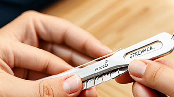
Understanding Tick Bites and Their Dangers
Why Proper Tick Removal is Crucial
Proper tick removal prevents pathogen transmission. When a tick remains attached, it can inject bacteria, viruses, or protozoa that cause Lyme disease, Rocky Mountain spotted fever, and other illnesses. Immediate, correct extraction stops this process before the pathogen is transferred into the bloodstream.
Incorrect technique often leaves mouthparts embedded in the skin. Retained fragments serve as a nidus for bacterial growth, provoke localized inflammation, and may require surgical intervention to eliminate. Additionally, squeezing the body increases pressure, forcing infected material deeper into the host.
Accurate removal with fine‑point tweezers, grasping the tick close to the skin, and pulling upward with steady pressure ensures the entire organism is extracted. This method minimizes tissue trauma and lowers the probability of secondary infection.
Key reasons for meticulous removal:
- Elimination of disease‑carrying vectors before transmission occurs.
- Prevention of residual mouthparts that can cause infection.
- Reduction of tissue damage and associated pain.
- Decrease of inflammation and healing time.
Potential Health Risks Associated with Tick Bites
Ticks transmit a range of pathogens that can cause acute illness, chronic disease, or fatal outcomes. Prompt extraction with fine‑point tweezers lowers transmission probability, yet several health threats remain.
- Borrelia burgdorferi – agent of Lyme disease; early signs include erythema migrans, fever, headache, fatigue; untreated infection may progress to arthritis, carditis, neuroborial involvement.
- Rickettsia rickettsii – causes Rocky Mountain spotted fever; rapid onset of high fever, rash, vascular injury; mortality rises without timely doxycycline therapy.
- Anaplasma phagocytophilum – produces human granulocytic anaplasmosis; symptoms comprise fever, myalgia, leukopenia; severe cases involve respiratory failure or organ dysfunction.
- Babesia microti – responsible for babesiosis; hemolytic anemia, jaundice, thrombocytopenia; co‑infection with Lyme disease worsens prognosis.
- Tick‑borne encephalitis virus – leads to meningitis or encephalitis; neurological sequelae may persist.
- Tick paralysis toxin – neurotoxic protein causing progressive weakness and respiratory failure; removal of the tick typically reverses paralysis within hours.
Transmission generally requires the tick to remain attached for several hours; Dermacentor and Ixodes species differ in minimum attachment times. Even brief feeding can introduce saliva‑borne allergens, provoking local dermatitis or systemic hypersensitivity. Secondary bacterial infection may develop at the bite site if the wound is not cleaned.
Monitoring after removal is essential. Persistent fever, expanding rash, joint pain, neurological deficits, or unexplained fatigue warrant immediate medical evaluation. Early antimicrobial treatment, guided by suspected pathogen and regional prevalence, reduces morbidity and prevents long‑term complications.
Essential Tools and Preparation
Gathering the Right Tweezers
When removing a tick, the first tool you must obtain is a pair of tweezers designed specifically for the task. Ordinary household tweezers often lack the precision and strength required, increasing the risk of breaking the tick’s mouthparts and leaving them embedded in the skin.
Select tweezers that meet the following criteria:
- Fine, pointed tips that can grasp the tick’s head without slipping.
- A flat or slightly curved jaw to maintain a secure grip on the tick’s body.
- Stainless‑steel construction to prevent rust and allow easy sterilization.
- A length of at least 5 cm to keep your hand away from the bite site while applying steady pressure.
- Textured or rubber‑coated handles to improve grip and reduce hand fatigue.
Before use, sterilize the tweezers with an alcohol wipe or boiling water. Inspect the tips for any bends or damage; replace them if the alignment is compromised. Properly chosen tweezers enable a clean, swift extraction that minimizes tissue trauma and reduces the chance of infection.
Additional Supplies for Safe Removal
When performing a tick extraction with tweezers, auxiliary items enhance safety and reduce infection risk. Disposable nitrile gloves protect the extractor’s hands from potential pathogens transmitted by the arthropod. A 70% isopropyl alcohol pad or povidone‑iodine swab should be ready to cleanse the bite site before and after removal, minimizing bacterial entry. A magnifying lens or handheld magnifier assists in locating the tick’s head, ensuring the grasp is as close to the skin as possible. A sterile container with a lid—such as a small zip‑lock bag or a screw‑cap tube—provides a secure place to store the removed tick for identification or disposal. After extraction, a clean gauze pad can apply gentle pressure to stop bleeding, followed by a fresh alcohol wipe to disinfect the area. Finally, a biohazard waste bag allows safe disposal of used gloves, swabs, and the tick itself, preventing environmental contamination.
Preparing the Environment and the Person
Prepare a clean, well‑lit area where the procedure will take place. Disinfect the surface with an alcohol‑based solution or a bleach dilution and allow it to dry. Keep a waste container nearby for the discarded tick and any used materials.
Ensure the individual is seated or lying down in a comfortable position that exposes the attachment site without causing strain. Ask the person to relax the skin around the tick; tension can increase the risk of the mouthparts breaking off. If the person is a child or has limited mobility, secure the limb gently with a soft strap or a rolled towel to maintain stability.
Gather the following items before beginning:
- Fine‑point, non‑slanted tweezers made of stainless steel
- Antiseptic wipes or swabs (70 % isopropyl alcohol)
- Disposable gloves (latex or nitrile)
- Small sealable bag or container for the tick
- Adhesive bandage or sterile gauze for post‑removal care
Don gloves, clean the tweezers with an antiseptic wipe, and verify that the lighting is sufficient to see the tick’s head and body clearly. Only after these preparations should the actual extraction commence.
Step-by-Step Tick Removal Process
Positioning for Optimal Access
Proper positioning is essential for clear visibility and steady control when extracting a tick with tweezers. Align the patient so the infested area is fully exposed and easily reachable; avoid clothing or hair that could obstruct the view. Use a well‑lit environment, preferably natural light or a focused lamp, to highlight the tick’s head and surrounding skin.
- Have the person sit or lie down in a comfortable position that keeps the target area level with the caregiver’s hands.
- Extend the limb or body part so the skin is taut; a gentle stretch with the opposite hand reduces skin movement.
- Position the tweezers at a 45‑degree angle to the skin surface, aiming directly at the tick’s mouthparts.
- Keep the dominant hand steady, resting the forearm on a firm surface if possible, to minimize tremor.
Maintain a calm posture; a relaxed grip on the tweezers prevents crushing the tick’s body and reduces the risk of pathogen release. After removal, clean the site and inspect the tick to confirm the head is intact.
Grasping the Tick Correctly
Grasping a tick correctly prevents the mouthparts from breaking off and reduces the risk of pathogen transmission. The tick’s head, or capitulum, must be captured as close to the skin as possible to ensure a clean removal.
- Use fine‑point, non‑slip tweezers; avoid blunt or rounded tips.
- Position the tweezers as close to the skin surface as the tick’s body allows.
- Apply steady, even pressure to pinch the tick’s head without crushing its abdomen.
- Maintain the grip while pulling straight upward with consistent force; do not twist or jerk.
- After extraction, disinfect the bite area and the tweezers with alcohol or an iodine solution.
Accurate placement of the tweezers and a firm, uninterrupted pull constitute the essential technique for safe tick removal from a human host.
Executing the Pulling Motion
When extracting a tick from a human host with tweezers, the pulling motion must be steady, straight, and uninterrupted to minimize attachment of the mouthparts.
- Position the tweezers as close to the skin as possible, grasping the tick’s head or mouthparts without crushing the body.
- Align the force vector directly toward the skin surface; any angular movement can cause the tick’s hypostome to break.
- Apply consistent pressure, maintaining grip while pulling outward in a single, smooth motion.
- Do not pause, wiggle, or rotate the instrument; these actions increase the risk of leaving fragments embedded.
- Release the grip once the tick detaches completely, then cleanse the bite area with antiseptic.
What to Do if Parts of the Tick Remain
When a tick is extracted with tweezers and fragments of the mouthparts stay embedded in the skin, prompt action reduces the risk of infection and inflammation.
First, cleanse the area with an antiseptic solution such as povidone‑iodine or alcohol. Apply gentle pressure with a sterile gauze pad to stop any minor bleeding.
Next, attempt to retrieve the remaining piece:
- Use a pair of fine‑pointed tweezers or a sterile needle.
- Grasp the visible fragment as close to the skin surface as possible.
- Pull upward with steady, even force; avoid twisting or squeezing, which can drive the fragment deeper.
- If the fragment is not visible or cannot be grasped, do not dig aggressively.
When removal is unsuccessful, follow these steps:
- Cover the site with a clean dressing.
- Monitor for signs of infection—redness expanding beyond the margin, swelling, warmth, or pus.
- Seek medical attention if any of those signs appear, if the tick was attached for more than 24 hours, or if the individual has a known allergy to tick bites or is immunocompromised.
After professional care, continue observation for at least two weeks. Document the date of the bite, the type of tick if known, and any symptoms that develop. Early reporting of fever, headache, rash, or joint pain can facilitate timely diagnosis of tick‑borne diseases.
Aftercare and Monitoring
Cleaning the Bite Area
After extracting the tick, disinfect the surrounding skin promptly. Use an antiseptic solution such as povidone‑iodine or chlorhexidine; apply a generous amount with a clean gauze pad, ensuring full coverage of the bite site. Allow the antiseptic to remain in contact for at least 30 seconds before wiping away excess fluid.
If a sterile swab is unavailable, a mild soap solution can serve as an interim cleanser. Wet the area, scrub gently for 10–15 seconds, and rinse with clean water. Pat the skin dry with a sterile cloth or disposable paper towel—avoid rubbing, which may irritate the wound.
Once the area is clean, cover it with a sterile, non‑adhesive dressing. Secure the dressing with medical tape, ensuring it does not constrict circulation. Change the dressing daily or whenever it becomes wet or contaminated.
Monitor the site for signs of infection: increasing redness, swelling, warmth, pus, or persistent pain. Should any of these symptoms appear, seek medical evaluation promptly.
Post-Removal Observation
After a tick is extracted with tweezers, monitor the bite site for at least 24 hours. Observe the skin for redness, swelling, or a rash that expands beyond the immediate area. Record any increase in size, change in color, or formation of a target‑shaped lesion, as these may indicate early infection.
Watch for systemic signs such as fever, chills, headache, muscle aches, or fatigue. If any of these symptoms appear within two weeks, seek medical evaluation promptly. Persistent or worsening local inflammation warrants professional assessment to rule out secondary bacterial infection.
Maintain the wound clean. Wash the area with mild soap and water twice daily, then apply a sterile, non‑adhesive dressing if the skin is broken. Avoid scratching or applying topical irritants that could obscure clinical signs.
Document the date of removal, the estimated duration of attachment, and the geographic region where the tick was found. This information assists healthcare providers in selecting appropriate diagnostic tests and prophylactic treatment, if necessary.
When to Seek Medical Attention
After removing a tick with fine‑pointed tweezers, monitor the bite site and the individual’s condition. Seek professional care if any of the following occurs:
- The tick was attached for more than 24 hours before removal.
- The bite area becomes increasingly red, swollen, or develops a rash that expands beyond the immediate circle of the bite.
- Flu‑like symptoms appear within two weeks: fever, headache, muscle aches, or fatigue.
- A bull’s‑eye rash (a clear center surrounded by a red ring) emerges, indicating possible Lyme disease.
- The person has a weakened immune system, chronic illness, or is pregnant.
- The tick could not be fully extracted, leaving mouthparts embedded in the skin.
If any of these signs are present, contact a healthcare provider promptly. Early evaluation and, if necessary, antibiotic treatment reduce the risk of infection and complications. Even in the absence of symptoms, a follow‑up appointment is advisable for individuals at high risk of tick‑borne diseases.
Prevention and Awareness
Tips for Avoiding Tick Bites
Ticks attach quickly and can transmit diseases within hours. Preventing a bite eliminates the need for later extraction and reduces health risks.
- Wear long sleeves and pants; tuck shirts into trousers and pants into socks.
- Choose light-colored clothing to spot ticks more easily.
- Apply EPA‑registered repellents containing DEET, picaridin, or IR3535 to skin and clothing.
- Treat outdoor gear, boots, and pet collars with permethrin; reapply according to label instructions.
- Perform thorough body checks after leaving wooded or grassy areas; inspect scalp, armpits, groin, and behind knees.
- Shower within 30 minutes of exposure; water flow dislodges unattached ticks.
- Keep lawns mowed short, remove leaf litter, and create a barrier of wood chips or gravel around play areas.
Consistent use of these measures dramatically lowers the chance of attachment, ensuring safer outdoor activities and reducing reliance on tick removal techniques.
Recognizing Different Tick Species
Recognizing the tick species attached to a patient guides the choice of removal technique and informs risk assessment for disease transmission.
Key morphological markers include overall length, color pattern, presence of a scutum, shape of the capitulum, and degree of engorgement. Size ranges from a few millimeters in unfed nymphs to over a centimeter in fully engorged adults. The scutum covers the dorsal surface in males and partially in females; its pattern distinguishes genera. The capitulum’s orientation (forward‑projecting versus angled) separates hard‑ticks from soft‑ticks.
- Deer tick (Ixodes scapularis) – small, reddish‑brown, darkened dorsal shield; legs shorter than body; common in eastern North America; vector of Lyme disease.
- Lone star tick (Amblyomma americanum) – adult females display a single white spot on the back; larger body; aggressive feeder; associated with ehrlichiosis and alpha‑gal allergy.
- American dog tick (Dermacentor variabilis) – brown, mottled scutum with white markings; found in grassy fields; transmitter of Rocky Mountain spotted fever.
- Rocky Mountain wood tick (Dermacentor andersoni) – darker, heavily patterned scutum; inhabits high‑altitude regions; carrier of Rocky Mountain spotted fever and tularemia.
- Brown dog tick (Rhipicephalus sanguineus) – reddish‑brown, oval body; thrives indoors and in warm climates; vector of canine ehrlichiosis and babesiosis.
Geographic distribution aligns with climate and host availability. Ixodes species dominate temperate forests, Amblyomma species favor humid, subtropical zones, while Dermacentor species concentrate in mountainous or prairie environments. Rhipicephalus persists in indoor settings worldwide.
Accurate identification dictates where to place tweezers: grip the tick as close to the skin as possible, avoid compressing the capitulum, and maintain steady pressure to prevent mouthpart rupture. Species with larger mouthparts, such as Amblyomma and Dermacentor, require careful handling to minimize tissue trauma. Recognizing the tick type before extraction reduces the likelihood of incomplete removal and subsequent infection.
