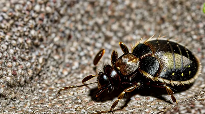
Understanding Tick Bites
What are Ticks and Why are Their Bites a Concern?
Ticks are small arachnids that attach to the skin of mammals, birds, and reptiles to obtain blood. Their mouthparts include barbed hypostome, enabling prolonged feeding for several days. During this period, saliva containing anticoagulants, immunomodulatory proteins, and potential pathogens is injected into the host.
Bite concerns arise from several factors:
- Transmission of bacterial, viral, and protozoal agents such as Borrelia burgdorferi (Lyme disease), Anaplasma phagocytophilum, Rickettsia spp., and tick‑borne encephalitis virus.
- Induction of local inflammation, erythema, and possible allergic reactions to tick saliva.
- Risk of secondary bacterial infection at the attachment site, especially if the skin is broken or the bite is scratched.
- Difficulty in detecting early-stage attachment, allowing pathogens to establish before the bite is noticed.
Understanding tick biology and the health risks associated with their bites informs appropriate wound management and preventive measures.
Identifying a Tick Bite
Common Symptoms
A tick bite may produce immediate local reactions that guide initial care. Redness, swelling, and tenderness around the attachment site are typical. A small central punctum often remains visible after the tick detaches.
Common local symptoms include:
- Erythema extending 2‑3 cm from the bite
- Mild to moderate pain or itching
- Small ulceration or crust formation
Systemic signs can emerge within days to weeks, indicating possible infection. Frequently observed manifestations are:
- Fever or chills
- Headache and muscle aches
- Fatigue or malaise
- Enlarged lymph nodes near the bite area
- Rash with a target‑like appearance, often expanding beyond the original site
Presence of any systemic symptom warrants prompt medical evaluation. Immediate consultation is also necessary if the bite site shows rapid expansion, necrosis, or persistent pain despite basic wound care. Early identification of these symptoms supports timely intervention and reduces the risk of complications.
Signs of a Tick Embedded
A tick that remains attached can be identified by several clinical clues.
- Small, raised papule at the bite site, often surrounded by a halo of erythema.
- Central punctum or “mouth‑part” opening from which the tick is anchored.
- Visible portion of the tick’s body, especially the abdomen, which may appear engorged.
- Localized swelling or edema that progresses despite gentle pressure.
- Persistent pruritus or mild pain that does not subside within a few hours.
- Presence of a “tick‑leg” pattern: two parallel lines of erythema indicating the tick’s legs.
If any of these signs are observed, immediate removal of the arthropod is required, followed by proper wound care and monitoring for infection or disease transmission.
Immediate Steps After a Tick Bite
Safe Tick Removal Techniques
Tools for Removal
Effective removal of a tick requires appropriate instruments that minimize tissue damage and ensure the entire organism is extracted.
• Fine‑pointed tweezers with a non‑slipping grip – ideal for grasping the tick close to the skin surface and applying steady, downward pressure.
• Small‑mouth forceps designed for medical use – provide a secure hold and allow precise manipulation without crushing the tick’s body.
• Dedicated tick removal devices (e.g., plastic loop or cartridge systems) – enable the tick to be lifted out in a single motion, reducing the risk of mouth‑part retention.
• Protective gloves – prevent direct contact with the arthropod and potential pathogen exposure.
After extraction, the bite area should be cleansed with an antiseptic solution such as povidone‑iodine or chlorhexidine. The removed tick must be placed in a sealed container for possible identification and pathogen testing. Failure to use a suitable tool can result in incomplete removal, increased inflammation, and heightened infection risk.
Step-by-Step Guide for Tick Removal
Tick removal must be performed promptly and precisely to minimise pathogen transmission.
- Gather required tools: fine‑point tweezers or a specialised tick‑removal device, disposable gloves, antiseptic solution, and a clean container for the specimen.
- Don gloves to avoid direct contact with the arthropod.
- Grasp the tick as close to the skin surface as possible, securing the head or mouthparts without crushing the body.
- Apply steady, upward traction; avoid twisting or jerking motions that could detach the mouthparts.
- Once the tick separates, place it in the container, cover, and label for possible laboratory analysis.
- Clean the bite area with antiseptic; cover with a sterile bandage if needed.
Post‑removal care includes monitoring the site for erythema, swelling, or necrosis. Document the date of removal and any observable changes.
Seek medical evaluation if:
- The bite area enlarges or develops a rash resembling a target.
- Flu‑like symptoms such as fever, headache, or muscle aches appear within weeks.
- The tick could not be removed intact, leaving mouthparts embedded.
Proper technique and vigilant follow‑up reduce the risk of tick‑borne disease.
Post-Removal Site Cleaning
Antiseptic Application
After the tick has been detached, the bite site requires immediate antiseptic treatment to lower the risk of bacterial infection.
Recommended antiseptics include:
- 70 % isopropyl alcohol
- 0.5 % povidone‑iodine solution
- Chlorhexidine gluconate (0.5 %–2 %)
Application procedure:
- Gently rinse the area with clean water to remove debris.
- Pat the skin dry with a sterile gauze pad.
- Apply a thin layer of the chosen antiseptic using a sterile swab or cotton ball.
- Allow the antiseptic to air‑dry; avoid covering the site with occlusive dressings unless additional protection is required.
Post‑application care:
- Re‑apply the antiseptic once daily for the first 48 hours if the wound remains moist or shows signs of irritation.
- Monitor for erythema, swelling, or discharge; seek medical evaluation if symptoms progress.
Proper antiseptic use constitutes a critical step in managing tick bite lesions and contributes to effective prevention of secondary infection.
What Not to Do After Removal
After a tick is detached, certain actions increase the risk of infection or delay healing.
- Do not apply heat, such as a flame or hot compress, to the bite area; thermal injury can damage surrounding tissue.
- Do not crush or squeeze the skin surrounding the attachment point; pressure may force mouthparts deeper and spread pathogens.
- Do not use harsh chemicals, including iodine, hydrogen peroxide, or alcohol, as they can irritate the wound and impede natural clotting.
- Do not cover the site with airtight dressings; occlusion creates a moist environment favorable to bacterial growth.
- Do not neglect observation; failing to monitor for redness, swelling, or fever postpones medical evaluation.
Following removal, gentle cleaning with mild soap and water, followed by a light, breathable bandage, supports optimal recovery. Regular inspection for abnormal signs remains essential.
Monitoring the Bite Site for Complications
What to Look For
Rash Development («Bullseye» Rash)
When a tick attaches to skin, the earliest cutaneous sign of infection often appears as a localized erythema with a concentric pattern. This presentation, termed the «Bullseye» Rash, develops in a minority of bites but signals possible transmission of Borrelia burgdorferi or other pathogens. Recognition of the rash guides immediate therapeutic decisions.
Typical characteristics of the rash include:
- Diameter of 5 cm or greater.
- Central clearing surrounded by a red halo.
- Appearance within 3–30 days after the bite.
- Absence of pain or itching in most cases.
If the rash is identified, prompt antimicrobial therapy is required. Recommended actions are:
- Initiate oral doxycycline (100 mg twice daily) for 10–21 days in adults; alternative agents for contraindications.
- Document the exact location and size of the lesion for follow‑up evaluation.
- Advise the patient to monitor for systemic symptoms such as fever, headache, arthralgia, or neurological changes.
- Schedule a reassessment within 48–72 hours to confirm rash regression and treatment tolerance.
In the absence of the characteristic lesion, routine wound care remains essential: clean the bite site with mild soap and water, apply an antiseptic, and observe for any delayed dermatologic changes. Early detection of the «Bullseye» Rash therefore constitutes a critical component of effective management of tick‑related skin exposures.
Swelling and Redness
Swelling and redness around a tick bite indicate a local inflammatory response. Immediate cleaning with mild soap and water reduces bacterial load and limits irritation. Applying a cold compress for 10‑15 minutes, several times a day, eases edema and discomfort.
If inflammation persists beyond 48 hours, an over‑the‑counter non‑steroidal anti‑inflammatory drug (NSANSA) can be administered according to dosing guidelines. Topical corticosteroid creams may be used for pronounced erythema, but only when contraindications are absent.
Monitoring is essential. Seek professional evaluation if any of the following occurs:
- Expansion of redness beyond the bite margin
- Pain intensifies or becomes throbbing
- Fever or chills develop
- Presence of a pustule or ulceration
- Persistent swelling after 72 hours
When medical assessment is warranted, clinicians may prescribe oral antibiotics targeting common skin pathogens, such as doxycycline or amoxicillin‑clavulanate, especially if secondary bacterial infection is suspected. Documentation of the bite site, including photographs, assists in tracking progression and informing treatment decisions.
Fever and Flu-like Symptoms
Fever and flu‑like symptoms may appear within days after a tick attachment, indicating possible systemic infection. Prompt assessment of temperature, chills, headache, myalgia, and fatigue is essential to differentiate benign reactions from early manifestations of tick‑borne diseases.
Evaluation should include:
- Measurement of body temperature at least twice daily.
- Documentation of symptom onset, intensity, and progression.
- Laboratory testing for serologic markers when fever exceeds 38 °C for more than 48 hours or when accompanying rash, joint pain, or neurologic signs develop.
Management of the bite site while addressing systemic signs involves:
- Removal of the tick with fine‑pointed tweezers, grasping close to the skin and pulling straight upward without crushing the mouthparts.
- Cleansing of the wound using antiseptic solution; avoidance of harsh chemicals that may irritate the skin.
- Application of a sterile dressing to protect against secondary bacterial infection.
- Administration of antipyretic agents such as acetaminophen or ibuprofen to control temperature and alleviate discomfort, respecting age‑appropriate dosing.
- Initiation of empiric antibiotic therapy (e.g., doxycycline 100 mg twice daily) when clinical suspicion for Lyme disease or other bacterial tick‑borne illnesses is high, especially in regions with known prevalence.
Continuous monitoring is required for at least two weeks after removal. Persistence or escalation of fever, emergence of new systemic manifestations, or worsening of the local lesion warrants referral to infectious‑disease specialists. Documentation of all observations supports accurate diagnosis and timely intervention.
When to Seek Medical Attention
Persistent Symptoms
Persistent symptoms after a tick bite demand prompt evaluation. Fever, expanding erythema, arthralgia, headache, or neurological signs indicate possible infection and require medical attention. Absence of immediate reaction does not exclude later disease emergence; monitoring for up to four weeks is essential.
Key actions include:
- Laboratory testing for Borrelia burgdorferi, Anaplasma, or Rickettsia when symptoms arise.
- Initiation of targeted antibiotic therapy according to identified pathogen and disease stage.
- Documentation of symptom progression and response to treatment at regular intervals.
- Referral to specialist care for persistent neurological or cardiac manifestations.
Resolution of symptoms should be reassessed after completion of therapy. Persistent or recurrent signs warrant repeat diagnostics and possible adjustment of antimicrobial regimen. Early recognition and systematic management minimize long‑term complications.
History of Tick-Borne Diseases in Your Area
The region’s record of tick‑borne illnesses extends over a century, beginning with sporadic cases of Rocky Mountain spotted fever reported in the early 1900s. Surveillance data from the 1930s show a rise in Lyme disease incidence following the identification of Borrelia burgdorferi in local tick populations. A notable surge occurred in the 1990s, when increased outdoor recreation correlated with higher numbers of anaplasmosis and babesiosis diagnoses. Since 2010, surveillance reports indicate a steady rise in co‑infection rates, reflecting expanding tick habitats due to climate change and land‑use alterations.
Key historical milestones:
- 1905 – First documented Rocky Mountain spotted fever case in the area.
- 1968 – Discovery of Ixodes species as primary vectors for Lyme disease.
- 1989 – Confirmation of Lyme disease as endemic, supported by serologic surveys.
- 1995 – Emergence of anaplasmosis cases linked to Anaplasma phagocytophilum.
- 2002 – First babesiosis outbreak recorded, highlighting Babesia microti transmission.
- 2015 – Detection of Borrelia miyamotoi infections, expanding the spectrum of tick‑borne pathogens.
Understanding this epidemiological evolution informs optimal management of a recent tick attachment. Immediate removal of the engorged tick with fine‑pointed tweezers, followed by cleansing of the bite site with antiseptic solution, reduces pathogen transmission risk. Observation for early signs of infection—fever, erythema migrans, or flu‑like symptoms—should continue for at least four weeks, given the documented latency periods of local pathogens. When systemic manifestations appear, prompt antimicrobial therapy, typically doxycycline for bacterial agents, aligns with established treatment protocols derived from regional disease patterns.
Preventing Tick Bites
Personal Protective Measures
Appropriate Clothing
After a tick attachment, immediate removal of clothing that covers the bite site is essential. Loose, breathable garments should replace tight or abrasive fabrics to prevent friction and allow visual inspection of the wound.
Key considerations for clothing post‑bite:
- Choose natural fibers such as cotton or linen that wick moisture and reduce skin irritation.
- Avoid synthetic blends that trap heat and humidity, creating an environment conducive to bacterial growth.
- Ensure sleeves, pant legs, or skirts are rolled up or shortened enough to keep the area fully visible during cleaning and monitoring.
- Wear light‑weight, non‑restrictive clothing to facilitate circulation and reduce swelling around the puncture.
Maintaining appropriate attire supports effective wound care, minimizes secondary infection risk, and simplifies ongoing assessment of any emerging symptoms.
Tick Repellents
Tick repellents reduce the risk of additional tick attachment after a bite has been removed, thereby limiting further exposure to pathogens.
Effective repellents contain active ingredients such as DEET, picaridin, IR3535, or oil of lemon eucalyptus. Formulations with at least 20 % DEET, 20 % picaridin, or 30 % lemon‑eucalyptus oil provide reliable protection for several hours.
When a tick is detached, the bite site should be cleaned with mild soap and antiseptic. After cleaning, a thin layer of repellent can be applied to the skin surrounding the wound to prevent subsequent bites. The repellent must not be placed directly into the ulcerated area to avoid irritation.
Safety considerations include avoiding products with high concentrations on infants, pregnant individuals, or persons with known skin sensitivities. Patch testing on a small area of intact skin before full application helps identify adverse reactions.
Key points for repellent use in bite‑site management:
- Choose a formulation with proven efficacy (≥20 % DEET or equivalent).
- Apply to intact skin around the bite, not into the wound.
- Re‑apply according to product guidelines, especially after sweating or washing.
- Observe for signs of irritation; discontinue if redness or itching develops.
Proper selection and application of repellents complement wound care, decreasing the likelihood of secondary tick bites and associated infections.
Environmental Control
Yard Maintenance
Proper decontamination of a tick‑induced wound requires immediate cleaning, antiseptic application, and monitoring for infection. Use mild soap and lukewarm water to remove debris, then apply a broad‑spectrum antiseptic. Cover the site with a sterile dressing and inspect daily for redness or swelling.
Maintaining the surrounding outdoor area reduces the likelihood of future bites and supports effective wound care. Regular landscaping practices limit tick habitats, decreasing exposure risk.
- Keep grass trimmed to a height of 5 cm or lower.
- Remove leaf litter, tall weeds, and brush from perimeters.
- Create a barrier of wood chips or gravel between lawn and wooded zones.
- Apply environmentally safe acaricides to high‑risk zones according to label instructions.
- Encourage natural predators, such as birds and ant‑eating insects, by providing appropriate habitats.
Implementing these measures complements medical treatment, minimizes re‑exposure, and promotes a safer environment for wound recovery.
Checking Pets for Ticks
Regular examination of companion animals is an essential component of preventing tick‑borne disease transmission to people. Ticks attach to pets before they may transfer to humans; early detection on animals reduces the likelihood of a bite site developing on a person.
Inspecting pets should be performed after each outdoor activity and before the animal enters the home. Use a fine‑toothed comb or gloved hand to run over the entire body, paying special attention to ears, neck, underbelly, and between toes. Remove any attached tick with tweezers, grasping close to the skin and pulling steadily upward. Dispose of the tick by placing it in alcohol or sealing it in a container for later identification.
Key points for an effective pet‑tick check:
- Conduct examinations daily during peak tick season.
- Wear disposable gloves to avoid direct contact with potential pathogens.
- Focus on hidden areas: armpits, groin, tail base, and between pads.
- Record findings: species, size, and attachment duration, if known.
- Clean the pet’s skin with antiseptic after removal; wash hands thoroughly.
Maintaining a regular schedule for pet grooming and using veterinarian‑recommended tick preventatives further diminishes the risk of humans encountering engorged ticks. Integrating pet checks with personal bite‑site care creates a comprehensive strategy for minimizing tick‑related health issues.
