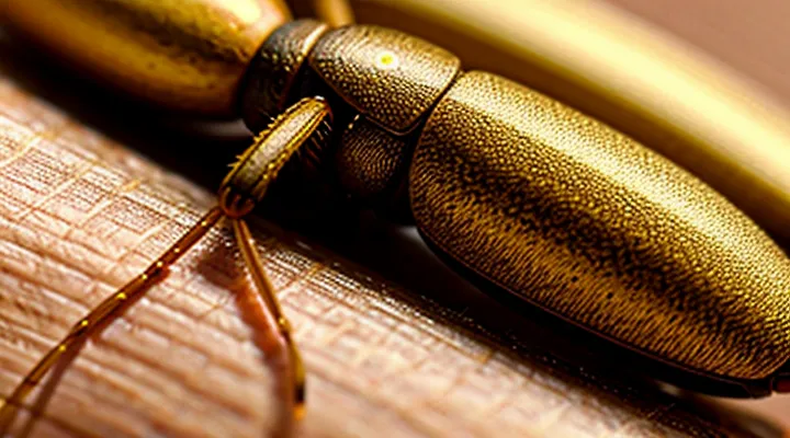
Understanding the Tick Lifecycle
Tick Development Stages
Larva Stage
The larval stage represents the first active phase after a tick hatches from its egg. Six-legged larvae emerge seeking a blood meal, typically from small mammals, birds, or occasionally humans. Upon contact with skin, the larva uses its chelicerae to pierce the epidermis, creating a minute opening that is often unnoticed. Saliva injected at this point contains anticoagulants and anesthetic compounds, preventing clotting and dulling the host’s sensation, which facilitates uninterrupted feeding.
Feeding duration for a larva lasts from several hours up to two days, during which the organism engorges on blood and expands dramatically. After detachment, the engorged larva drops to the ground, molts, and progresses to the eight-legged nymph stage. The transition from larva to nymph is essential for the tick’s life cycle and determines its capacity to transmit pathogens to subsequent hosts.
Key characteristics of the larval attachment process:
- Six legs, measuring 0.5–1 mm in length.
- Rapid insertion using specialized mouthparts.
- Salivary secretion containing anticoagulant and anesthetic agents.
- Feeding period of 12–48 hours before detachment.
- Immediate drop-off and molting to nymph after engorgement.
Nymph Stage
The nymph stage follows larval molting and precedes adulthood. Nymphs measure 0.5–1 mm, lack visible eyes, and possess a soft, pale cuticle that blends with human skin. Their small size enables unnoticed migration across the surface of the body.
Attachment occurs when a nymph climbs onto a host, senses heat and carbon‑dioxide, and inserts its hypostome into the epidermis. Salivary secretions containing anesthetic and anticoagulant compounds suppress pain and clotting, allowing the parasite to feed for 3–5 days without detection. During this period the tick’s mouthparts remain anchored while the body expands as blood is ingested.
Early identification reduces pathogen transmission risk. Signs of nymphal attachment include:
- A tiny, dark speck that may appear as a freckle
- Localized redness or a small, raised bump
- Absence of itching in the initial hours
Prompt removal with fine‑tipped tweezers, grasping the tick close to the skin and pulling straight upward, eliminates the feeding parasite and minimizes the chance of disease transfer. After extraction, the bite site should be cleansed with antiseptic and monitored for evolving inflammation.
Adult Stage
The adult stage represents the final developmental phase of a tick, during which the organism possesses fully developed mouthparts and a reproductive system capable of producing eggs. At this point the tick is capable of long‑distance movement and actively searches for a suitable host.
Host‑seeking behavior, commonly called questing, relies on sensory structures that detect carbon dioxide, heat, and vibrations emitted by potential mammals. When a human passes within reach, the tick climbs onto the skin surface and initiates attachment.
Attachment proceeds through a series of actions:
- The hypostome, a barbed feeding tube, penetrates the epidermis.
- Salivary secretions containing anticoagulants and immunomodulatory compounds are injected to inhibit clotting and suppress host defenses.
- A proteinaceous cement is deposited around the mouthparts, securing the tick for several days of feeding.
During the feeding period the tick expands dramatically, becoming visible as a swollen, dark nodule. Engorgement typically lasts from three to seven days, after which the tick detaches to lay eggs.
The adult’s prolonged attachment provides ample opportunity for the transmission of pathogens such as Borrelia burgdorferi or Rickettsia spp. Prompt identification and removal of the attached adult tick reduces the risk of infection and interrupts the life cycle of the parasite.
The Tick's Search for a Host
Habitat Preferences of Ticks
Ticks thrive in environments that provide stable humidity, moderate temperature, and access to suitable hosts. Dense low vegetation, such as grasslands, shrubbery, and forest understory, retains moisture and shelters questing ticks. Leaf litter and moss layers create microclimates with relative humidity above 80 %, preventing desiccation. Seasonal temperature ranges between 7 °C and 30 °C support active development stages; extreme heat or cold suppress activity.
Preferred hosts influence habitat selection. Small mammals (e.g., rodents, shrews) occupy ground‑level burrows and dense cover, attracting immature ticks. Larger mammals (e.g., deer, livestock) frequent edge habitats where forest meets pasture, drawing adult ticks. Domestic animals often graze in fields adjacent to woodlands, creating a bridge between natural habitats and human‑occupied areas.
Human exposure results from overlap of these habitats with recreational or occupational spaces. Key factors include:
- Trails and lawns bordering wooded areas where questing ticks wait on vegetation.
- Gardens with tall grasses, leaf piles, or stone walls retaining moisture.
- Pasture lands used for livestock, where ticks transfer from animals to people handling them.
- Recreational parks with mixed woodland and open fields, offering suitable microclimates for tick activity.
Understanding these ecological preferences enables targeted management, such as maintaining short grass along pathways, removing leaf litter near homes, and limiting wildlife access to residential yards, thereby reducing the likelihood of ticks attaching to human skin.
Host-Seeking Behavior
Ticks locate potential hosts through a sequence of sensory-driven actions. Their quest begins with questing, a behavior in which the arthropod climbs vegetation and extends its forelegs to detect environmental cues. The primary stimuli include:
- Carbon dioxide: Elevated levels in exhaled breath trigger hygroreceptors that signal a nearby vertebrate.
- Heat: Infrared-sensitive sensilla sense temperature gradients, directing movement toward warm bodies.
- Odorants: Volatile compounds such as ammonia, lactic acid, and certain skin lipids activate chemoreceptors, refining host identification.
- Vibrations: Subtle movements of foliage caused by passing animals are registered by mechanoreceptors, prompting the tick to adjust its position.
Upon detecting these signals, the tick lowers its legs, grasps the host’s skin, and begins the attachment process. Salivary secretions, rich in anti‑coagulants and anesthetics, facilitate painless penetration of the epidermis, allowing the parasite to embed its mouthparts and commence blood feeding. This cascade of host‑seeking mechanisms explains how ticks transition from the environment to the surface of human skin.
Sensory Cues for Locating a Host
Ticks locate potential hosts by interpreting a limited set of environmental signals that indicate the presence of a warm‑blooded animal. Detection of these signals triggers questing behavior, the final step before the tick grasps the skin and begins feeding.
- Carbon dioxide (CO₂) exhaled by the host creates a concentration gradient that ticks sense through chemoreceptors in the Haller’s organ.
- Heat emitted from the body surface produces a thermal gradient detectable by thermoreceptors on the forelegs.
- Mechanical vibrations generated by walking or breathing are perceived by mechanosensory setae, informing the tick of movement nearby.
- Host‑derived odorants such as ammonia, lactic acid, and certain fatty acids are captured by olfactory sensilla, providing chemical confirmation of a suitable target.
- Relative humidity influences questing height; higher moisture levels near the skin enhance the likelihood of attachment.
The Haller’s organ, situated on the first pair of legs, houses the majority of these receptors. Each modality contributes to a multimodal assessment; the tick integrates the inputs through central neural circuits and initiates a rapid response once a threshold combination is reached. Upon contact, the tick inserts its mouthparts into the epidermis, establishing the feeding site that marks its appearance on human skin.
The Tick's Attachment Process
Finding a Suitable Spot on the Skin
Ticks locate attachment sites by scanning the host’s surface for areas that meet specific physical criteria. The selection process relies on sensory organs that detect temperature gradients, carbon‑dioxide emissions, and tactile cues.
Key characteristics of a suitable spot include:
- Thin epidermal layer that allows easy penetration of the mouthparts.
- Minimal hair or fur, reducing obstruction during attachment.
- Warm, moist microenvironment that supports engorgement.
- Surface that provides stable contact, such as elbows, knees, waistline, or behind the ears.
During questing, a tick extends its front legs and monitors the air for exhaled carbon‑dioxide. Upon detecting a gradient, it steps onto the skin and evaluates the local texture. If the epidermis is thin and hair density low, the tick inserts its hypostome and begins feeding.
The choice of an optimal site directly influences attachment duration, blood intake, and pathogen transmission potential. Consequently, the tick’s ability to identify a favorable location underlies its success as an ectoparasite.
The Role of the Hypostome
The hypostome is the central oral apparatus that enables a tick to embed within human skin. Its barbed, serrated structure penetrates the epidermis and dermis, creating a stable anchorage that resists host grooming and movement. The attachment point forms a permanent channel through which the tick accesses blood vessels.
Anatomically, the hypostome consists of:
- A rigid, cone‑shaped base that supports the feeding tube.
- Microscopic hooks that interlock with collagen fibers.
- Salivary glands that release anticoagulants and immunomodulators directly into the wound.
During feeding, the hypostome performs three critical functions:
- Mechanical anchoring – the hooks lock into tissue, preventing dislodgement.
- Pathway creation – the feeding tube remains open, allowing continuous blood flow.
- Chemical mediation – saliva injected via the hypostome suppresses clotting and local immune responses, facilitating prolonged attachment.
The combination of physical penetration and biochemical modulation makes the hypostome the primary driver of successful tick colonization on human skin.
Secretion of Cement-Like Substance
Ticks attach to the skin by inserting their hypostome and releasing a proteinaceous, cement‑like secretion that hardens within seconds. The material forms a stable bond between the mouthparts and the epidermal surface, preventing dislodgement during host movement.
The secretion consists mainly of glycine‑rich proteins, lipids, and polymeric polysaccharides. These components polymerize upon exposure to the host’s temperature and pH, creating a viscoelastic matrix that adheres to both cuticular structures of the tick and the keratinized layers of the skin.
Secretion begins immediately after the hypostome penetrates the epidermis. Within the first minute, the cement solidifies enough to support the tick’s weight. Continuous excretion maintains the bond for the duration of feeding, which may last from several days to weeks, depending on the species and life stage.
The cement layer masks tick antigens from the host’s immune cells, reducing inflammatory responses and delaying detection. Its chemical composition interferes with fibrinolysis, allowing the feeding site to remain open without clot formation.
Key points for clinicians and researchers:
- Rapid polymerization ensures attachment within seconds.
- Protein‑lipid matrix resists mechanical removal.
- Immunomodulatory properties facilitate prolonged feeding.
- Disruption of cement formation is a target for anti‑tick interventions.
Feeding Mechanism of the Tick
Anesthesia and Anticoagulation
When a tick embeds its mouthparts in the epidermis, the host’s skin experiences a localized inflammatory reaction. The procedure to detach the arthropod often requires two pharmacologic considerations: pain control and hemostasis.
Local anesthetic agents, such as lidocaine or prilocaine, are injected subcutaneously around the tick’s attachment site. The drug blocks sodium channels in peripheral nerve fibers, eliminating nociceptive signals during the removal. Adequate anesthesia prevents involuntary muscle contraction that could fragment the tick’s hypostome, reducing the risk of retained mouthparts.
Anticoagulant therapy influences bleeding risk at the bite location. Patients receiving systemic anticoagulants (e.g., warfarin, direct oral anticoagulants) exhibit prolonged clotting times, which may increase oozing after the tick is extracted. Management guidelines include:
- Verify the patient’s anticoagulation status before the procedure.
- Apply a pressure dressing immediately after removal to control hemorrhage.
- If bleeding persists, consider topical hemostatic agents (e.g., thrombin spray) while monitoring coagulation parameters.
Combining precise local anesthesia with careful anticoagulation management minimizes procedural discomfort and hemorrhagic complications, ensuring complete removal of the tick’s mouthparts and reducing the likelihood of secondary infection.
Blood Meal Intake
Ticks attach to the host by inserting their specialized chelicerae and hypostome into the epidermis. The hypostome, equipped with backward‑pointing barbs, anchors the arthropod, preventing dislodgement during blood acquisition. Salivary glands release anticoagulants, vasodilators, and immunomodulatory proteins, which maintain blood flow and suppress the host’s immediate inflammatory response. The tick then creates a feeding pool by drawing plasma and erythrocytes through a porous mouthpart called the canal. Blood is stored in the midgut, where it is gradually concentrated as the tick swells.
Key aspects of the blood‑meal process:
- Attachment – barbed hypostome secures the tick within the dermal layer.
- Saliva injection – anticoagulants (e.g., apyrase) and anti‑inflammatory agents facilitate uninterrupted feeding.
- Ingestion – blood enters the foregut, passes into the midgut, and is incrementally processed.
- Engorgement – the tick’s body expands up to several times its unfed size, creating a visible swelling at the attachment site.
- Detachment – after completing the meal, the tick releases its grip and drops off, leaving behind a small puncture scar.
The visible mark on human skin results from the combination of the puncture wound, the localized inflammatory reaction to tick saliva, and the mechanical distension caused by the engorged tick. This process completes the tick’s blood‑meal intake, enabling pathogen transmission and subsequent development.
Duration of Feeding
Ticks attach to the host’s epidermis and remain attached for a defined feeding period that varies by species, life stage, and sex. The feeding interval determines the amount of blood ingested and the window for pathogen transmission.
- Larvae – typically 2 – 5 days; engorgement occurs gradually, reaching maximal weight near the end of the period.
- Nymphs – generally 3 – 7 days; early phase involves slow uptake, followed by rapid expansion during the final 24–48 hours.
- Adult females – 5 – 10 days; initial 3–4 days feature low-volume ingestion, then a surge of blood intake leading to full engorgement and egg development.
- Adult males – often 1 – 2 days; feed minimally, primarily to sustain activity while seeking mates.
Feeding proceeds through three stages: (1) attachment and cement secretion, (2) slow feeding with limited blood flow, and (3) rapid engorgement where the tick’s body can increase severalfold in size. The duration of the rapid phase correlates with the likelihood of transmitting agents such as Borrelia or Rickettsia, because pathogens require time to migrate from the tick’s salivary glands to the host’s bloodstream. Consequently, prompt removal within the first 24 hours markedly reduces infection risk.
Post-Attachment Considerations
Factors Affecting Tick Detachment
Ticks remain attached to human skin until they complete a blood meal, detach, or are mechanically removed. Detachment timing varies because several biological and environmental variables influence the attachment strength and feeding behavior.
- Engorgement level – As the tick’s abdomen expands, the cuticle stretches, reducing the grip of the mouthparts and facilitating natural release.
- Salivary composition – Enzymes and anticoagulants in tick saliva modulate host tissue responses; higher concentrations of proteolytic enzymes weaken the cement-like attachment matrix.
- Host immune response – Local inflammation and histamine release can cause tissue swelling that disrupts the anchoring apparatus, prompting earlier detachment.
- Temperature and humidity – Elevated ambient temperature accelerates metabolic rate, shortening feeding duration; low humidity may desiccate the cement, decreasing adhesion.
- Host grooming behavior – Mechanical disturbance from scratching or clothing friction can dislodge the tick before full engorgement.
Understanding these determinants helps clinicians anticipate the window for safe removal and informs public‑health strategies aimed at reducing tick‑borne disease transmission.
Potential Health Risks
Ticks embed their mouthparts into the epidermis and dermis to obtain a blood meal, creating a small, often unnoticed puncture. During this feeding period, pathogens can be transmitted from the arthropod to the host’s bloodstream.
Potential health risks include:
- Bacterial infections such as Lyme disease, caused by Borrelia burgdorferi.
- Rickettsial illnesses, including Rocky Mountain spotted fever.
- Viral diseases like tick-borne encephalitis.
- Protozoan infections such as babesiosis.
- Anaphylactic reactions to tick saliva in sensitized individuals.
Prompt removal of the attached arthropod reduces the likelihood of pathogen transmission. After extraction, a thorough inspection of the bite site and a medical assessment are advisable, especially if flu‑like symptoms, rash, or neurological signs develop within days to weeks. Early diagnosis and appropriate antimicrobial therapy significantly improve outcomes.
Prevention Strategies
Ticks locate hosts by detecting carbon dioxide, heat, and movement. Preventing their contact with skin requires eliminating exposure opportunities and removing attached specimens promptly.
- Wear long sleeves and trousers; tuck shirts into pants and pant legs into socks.
- Apply EPA‑registered repellents containing DEET, picaridin, IR3535, or oil of lemon eucalyptus to exposed skin and clothing.
- Treat outdoor clothing and gear with permethrin; reapply after washing.
- Conduct thorough body checks after leaving wooded or grassy areas; focus on scalp, armpits, groin, and behind knees.
- Shower within two hours of returning from a tick‑prone environment; water pressure helps dislodge unattached ticks.
- Maintain yard by mowing lawns, clearing leaf litter, and creating a barrier of wood chips or gravel between vegetation and play areas.
- Reduce wildlife hosts by installing fencing, using deer‑exclusion devices, and managing bird feeders that attract rodents.
If a tick is found attached, grasp the head with fine‑point tweezers, pull upward with steady pressure, and clean the bite site with antiseptic. Prompt removal minimizes pathogen transmission risk.
