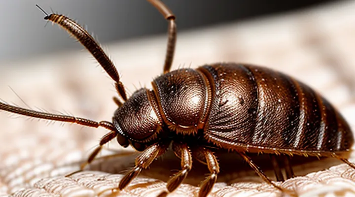
The Anatomy of a Bedbug's Mouthparts
The Proboscis: A Specialized Feeding Tube
The Stylets: Piercing and Sucking Mechanisms
Bedbugs possess a specialized feeding apparatus composed of two slender, needle‑like stylets housed within the proboscis. During a blood‑sucking episode, the insect inserts these stylets sequentially into the host’s skin.
The first stylet, the mandibular tube, acts as a mechanical penetrator. It creates a narrow channel by cutting through the epidermis and dermis, allowing the second stylet to follow. The second stylet, the maxillary tube, functions as a fluid conduit. Its internal lumen connects to a salivary duct that delivers anticoagulant enzymes, preventing clot formation as blood flows toward the bug’s foregut.
Key steps of the process:
- Penetration: Mandibular stylet pierces the skin, establishing a pathway.
- Saliva injection: Maxillary stylet releases anti‑hemostatic compounds.
- Suction: Blood is drawn through the maxillary lumen into the alimentary canal.
- Withdrawal: Both stylets retract once feeding ceases, leaving a microscopic puncture site.
The coordinated action of the two stylets enables rapid extraction of host blood while minimizing detection, a hallmark of the bedbug’s feeding strategy.
The Biting Process: A Step-by-Step Guide
Locating a Host: Sensory Cues
Bedbugs must identify a suitable blood source before they can inject saliva and obtain a meal. Successful host location depends on a combination of sensory inputs that guide the insect toward a resting human or animal.
- Thermal detection: Specialized receptors on the antennae sense temperature gradients, allowing the bug to move toward the warmth emitted by a living host.
- Carbon‑dioxide perception: Labial palps contain chemoreceptors that detect elevated CO₂ levels, a reliable indicator of respiration.
- Chemical cues: Cutaneous odors and skin secretions contain volatile compounds that bind to olfactory receptors, providing directional information.
- Mechanical vibrations: Minute movements of a sleeping host generate substrate vibrations, which are sensed by mechanoreceptors on the legs.
These cues are processed simultaneously. An increase in temperature and CO₂ concentration triggers rapid locomotion, while odor gradients refine the trajectory. When the insect detects a sufficient combination of signals, it initiates a search pattern that culminates in direct contact with the skin, at which point the feeding apparatus is deployed.
Understanding the sensory strategy clarifies how bedbugs transition from passive hideouts to active feeders, informing control measures that disrupt cue detection, such as temperature modulation or CO₂ masking.
Penetration of the Skin: Anesthesia and Anticoagulants
Bedbugs access the host’s bloodstream by inserting a slender, needle‑like mouthpart called a proboscis. The proboscis pierces the epidermis and dermis within seconds, creating a channel that remains open while the insect feeds.
During penetration, the bug injects a complex saliva that serves two primary functions. First, the saliva contains anesthetic compounds—principally small neurotoxins such as nitrophenols—that temporarily block sensory nerve endings. This prevents the host from detecting the puncture, allowing uninterrupted blood intake. Second, the saliva delivers anticoagulant agents, notably apyrase and other phosphatases, which degrade ADP and inhibit platelet aggregation. These substances keep the blood flowing freely through the feeding canal.
Key components of the salivary mixture:
- Anesthetic agents – reduce pain perception by interfering with voltage‑gated sodium channels.
- Anticoagulants – prevent clot formation by hydrolyzing ADP and disrupting fibrinogen conversion.
- Digestive enzymes – facilitate blood liquefaction for easier ingestion.
The coordinated action of anesthetic and anticoagulant factors ensures that the wound remains painless and unclotted throughout the feeding period, typically lasting five to ten minutes. After the bug withdraws, the anesthetic effect dissipates, and the anticoagulant activity ceases as the host’s hemostatic mechanisms resume.
Blood Meal Acquisition: Duration and Volume
Bedbugs locate a host through heat, carbon‑dioxide, and skin odors, then insert their elongated mouthparts into the epidermis. The feeding episode proceeds rapidly; adult insects complete a blood meal in 3–10 minutes, while nymphs require 2–5 minutes. Feeding duration correlates with the insect’s developmental stage and ambient temperature: higher temperatures shorten the interval by up to 30 percent.
The volume of blood ingested per meal is limited by the insect’s body size. Typical values are:
- First‑instar nymph: 0.2–0.3 µL
- Fifth‑instar nymph: 0.8–1.0 µL
- Adult female: 1.5–2.5 µL
- Adult male: 1.0–1.8 µL
A single blood meal supplies enough protein and lipids for egg production in females and sustains metabolism for several days. After feeding, the abdomen expands visibly, and the insect retreats to a concealed harbor where digestion proceeds. Re‑feeding intervals range from 4 to 7 days under optimal conditions, extending to weeks when hosts are scarce.
Factors Influencing Bedbug Bites
Feeding Frequency: Nocturnal Habits
Bedbugs are primarily active after darkness falls, seeking a host when people are stationary and undisturbed. Their feeding cycle aligns with the host’s sleep schedule, allowing the insect to attach, insert its proboscis, and withdraw blood with minimal detection.
Feeding frequency varies with developmental stage and environmental conditions:
- First‑instar nymphs: feed every 3–5 days.
- Later nymphal stages: require a meal roughly every 5–7 days.
- Adult females: typically feed every 5–10 days; under cooler temperatures the interval can extend to 2 weeks.
- Adult males: may feed less often, sometimes every 7–14 days, depending on energy reserves.
The nocturnal pattern is driven by the insect’s phototactic response; light exposure suppresses activity, while darkness triggers host‑seeking behavior. Temperature influences metabolic rate: higher ambient temperatures accelerate digestion, shortening the inter‑meal interval, whereas low temperatures prolong it. Bedbugs locate a host through carbon‑dioxide exhalation and body heat, then navigate the sleeping surface using tactile cues before delivering a bite.
Bite Location: Exposed Skin Preferences
Bed bugs locate their targets by detecting carbon dioxide, heat, and skin odor. After a host is identified, the insect positions itself on areas where the skin is uncovered and easily accessible. Preference for exposed regions stems from reduced physical barriers and higher surface temperature, facilitating rapid blood extraction.
Typical bite sites include:
- Hands, wrists, and forearms
- Neck and face, especially around the jawline
- Ankles and lower legs
- Upper arms and shoulders when clothing is short‑sleeved
- Feet, particularly when shoes are removed
These locations share common characteristics: minimal clothing coverage, thin epidermis, and proximity to blood vessels close to the surface. The insect inserts its elongated proboscis at an angle that allows penetration through the stratum corneum with minimal resistance. Salivary enzymes are injected simultaneously, preventing clotting and numbing the area, which often results in delayed awareness of the bite.
In environments where clothing covers most of the body, bed bugs will exploit any gaps—such as folds, seams, or openings—by extending their reach to the nearest exposed skin. Consequently, bite distribution frequently reflects the pattern of uncovered body parts rather than a random spread across the entire surface.
Host Response: Variability in Reactions
Bedbugs inject saliva containing anticoagulants and anesthetic compounds while piercing the epidermis with their elongated mouthparts. The saliva triggers an immune response that varies widely among individuals. Some people develop immediate erythema and pruritus, while others exhibit delayed, barely perceptible lesions.
Key determinants of reaction intensity include:
- Genetic predisposition influencing histamine release.
- Prior exposure leading to sensitization or tolerance.
- Age, with children often showing stronger inflammatory signs.
- Skin condition, such as eczema, which can amplify local irritation.
- Medications that modulate immune activity, for example antihistamines or corticosteroids.
The initial bite creates a micro-wound that releases cytokines, recruiting mast cells and neutrophils. In sensitized hosts, IgE antibodies bind to mast cells, causing rapid degranulation and pronounced swelling. Non‑sensitized individuals may experience a muted response, with lesions appearing only after several days as T‑cell–mediated inflammation develops.
Repeated infestations can shift the pattern of response. Some hosts become hyper‑reactive, displaying larger, more painful welts, whereas others develop hypo‑reactivity, showing minimal visible signs despite ongoing feeding. This variability complicates detection and underscores the need for thorough inspection in environments where bedbugs are suspected.
Distinguishing Bedbug Bites from Other Insect Bites
Appearance and Pattern of Bites
Bedbug bites appear as small, raised, erythematous papules that may develop into pruritic wheals. The initial lesion is usually a pinpoint red dot, often expanding to a 2‑5 mm patch with a central punctum where the insect’s proboscis entered. In some individuals, the reaction intensifies to a larger, swollen area with surrounding erythema and occasional vesiculation.
Typical distribution patterns include:
- Linear or zig‑zag arrangements, reflecting the insect’s movement along the skin while feeding.
- Clusters of three to five bites grouped within a 2‑inch radius, commonly termed “breakfast, lunch, and dinner” formations.
- Isolated spots on exposed areas such as the face, neck, arms, and hands; however, bites frequently occur on concealed regions like the torso, waistline, and legs where the bedbug can access skin during sleep.
The timing of lesion development varies; erythema may emerge within minutes to several hours after feeding, while a delayed hypersensitivity response can appear after 24–48 hours, producing larger, more inflamed plaques. Absence of a bite mark does not rule out exposure, as some individuals exhibit minimal or no cutaneous reaction.
Common Misidentifications: Mosquitoes, Fleas, and Spiders
Bedbugs feed by inserting a slender, needle‑like proboscis through the epidermis, releasing saliva that contains anticoagulants and anesthetic compounds. The saliva prevents blood clotting and dulls the immediate sensation of the bite, allowing the insect to engorge for several minutes before withdrawing its mouthparts. The delayed immune response to the salivary proteins produces a raised, red, often clustered wel‑worn lesion that may itch for days.
The following points clarify why bedbug bites are frequently confused with those of other arthropods:
- Mosquitoes – puncture wounds are typically isolated, raised bumps with a central punctum; they appear shortly after exposure and often occur on exposed skin such as arms and legs. Bedbug lesions cluster in linear or zig‑zag patterns along areas covered by clothing.
- Fleas – bites are small, hard, red papules surrounded by a halo of redness; they commonly affect the ankles and lower legs and are associated with a sudden, sharp itch. Bedbug marks are larger, smoother, and can appear on the torso, neck, and face.
- Spiders – spider bites may produce a necrotic center, ulceration, or a painful, throbbing sensation, sometimes accompanied by systemic symptoms. Bedbug lesions lack necrosis, are not painful at the moment of feeding, and do not progress to ulceration.
Recognizing these distinctions helps avoid misdiagnosis and ensures appropriate pest‑control measures are applied.
Preventing and Managing Bedbug Bites
Early Detection: Signs of Infestation
Early detection of a bed‑bug problem relies on observable evidence rather than speculation. The insects leave a trail of clues that, when recognized promptly, can prevent widespread feeding and the associated skin reactions.
- Small, red welts clustered in linear or zig‑zag patterns, often on exposed skin such as forearms, neck, or face.
- Dark, rust‑colored spots on bedding, mattresses, or furniture; these are fecal deposits left after feeding.
- Tiny, translucent shells or exuviae shed during molting, typically found along seams, cracks, or behind baseboards.
- Live insects or eggs, measuring 1–5 mm, hidden in mattress folds, box‑spring seams, or the crevices of headboards.
- Unexplained, persistent itching that intensifies at night, coinciding with the insects’ nocturnal feeding schedule.
In addition to visual cues, a subtle, sweetish odor may develop in heavily infested areas, detectable after prolonged exposure. Inspecting sleeping surfaces, surrounding furniture, and adjacent wall voids weekly can reveal these indicators before the population expands.
Prompt identification enables targeted interventions, reducing the frequency of bites and limiting the spread of the pests throughout the dwelling.
Treatment for Bites: Symptomatic Relief
Bedbugs inject saliva containing anticoagulants and anesthetic compounds while feeding, which triggers localized skin reactions. The resulting lesions are typically red, raised, and itchy, sometimes developing small blisters or swelling.
Symptomatic relief focuses on reducing inflammation, alleviating itching, and preventing secondary infection. Effective measures include:
- Applying a cold compress for 10–15 minutes to diminish swelling and numb the area.
- Using over‑the‑counter topical corticosteroids (e.g., hydrocortisone 1 %) to suppress inflammatory response.
- Administering oral antihistamines such as cetirizine or diphenhydramine to control pruritus.
- Cleaning the bite with mild soap and water, then covering with a sterile bandage if abrasion occurs.
- Applying calamine lotion or a 1 % zinc oxide paste to dry out exudate and soothe irritation.
If lesions worsen, exhibit signs of infection, or persist beyond a week, seek medical evaluation for possible prescription therapy.
Professional Pest Control: Eradication Strategies
Bedbugs inject a small amount of anticoagulant saliva while piercing the skin, which triggers the characteristic welts. Effective professional control targets both the insects and the conditions that allow them to feed and reproduce.
- Conduct a thorough visual inspection, focusing on seams, mattress edges, and upholstered furniture; document all findings with a calibrated detection device.
- Apply regulated residual insecticides to cracks, crevices, and harborages; rotate active ingredients to prevent resistance.
- Perform whole‑room heat treatment, raising ambient temperature to 50 °C (122 °F) for a minimum of 90 minutes to achieve 100 % mortality across all life stages.
- Use focused steam applications on bedding, luggage racks, and baseboards; maintain steam temperature above 70 °C (158 °F) for at least 30 seconds per area.
- Install encasements on mattresses and box springs that meet industry standards for bedbug exclusion; replace or repair damaged sections promptly.
- Deploy passive monitors (e.g., interceptors) beneath legs of furniture and active pheromone traps in high‑risk zones; inspect and replace devices weekly.
- Implement an integrated pest management plan that combines sanitation, clutter reduction, and regular follow‑up inspections at 2‑week intervals for the first month, then monthly for six months.
Each component must be documented, and treatment efficacy verified through post‑treatment monitoring before declaring the infestation eliminated.
