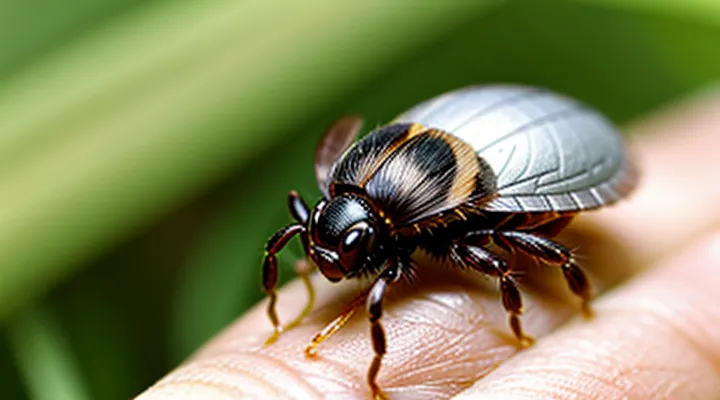
Introduction to Tick Bites
Why Safe Tick Removal is Crucial
Ticks attach to the skin for several days before detaching naturally. During this period they may transmit bacteria, viruses, or parasites. Early and correct extraction limits the window for pathogen transfer.
Improper removal—crushing the body, pulling with excessive force, or using unsterile tools—can force saliva and infected fluids deeper into the host tissue. This action increases the probability that disease‑causing agents enter the bloodstream.
Consequences of unsafe extraction include:
- Higher incidence of Lyme disease, Rocky Mountain spotted fever, or anaplasmosis.
- Extended local inflammation and secondary infection at the bite site.
- Potential for chronic joint, neurological, or cardiac complications.
Adhering to recommended techniques—gripping the tick close to the skin with fine‑point tweezers, applying steady upward pressure, and disinfecting the area afterward—prevents mouthpart breakage and reduces pathogen exposure. Proper disposal of the removed tick further eliminates the risk of accidental re‑attachment or contamination.
Risks Associated with Improper Removal
Improper removal of a tick can lead to several medical complications.
• Pathogen transmission increases when the mouthparts remain embedded; bacteria such as Borrelia or viruses may enter the bloodstream through the exposed wound.
• Skin trauma occurs if forceful pulling or crushing is applied, resulting in lacerations, hemorrhage, or scarring.
• Allergic reactions may develop from contact with tick saliva or from the inflammatory response to retained fragments.
• Secondary bacterial infection is possible when the puncture site is not cleaned promptly, providing a portal for opportunistic microbes.
• Incomplete extraction leaves tick parts in the dermis, which can act as a nidus for chronic inflammation and may complicate diagnostic evaluation.
Each risk underscores the necessity of adhering to established extraction techniques that minimize tissue damage and ensure complete removal of the parasite.
Preparation for Tick Removal
Essential Tools for Safe Removal
Fine-Tipped Tweezers
Fine‑tipped tweezers deliver the precision needed for safe tick extraction from human skin. Their narrow, pointed jaws allow a firm grip on the tick’s head without compressing the body, reducing the risk of pathogen release.
The tool’s design minimizes tissue damage. By grasping the tick as close to the skin surface as possible, the mouthparts are removed intact, preventing the head from breaking off and remaining embedded.
Steps for removal using fine‑tipped tweezers:
- Disinfect the tweezers with alcohol before use.
- Grasp the tick’s mouthparts directly, avoiding contact with the abdomen.
- Apply steady, upward pressure; pull straight out without twisting or jerking.
- After extraction, clean the bite area with antiseptic.
- Preserve the tick in a sealed container for identification if needed.
- Monitor the site for signs of infection over the following weeks.
Proper handling of fine‑tipped tweezers ensures complete removal, limits trauma, and lowers the chance of disease transmission.
Antiseptic Wipes or Rubbing Alcohol
Antiseptic wipes and rubbing alcohol are essential for post‑removal skin care after extracting a tick. After the tick is grasped with fine‑pointed tweezers and pulled straight upward, the bite site should be disinfected to reduce bacterial contamination.
- Choose a sterile wipe impregnated with an approved antiseptic or a cotton ball saturated with 70 % isopropyl alcohol.
- Apply the wipe directly to the puncture wound, covering the area for several seconds.
- Allow the surface to air‑dry; do not rinse immediately, as drying enhances antimicrobial action.
- Dispose of the used wipe in a sealed container to prevent cross‑contamination.
Rubbing alcohol can also be used to cleanse the tweezers before and after the procedure, ensuring the instrument remains free of pathogens. Consistent use of these disinfectants lowers the risk of secondary infection and supports proper wound healing.
Airtight Container or Ziploc Bag
After extraction, the parasite must be secured to prevent accidental release and to enable later identification. An airtight container or a sealable plastic bag creates a closed environment that isolates the specimen from the surrounding area.
Steps for handling the removed parasite:
- Transfer the organism directly into the container or bag without crushing it.
- Expel excess air and close the seal tightly, ensuring no gaps remain.
- Attach a label indicating the date, body site, and geographic location of the bite.
- Store the sealed unit in a refrigerator (4 °C) if identification is required within a few days; otherwise, freeze at –20 °C for long‑term preservation.
- When disposal is final, place the sealed unit in a fire‑safe container and incinerate, or follow local regulations for biological waste.
The sealed package also serves as a safe transport vessel for laboratory analysis, eliminating the risk of the creature escaping during transit.
Preparing the Area Around the Tick
Preparing the area around the attached parasite is essential for safe extraction. Clean the skin, improve visibility, and arrange necessary tools before attempting removal.
- Disinfect the surrounding skin with an alcohol swab or antiseptic wipe.
- Apply a magnifying lens if the parasite is small or located in a low‑visibility region.
- Ensure bright illumination; a LED lamp or natural daylight reduces errors.
- Place a sterile gauze pad or cloth to hold the skin taut, preventing movement.
- Keep fine‑tipped tweezers or a dedicated removal device within immediate reach.
After these steps, the environment is ready for precise and hygienic extraction of the «tick».
Step-by-Step Guide to Safe Tick Removal
Grasping the Tick Correctly
Avoiding Squeezing the Tick's Body
When a tick’s body is squeezed, its internal contents—including saliva, blood, and potential pathogens—can be forced into the wound. This increases the risk of disease transmission and complicates subsequent treatment.
To prevent compression of the tick’s abdomen, follow these steps:
- Use fine‑pointed, non‑slipping tweezers; avoid thumb‑fingers that may crush the tick.
- Position the tweezers as close to the skin as possible, grasping the tick’s head or mouthparts.
- Apply steady, even pressure while pulling straight upward; do not twist or jerk the instrument.
- Release the tick onto a clean surface; do not press it against the skin or any surface.
After removal, cleanse the area with antiseptic, then monitor for signs of infection or rash. If symptoms develop, seek medical evaluation promptly.
Pulling the Tick Out
Steady, Upward Motion
The removal of a tick from human skin depends on applying a constant upward pull without twisting. The motion must remain steady, maintaining tension directly away from the skin surface, which prevents the mouthparts from breaking off and remaining embedded.
• Grasp the tick as close to the skin as possible with fine‑point tweezers.
• Position the tweezers so the force aligns with the tick’s body axis.
• Apply a slow, continuous upward pressure, keeping the motion smooth and uninterrupted.
• Continue the pull until the tick releases entirely.
• Disinfect the bite area and the tweezers after extraction.
A smooth, uninterrupted lift minimizes tissue damage and reduces the risk of pathogen transmission. The method relies solely on the principle of «steady, upward motion», ensuring the tick is removed in one piece.
Avoiding Twisting or Jerking
When extracting a tick, the primary objective is to detach the parasite without compressing its body. Applying a steady, vertical force with fine‑point tweezers prevents the mouthparts from breaking off and remaining embedded in the skin.
- Grip the tick as close to the skin surface as possible.
- Pull upward with constant pressure; avoid any rotational movement.
- Do not jerk the instrument; sudden motions increase the risk of mouthpart fracture.
- After removal, cleanse the site with antiseptic and monitor for signs of infection.
Maintaining a smooth, linear traction eliminates the need for twisting, reduces tissue trauma, and ensures the entire organism is removed intact.
What to Do if Parts of the Tick Remain
When a tick’s mouthparts stay embedded in the skin, the remaining tissue can cause irritation, infection, or transmit disease. Prompt, proper action reduces these risks.
- Use fine‑point tweezers to grasp the visible portion of the mouthpart as close to the skin as possible.
- Apply steady, downward pressure to extract the fragment without squeezing the surrounding tissue.
- Avoid digging, burning, or using chemicals on the embedded piece; these methods increase tissue damage and infection risk.
- After removal, cleanse the area with antiseptic solution and cover with a clean bandage.
If the fragment cannot be removed easily, or if bleeding, redness, or swelling develops, seek professional medical care. A health‑care provider can excise the residual part safely and assess the need for prophylactic antibiotics or tick‑borne disease testing. Continuous monitoring of the site for several days is advisable; any worsening symptoms warrant immediate evaluation.
Aftercare and Monitoring
Cleaning the Bite Area
After a tick has been detached, the bite site must be decontaminated to lower the chance of bacterial entry. Immediate cleaning removes residual saliva and potential pathogens that remain on the skin.
- Wash hands thoroughly with soap and water before touching the wound.
- Rinse the bite area with running water, then cleanse with a mild antiseptic such as povidone‑iodine or chlorhexidine; apply the solution with a clean gauze pad.
- Pat the skin dry with a sterile cloth; avoid rubbing, which can irritate the tissue.
- Cover the spot with a sterile, non‑adhesive dressing only if bleeding occurs; otherwise, leave the area exposed to air.
Observe the site for redness, swelling, or a rash over the next several days. Any signs of infection or a persistent erythematous ring warrant medical evaluation. Regular monitoring ensures prompt treatment if complications arise.
Disposing of the Tick
Preserving the Tick for Identification
Preserving the removed tick enables accurate species determination and assessment of disease transmission risk. Proper handling prevents degradation of morphological features and preserves any pathogen material that may be present.
- Place the tick in a small, sealable container such as a vial or zip‑lock bag.
- If the specimen is still alive, keep it moist with a drop of saline solution; otherwise, allow it to air‑dry on a paper towel.
- Label the container with the date of removal, anatomical site of attachment, and any relevant exposure details.
- Store the sealed container at room temperature for up to 24 hours; for longer periods, refrigerate at 4 °C to inhibit bacterial growth.
When the tick is ready for analysis, submit the sealed container to a qualified laboratory or public health authority. Include the accompanying label information to facilitate precise identification and appropriate medical follow‑up.
Monitoring for Symptoms of Tick-Borne Illnesses
Common Symptoms to Watch For
After a tick attachment, monitor the skin and systemic condition for specific indicators that may signal infection. Early detection relies on recognizing patterns that differ from normal post‑removal irritation.
Key signs to observe include:
- Localized expanding redness with a clear center, often described as «erythema migrans».
- Persistent fever exceeding 38 °C (100.4 °F).
- Severe headache, especially when accompanied by neck stiffness.
- Muscle or joint pain that does not resolve within a few days.
- Unexplained fatigue or malaise.
- Swollen or tender lymph nodes near the bite site.
- Nausea, vomiting, or abdominal discomfort.
- Rash with spots or a mottled appearance, characteristic of certain rickettsial infections.
The presence of any combination of these symptoms warrants immediate medical evaluation. Prompt laboratory testing can confirm the specific pathogen and guide appropriate antimicrobial therapy. Continuous observation for at least two weeks after removal improves the likelihood of early intervention.
When to Seek Medical Attention
After a tick is removed, immediate medical evaluation is required if any of the following conditions are present.
- The tick remains attached or a portion of its mouthparts is visible in the skin.
- The bite site shows increasing redness, swelling, warmth, or pus formation.
- The individual develops fever, chills, headache, muscle aches, or fatigue within weeks of the bite.
- A rash appears, especially one resembling a bull’s‑eye (central clearing with a red outer ring) or any other unusual skin eruption.
- Signs of an allergic reaction occur, such as hives, difficulty breathing, swelling of the face or throat, or rapid pulse.
- The bite is located on the scalp, face, or other hard‑to‑reach area where complete removal cannot be confirmed.
- The tick was attached for more than 24 hours, increasing the risk of pathogen transmission.
Prompt consultation with a healthcare professional ensures appropriate testing, prophylactic treatment, and management of potential complications. Early intervention reduces the likelihood of severe disease progression.
Prevention of Tick Bites
Personal Protective Measures
When a tick attaches to skin, direct contact with the parasite poses a risk of pathogen transmission. Reducing exposure requires specific protective actions before, during, and after extraction.
- Wear disposable nitrile or latex gloves to create a barrier between hands and the arthropod.
- Select fine‑pointed, non‑toothed tweezers; grip the tick as close to the skin surface as possible.
- Disinfect the extraction site with an alcohol‑based solution or iodine before grasping the tick.
- Apply steady, upward pressure; avoid twisting or squeezing the body to prevent rupture of the abdomen.
- Place the removed tick in a sealed container with alcohol for later identification if needed.
- Clean hands thoroughly with soap and water after glove removal, then discard gloves according to biohazard protocols.
Additional precautions include wearing long‑sleeved shirts and trousers when in tick‑infested areas, tucking clothing into socks, and performing full‑body inspections after outdoor exposure. Regularly launder clothing at high temperatures to eliminate any unnoticed specimens. These measures collectively minimize direct contact and lower the probability of disease transmission during tick removal.
Tick-Proofing Your Environment
Tick‑proofing an environment reduces the likelihood of human exposure and simplifies safe removal when contact occurs.
Effective landscape management includes:
- Maintaining grass at a height of 2‑3 inches to limit tick habitat.
- Removing leaf litter, tall weeds, and brush from perimeters.
- Applying acaricides to borders of lawns, gardens, and wooded edges according to label instructions.
Indoor safeguards consist of:
- Sealing cracks and gaps in foundations, doors, and windows to prevent tick entry.
- Installing screens on vents and utility openings.
- Using vacuum cleaners with HEPA filters to capture ticks that may hitchhike indoors.
Pet‑focused measures involve:
- Administering veterinary‑approved tick preventatives regularly.
- Bathing and grooming pets weekly, checking for attached ticks after outdoor activity.
- Restricting animal access to high‑risk zones such as dense underbrush.
Routine monitoring and maintenance support long‑term protection:
- Conducting monthly inspections of yards and structures for tick signs.
- Rotating treated zones to avoid resistance buildup.
- Recording observations to adjust control strategies promptly.
Implementing these practices establishes a hostile environment for ticks, thereby decreasing the probability of attachment and facilitating safer removal if exposure occurs.
