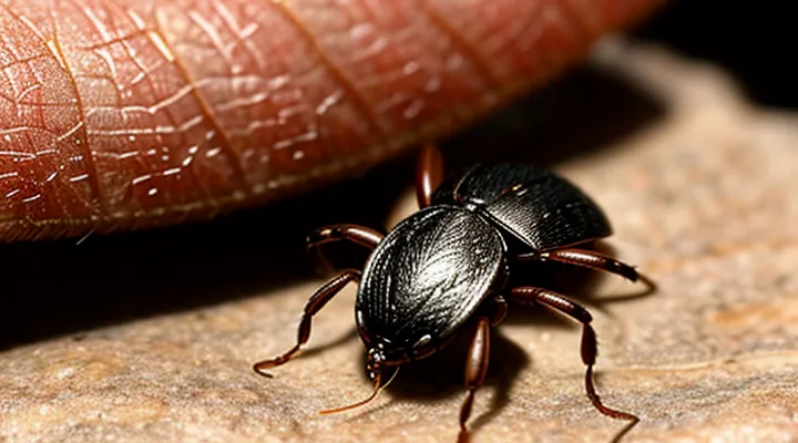
«Understanding Tick Bites»
«Identifying a Tick Bite»
«Visual Confirmation»
Visual confirmation is a critical step after extracting a tick from a person. It ensures that the parasite has been entirely removed and reduces the risk of infection.
After removal, inspect the bite site with adequate lighting. Look for any remaining parts of the tick, especially the mouthparts, which can remain embedded in the skin. Any visible fragment warrants immediate re‑extraction.
Key points for visual verification:
- Confirm that the tick’s body is intact; a split or missing segment suggests incomplete removal.
- Observe the skin for a small, dark spot indicative of a retained mouthpart.
- Use a magnifying glass or a dermatoscope if the area is difficult to see.
- Clean the area with antiseptic solution and re‑examine after a few minutes to detect any residual debris.
If any portion of the tick is still visible, repeat the extraction with fine‑point tweezers, grasping as close to the skin as possible. After successful removal, apply a sterile bandage and monitor the site for signs of infection, such as redness, swelling, or a rash.
«Visual confirmation» therefore serves as the final safeguard, verifying complete tick extraction and supporting prompt medical attention when necessary.
«Common Symptoms»
Ticks attach to the skin to feed on blood, and their bite can produce a range of clinical signs. Recognizing these manifestations enables timely intervention and reduces the risk of disease transmission.
«Common Symptoms» of a tick bite include:
- Localized redness or a small, raised bump at the attachment site
- Expanding erythema‑migrans, often described as a target‑shaped rash, typically appearing 3‑30 days after the bite
- Mild to moderate fever, sometimes accompanied by chills
- Headache, which may be persistent or intermittent
- Muscular or joint pain, frequently reported as generalized aches
- Fatigue or a feeling of malaise that develops within days of exposure
- Swollen or tender lymph nodes near the bite or in regional areas
The appearance of any of these signs, especially a spreading rash or fever, warrants professional evaluation. Prompt removal of the tick and appropriate medical treatment mitigate complications such as Lyme disease, Rocky Mountain spotted fever, or other tick‑borne infections.
«Safe Tick Removal Methods»
«Preparation for Removal»
«Gathering Necessary Tools»
Effective tick extraction requires specific instruments that minimize tissue damage and reduce infection risk. Selecting appropriate tools before beginning the procedure ensures a swift, controlled removal.
- «Fine‑point tweezers» or small curved forceps, stainless‑steel, non‑slipping grip
- Disposable nitrile gloves to prevent direct contact with the parasite
- Antiseptic solution (e.g., 70 % isopropyl alcohol or povidone‑iodine) for skin preparation and post‑removal cleaning
- Magnifying glass or portable loupe to enhance visibility of the attachment site
- Sealable plastic container with a lid or a puncture‑proof vial for safe disposal of the tick
Prepare the work area by disinfecting the skin around the attachment point, donning gloves, and arranging the listed items within easy reach. After removal, apply antiseptic to the bite site, seal the tick in the container, and discard it according to local health regulations.
«Hand Hygiene»
Effective hand hygiene is essential when extracting a tick from a person’s skin. Clean hands reduce the risk of introducing pathogens from the tick’s mouthparts into the wound.
Before handling the tick, wash hands thoroughly with soap and water for at least 20 seconds. Apply an alcohol‑based hand rub if soap is unavailable. Wear disposable gloves if possible; gloves provide an additional barrier against bacterial contamination.
During removal, use fine‑pointed tweezers to grasp the tick as close to the skin as possible. Pull upward with steady, even pressure, avoiding squeezing the body. After the tick is detached, place it in a sealed container for identification if needed.
Immediately after extraction, discard gloves and perform hand hygiene again. Wash hands with soap and water, then apply an antiseptic solution to the bite site. Repeat hand washing before any subsequent medical tasks.
Key steps for optimal hand hygiene in tick removal:
- Wash hands with soap and water (≥20 seconds).
- Use alcohol‑based hand rub if soap unavailable.
- Wear disposable gloves when feasible.
- Discard gloves and wash hands again after removal.
- Apply antiseptic to the bite area.
Consistent adherence to these practices minimizes infection risk and supports safe tick extraction.
«The Proper Removal Technique»
«Grasping the Tick»
Effective removal of a tick begins with a secure grip. The goal is to isolate the parasite’s mouthparts without compressing its body, which could cause the release of infectious fluids.
To achieve a proper grasp, follow these steps:
- Use fine‑point tweezers or a specialized tick‑removal tool; avoid blunt instruments.
- Position the tweezers as close to the skin as possible, targeting the head region.
- Apply steady, gentle pressure to lift the tick straight upward; do not twist or jerk.
- Maintain continuous traction until the entire organism separates from the host.
- After extraction, disinfect the bite site with an antiseptic solution.
Improper handling, such as squeezing the abdomen, increases the risk of pathogen transmission. Secure, vertical traction ensures the mouthparts remain intact and the bite heals without complications.
«Gentle, Steady Pull»
The method known as «Gentle, Steady Pull» minimizes tissue damage while detaching the parasite. By applying constant, moderate pressure to the mouthparts, the tick disengages without crushing the body, reducing the risk of pathogen transmission.
Steps for implementation:
- Position fine‑point tweezers as close to the skin as possible, grasping the tick’s head.
- Apply a smooth, even force directly outward, avoiding jerky movements.
- Maintain traction until the entire organism separates from the host.
- Disinfect the bite site with an antiseptic solution.
- Store the removed tick in a sealed container for possible identification.
«Avoiding Common Mistakes»
Removing a tick safely requires precision; errors can increase infection risk and leave mouthparts embedded.
Common mistakes include:
- Grasping the tick with fingers or tweezers that compress the body.
- Pulling upward with a jerking motion.
- Leaving the head in the skin.
- Applying chemicals, heat, or oils to force detachment.
- Delaying removal after discovery.
To avoid these errors, follow a strict protocol:
- Choose fine‑pointed, non‑slipping tweezers; position them as close to the skin as possible, holding the tick’s head.
- Apply steady, gentle pressure outward, maintaining alignment with the skin to prevent tearing.
- Inspect the site after extraction; ensure no fragments remain, then cleanse with antiseptic.
- Discard the tick in a sealed container; avoid crushing it.
- Record the removal date and monitor the bite area for signs of infection over the next several weeks.
«Post-Removal Care»
«Wound Cleaning and Disinfection»
After a tick is detached from the skin, the bite site requires immediate cleaning and disinfection to prevent secondary infection. The area should be flushed with running water for at least 30 seconds. Gentle mechanical removal of debris using a sterile gauze pad eliminates residual saliva and tick fragments.
The following steps ensure optimal wound care:
- Irrigate the puncture with sterile saline or clean tap water.
- Apply a mild antiseptic solution (e.g., 0.5 % povidone‑iodine or 2 % chlorhexidine) using a sterile swab.
- Allow the antiseptic to remain in contact for 2–3 minutes before drying.
- Cover the site with a sterile, non‑adhesive dressing if bleeding persists.
- Monitor the wound daily for signs of erythema, swelling, or purulent discharge; seek medical attention if such symptoms develop.
Avoid using harsh chemicals such as hydrogen peroxide or alcohol directly on the wound, as they may delay tissue healing. Replace the dressing at least once daily or when it becomes wet or soiled. Proper wound management reduces the risk of bacterial colonization and supports rapid recovery after tick removal.
«Monitoring the Bite Area»
After a tick is removed, the bite area requires continuous observation. Any deviation from normal skin appearance may indicate infection or disease transmission.
- Redness extending beyond the immediate site
- Swelling or a raised bump
- Development of a rash, especially a concentric “bull’s‑eye” pattern
- Fever, chills, fatigue, muscle or joint pain
Monitoring should extend for at least fourteen days. Record temperature daily and note any new skin changes. Photograph the area at 24‑hour intervals to document progression.
If any listed signs appear, obtain medical evaluation promptly. Early treatment reduces the risk of complications such as Lyme disease or bacterial infection.
«When to Seek Medical Attention»
«Incomplete Removal»
Incomplete removal occurs when the mouthparts of a tick remain embedded in the skin after the body is pulled off. Retained mandibles can continue to feed, release pathogens, and provoke local inflammation.
Residual mouthparts often cause a small, raised bump that may itch, swell, or become tender. If the area enlarges, oozes, or develops a rash, the risk of infection increases.
Identification relies on visual inspection and gentle palpation. A visible fragment, a pinpoint puncture, or persistent irritation after removal suggests that the tick was not fully extracted.
To address incomplete removal:
- Disinfect the site with an antiseptic solution.
- Apply fine‑point tweezers or a sterile needle to grasp the exposed tip of the mouthpart.
- Pull straight upward with steady pressure, avoiding twisting.
- Clean the area again after extraction and apply a sterile bandage.
Professional evaluation is warranted if:
- The fragment cannot be accessed safely.
- The bite site shows signs of spreading redness, fever, or flu‑like symptoms.
- Uncertainty exists about the tick species or duration of attachment.
Prompt, complete extraction reduces the likelihood of tick‑borne disease transmission and minimizes tissue damage.
«Signs of Infection»
Monitoring the area after a tick is extracted is essential. Recognizing «Signs of Infection» enables timely medical intervention.
Typical indicators include:
- Redness that spreads beyond the bite site
- Swelling or a palpable lump
- Increased warmth around the area
- Pus or other discharge
- Fever, chills, or night sweats
- Headache, fatigue, or muscle aches
- Joint pain or stiffness
- Emerging rash, particularly a concentric “bull’s‑eye” pattern
These symptoms may appear within 24 hours to several weeks post‑removal. Persistent or worsening signs warrant professional evaluation, as they can signal bacterial infection, Lyme disease, or other tick‑borne illnesses. Prompt treatment reduces complications and promotes recovery.
«Symptoms of Tick-Borne Illnesses»
«Lyme Disease Indicators»
Removing a tick promptly reduces the risk of infection, yet recognizing early signs of Lyme disease remains essential for timely treatment. After extraction, monitor the bite site and overall health for specific indicators that suggest Borrelia burgdorferi transmission.
Key clinical signs include:
- Erythema migrans – a expanding, red skin lesion often resembling a bull’s‑eye pattern, appearing within 3‑30 days.
- Flu‑like symptoms – fever, chills, headache, fatigue, muscle and joint aches without an obvious cause.
- Neurological manifestations – facial palsy, meningitis‑like headache, or peripheral neuropathy.
- Cardiac involvement – irregular heartbeat or heart block, typically occurring weeks after the bite.
- Arthritic symptoms – intermittent joint swelling, especially in the knees, emerging weeks to months later.
Absence of a rash does not exclude infection; laboratory testing (ELISA followed by Western blot) confirms seroconversion when clinical suspicion persists. Immediate consultation with a healthcare provider is advised if any of the above signs develop, even after proper tick removal.
«Other Potential Illnesses»
Ticks can transmit a range of pathogens beyond the immediate concern of the bite site. Awareness of these additional health risks is essential for anyone who has removed a tick.
- Lyme disease – infection by Borrelia burgdorferi, characterized by erythema migrans, joint pain, and neurological symptoms.
- Rocky Mountain spotted fever – caused by Rickettsia rickettsii, presenting with fever, rash, and headache.
- Anaplasmosis – Anaplasma phagocytophilum infection, leading to fever, muscle aches, and leukopenia.
- Ehrlichiosis – Ehrlichia chaffeensis infection, similar clinical picture to anaplasmosis.
- Babesiosis – protozoan Babesia microti infection, causing hemolytic anemia and fever.
- Tularemia – Francisella tularensis transmission, resulting in ulcerative lesions and lymphadenopathy.
- Powassan virus disease – rare flavivirus infection, potentially causing encephalitis.
- Tick‑borne relapsing fever – Borrelia spp. infection, marked by recurrent fever episodes.
Medical evaluation should follow any tick encounter, especially when symptoms such as fever, rash, joint pain, or neurological changes appear within weeks. Laboratory testing can confirm specific infections, guiding appropriate antimicrobial or antiviral therapy. Prompt treatment reduces the likelihood of severe complications and long‑term sequelae.
«Tick Bite Prevention»
«Protective Clothing»
Protective clothing serves as a primary barrier against tick attachment during outdoor activities. Fabric that is tightly woven, such as denim or synthetic blends, reduces the likelihood of arthropod penetration. Light-colored garments improve visual detection of attached ticks, facilitating prompt removal.
Key features of effective protective apparel include:
- Long sleeves and full-length trousers, preferably tucked into socks or boots.
- Elastic cuffs or gaiters that seal the gap between clothing and skin.
- Insect‑repellent treatments applied to fabric, such as permethrin, which remain active after several washes.
- Seamless or smooth interior surfaces that discourage tick movement.
When a tick is discovered, the clothing should be examined for additional specimens before removal. Gloves, preferably disposable, protect the hands from direct contact with the tick’s mouthparts. Using fine‑pointed tweezers, grasp the tick as close to the skin as possible and extract with steady, upward pressure. After removal, cleanse the bite site with antiseptic and inspect the surrounding clothing for any displaced ticks.
Maintaining the integrity of «protective clothing» through regular laundering and re‑application of repellents sustains its defensive capability. Replacing worn or damaged garments eliminates gaps that could facilitate tick attachment.
«Insect Repellents»
Insect repellents play a decisive role in reducing the risk of tick attachment, thereby simplifying subsequent extraction procedures. Effective repellents contain active ingredients that deter questing ticks from climbing onto human skin.
Key active compounds include:
- DEET (N,N‑diethyl‑m‑toluamide) – broad‑spectrum efficacy, long‑lasting protection.
- Picaridin – comparable potency to DEET, lower odor profile.
- Permethrin – applied to clothing, creates a contact‑kill barrier.
- IR3535 – moderate efficacy, suitable for sensitive skin.
Application recommendations:
- Apply liquid or spray formulations to exposed skin at least 30 minutes before exposure; reapply according to product specifications, typically every 6–8 hours.
- Treat socks, trousers, and shirts with permethrin; allow treated garments to dry completely before wearing.
- Avoid applying repellents to broken skin or near eyes; wash hands after handling.
Following successful tick removal, re‑application of a skin‑safe repellent can prevent additional attachment during the same outing. Maintaining a routine of repellent use in tick‑endemic areas markedly lowers the incidence of bites and the need for emergency extraction.
«Regular Tick Checks»
Regular tick checks are a critical component of effective tick management. Systematic examination of the skin reduces the likelihood that a feeding tick remains attached long enough to transmit pathogens.
A practical routine includes the following steps:
- Perform a full‑body inspection within 24 hours after outdoor exposure. Use a hand mirror for hard‑to‑see areas such as the scalp, behind the ears, underarms, and groin.
- Examine each body region methodically, moving from head to toe. Pay special attention to moist folds and hair‑covered zones where ticks commonly attach.
- Use fine‑toothed tweezers or a tick‑removal tool to grasp any visible tick as close to the skin surface as possible. Pull upward with steady, even pressure, avoiding twisting or crushing the parasite.
- After removal, cleanse the bite site with soap and water or an antiseptic solution. Store the tick in a sealed container for identification if symptoms develop.
- Document the date, location of the bite, and removal method. Record any subsequent signs of infection for prompt medical evaluation.
Conduct these checks daily during peak tick season and after any activity in wooded or grassy habitats. Consistent implementation of «Regular Tick Checks» enhances early detection, facilitates safe extraction, and minimizes health risks associated with tick‑borne diseases.
