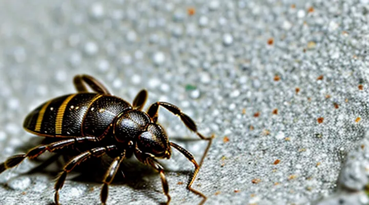
Understanding the Risks of Tick Bites
Why Proper Removal Matters
Potential Health Complications
Ticks attached to the skin can introduce a range of medical issues if removal is improper. The most significant complications include:
- Transmission of bacterial infections such as Lyme disease, caused by Borrelia burgdorferi, which may develop weeks after the bite.
- Viral illnesses like tick‑borne encephalitis, presenting with fever, headache, and neurological symptoms.
- Anaplasmosis and ehrlichiosis, leading to fever, muscle aches, and possible organ dysfunction.
- Babesiosis, a malaria‑like disease that can cause hemolytic anemia, especially in immunocompromised patients.
- Localized skin infection at the bite site, often due to secondary bacterial invasion, resulting in redness, swelling, and pus formation.
- Allergic reactions to tick saliva or to medications used during removal, ranging from mild urticaria to severe anaphylaxis.
- Development of a granuloma or persistent nodule if mouthparts remain embedded, which may require surgical excision.
Prompt, complete extraction reduces the likelihood of these outcomes. If symptoms such as fever, rash, joint pain, or neurological changes appear after a bite, immediate medical evaluation is essential. Early antibiotic therapy can prevent progression of bacterial infections, while antiviral or supportive care may be required for viral syndromes. Monitoring the removal site for signs of infection or retained fragments is a critical component of post‑removal care.
Preventing Secondary Infections
When a tick is detached, the skin surrounding the bite remains vulnerable to bacterial invasion. Immediate cleaning eliminates surface contaminants and reduces the risk of secondary infection.
- Wash the area with antiseptic soap or a mild disinfectant for at least 30 seconds. Rinse thoroughly with clean water.
- Apply an alcohol‑based solution or povidone‑iodine to the puncture site. Allow it to dry before covering.
- Use a sterile adhesive bandage only if the wound bleeds; otherwise keep the area exposed to air to promote drying.
Monitor the site for signs of infection: increasing redness, swelling, warmth, pus, or escalating pain. If any of these symptoms develop, seek medical evaluation promptly. Antibiotic therapy may be required, especially if the bite was deep or the individual has compromised immunity.
Avoid scratching or manipulating the wound, as this can introduce additional microbes. Keep the area clean, dry, and protected from friction for several days. Regularly replace bandages with fresh sterile dressings if they become wet or soiled.
Vaccination against tetanus should be up to date; a booster may be indicated if the bite penetrates deeply or if the last immunization exceeds ten years. Maintaining overall skin health and promptly addressing any irritation further diminishes the chance of secondary infection after tick removal.
Essential Tools for Tick Removal
Recommended Items
Effective tick extraction requires specific supplies. Use items that allow firm grip, precise control, and immediate wound care.
- Fine‑point tweezers (flat or slanted tip) designed to grasp the tick’s head without crushing the body.
- Small, sterile needle or pin for lifting the tick if its mouthparts are embedded in difficult locations.
- Antiseptic wipes or alcohol pads to clean the bite area before and after removal.
- Disposable gloves to prevent direct contact with the tick and reduce infection risk.
- Sealable container (plastic bag or vial) with a small amount of 70 % isopropyl alcohol for preserving the tick if identification is needed.
- Hydrocortisone cream or a mild topical antibiotic ointment for post‑removal skin irritation.
- Sharp, clean scissors (optional) for trimming excess skin if the tick’s mouthparts remain embedded after extraction.
Having these items readily available ensures the tick is removed promptly, minimizing the chance of pathogen transmission and reducing tissue damage.
Items to Avoid
When extracting a tick, using inappropriate tools or techniques can damage the parasite, increase the chance of pathogen transmission, and cause skin injury. Avoid actions that compromise the integrity of the tick’s mouthparts or that introduce contaminants to the wound.
- Do not crush, squeeze, or mutilate the tick’s body; this may release infectious fluids.
- Do not use fingers alone to pull the tick; lack of grip can cause the head to break off.
- Do not employ hot objects (e.g., matches, heated needles) to burn or melt the tick; heat can force saliva deeper into the skin.
- Do not apply petroleum jelly, nail polish, alcohol, or other chemicals to the tick; these substances do not detach the parasite and may irritate the skin.
- Do not use tweezers with blunt or rounded tips; insufficient grip can cause the tick to detach incompletely.
- Do not twist, jerk, or yank the tick forcefully; such motion often results in a fragmented mouthpart remaining embedded.
- Do not delay removal; prolonged attachment increases pathogen transmission risk.
Selecting the correct method—fine‑point tweezers, steady traction, and immediate cleaning—eliminates the hazards associated with the items listed above.
Step-by-Step Tick Removal Procedure
Preparation
Locating the Tick
When a tick attaches, it embeds its mouthparts in the skin, often leaving only the body visible. Begin by exposing the area with a bright light; a magnifying glass can improve visibility. Look for a small, rounded mass that may be brown, black, or gray, ranging from a few millimeters to a centimeter in length. The head may be obscured, but the tick’s body will appear raised above the skin surface.
If the bite site is in a hair‑covered region, part the hair away from the skin with a fine comb or a disposable brush. Run the comb from the skin outward, clearing hair that could conceal the parasite. In areas with dense fur or clothing fibers, gently press a piece of transparent adhesive tape against the skin; the tick may adhere to the tape, revealing its position.
When the tick is located, note the exact spot before attempting removal. Marking the site with a non‑permanent skin marker or recording the body region helps ensure precise handling and reduces the risk of leaving mouthparts behind.
Cleaning the Area
After extracting a tick, the surrounding skin must be disinfected to prevent infection. Begin by washing the area with mild soap and running water, removing any residual blood or debris. Rinse thoroughly and pat dry with a clean towel.
Apply an antiseptic solution—such as 70 % isopropyl alcohol, povidone‑iodine, or a chlorhexidine wipe—directly onto the bite site. Allow the disinfectant to remain for at least 30 seconds before removing excess fluid with a sterile gauze pad.
If irritation or redness develops, repeat the antiseptic application twice daily for 24–48 hours. Monitor the area for signs of infection (increasing pain, swelling, pus) and seek medical attention if symptoms worsen.
Cleaning protocol
- Soap and water wash – 15 seconds.
- Rinse and dry with sterile material.
- Apply antiseptic – cover fully.
- Hold for 30 seconds, then blot.
- Re‑apply as needed for 2 days.
The Removal Itself
Grasping the Tick
Use fine‑tipped tweezers or a specialized tick‑removal tool. Grip the tick as close to the skin as possible, avoiding the body of the parasite. Apply steady pressure to pull straight upward; do not twist, jerk, or squeeze the tick’s abdomen, which can force infected fluids into the wound.
- Choose tweezers with narrow, pointed tips.
- Position the tips at the tick’s head, just below the skin surface.
- Clamp firmly, ensuring the mouthparts are captured.
- Pull upward with constant, even force until the tick releases.
- Disinfect the bite area and wash hands after removal.
Pulling Technique
The pulling technique removes a tick by grasping the mouthparts and applying steady traction without twisting or crushing the body.
Before beginning, wash hands, wear disposable gloves if available, and expose the attachment site with a clean towel or gauze. Use fine‑pointed, flat‑tipped tweezers or a specialized tick‑removal tool; avoid using fingers or blunt instruments.
- Position the tweezers as close to the skin as possible, gripping the tick’s head or the part embedded in the epidermis.
- Apply gentle, continuous pressure upward, maintaining alignment with the body’s axis.
- Continue pulling until the entire organism separates from the skin.
- Release the grip immediately after detachment; do not squeeze the tick’s abdomen.
After extraction, place the tick in a sealed container for identification if needed, then disinfect the bite area with an antiseptic solution. Observe the site for several days; seek medical advice if redness, swelling, or flu‑like symptoms develop.
Aftercare
Cleaning the Bite Site
After extracting a tick, the bite area requires immediate attention to reduce infection risk. First, wash hands thoroughly with soap and water before touching the site. Then cleanse the skin using mild antibacterial soap, applying gentle friction for at least 20 seconds. Rinse completely and pat dry with a clean disposable towel.
Apply a topical antiseptic such as povidone‑iodine, chlorhexidine, or an alcohol‑based solution. Allow the antiseptic to remain on the skin for the recommended contact time (typically 30 seconds to one minute) before covering the area. If a sterile dressing is needed, use a non‑adhesive gauze pad secured with hypoallergenic tape.
Monitor the site for signs of inflammation—redness expanding beyond a few millimeters, swelling, warmth, or pus formation. Should any of these symptoms appear, seek medical evaluation promptly.
Key steps for post‑removal care
- Hand hygiene before handling the wound
- Soap‑water cleaning, 20 seconds of gentle scrubbing
- Rinse and dry with sterile material
- Apply antiseptic, observe recommended dwell time
- Cover with sterile dressing if necessary
- Observe for infection indicators and consult a professional if they develop
Completing these actions promptly minimizes bacterial entry and supports optimal healing after a tick bite.
Monitoring for Symptoms
After a tick is extracted, observe the bite site and the patient for at least four weeks. Early detection of infection relies on recognizing specific signs rather than waiting for severe illness.
Watch for the following symptoms:
- Redness or expanding rash, especially a target‑shaped lesion.
- Fever, chills, or muscle aches without an obvious cause.
- Persistent headache, fatigue, or joint pain.
- Swelling of lymph nodes near the bite area.
- Unusual nausea, vomiting, or abdominal discomfort.
If any of these manifestations appear, seek medical evaluation promptly. Documentation of the tick’s appearance, removal date, and geographic location can aid clinicians in selecting appropriate diagnostic tests and treatment. Continuous monitoring reduces the risk of delayed diagnosis and complications associated with tick‑borne diseases.
What Not to Do When Removing a Tick
Common Mistakes
Incorrect Removal Methods
Improper techniques increase the chance of infection, cause the tick’s mouthparts to stay embedded, or trigger the release of disease‑carrying fluids.
- Burning the tick with a match, lighter, or hot surface. Heat damages the tick’s body, often leaving the head lodged in the skin and may spread pathogens.
- Applying chemicals such as petroleum jelly, nail polish remover, or insecticide. These substances do not detach the parasite and can irritate the surrounding tissue.
- Squeezing or crushing the tick with fingers or tweezers. Pressure forces saliva and infected fluids into the bite site, raising the risk of disease transmission.
- Pulling the tick with excessive force or twisting motions. Aggressive traction can break the tick, leaving the capitulum embedded and requiring surgical removal.
- Using folk remedies like a single‑pointed needle, a hair dryer, or a hot water soak. These methods are ineffective and may cause additional skin injury.
Each of these practices fails to achieve a clean extraction and may worsen health outcomes. The only reliable approach involves grasping the tick close to the skin with fine‑pointed tweezers and applying steady, even pressure to remove it whole.
Using Unsuitable Substances
Removing a tick from human skin requires a method that minimizes the chance of the parasite’s mouthparts breaking off and reduces the risk of pathogen transmission. Applying substances that are not designed for this purpose interferes with those goals.
Commonly used inappropriate agents include:
- Petroleum‑based products (e.g., Vaseline, mineral oil). They cause the tick to swallow fluid, potentially leading to deeper attachment and increased chance of mouthpart loss during extraction.
- Alcohol or iodine solutions. Immediate exposure irritates the tick, prompting it to release saliva that may contain infectious agents.
- Heat sources such as a candle flame or hot match. Sudden temperature change can trigger salivation and increase pathogen transfer.
- Nail polish remover, acetone, or other solvents. These chemicals damage skin tissue, obscure the tick’s position, and do not facilitate removal of the entire organism.
The physiological response of the tick to these substances often results in prolonged feeding, heightened engorgement, and a higher probability of the exoskeleton remaining embedded. Moreover, chemical irritation can cause the tick’s salivary glands to discharge additional infectious material into the bite site.
For safe extraction, use fine‑pointed tweezers to grasp the tick as close to the skin as possible and apply steady, upward pressure. Do not twist, jerk, or compress the body. After removal, cleanse the area with soap and water, then monitor for signs of infection. Avoid any of the listed unsuitable agents, as they compromise the effectiveness of the procedure and increase health risks.
When to Seek Medical Attention
Signs of Complications
After a tick is taken out, monitor the bite site and overall health for any abnormal developments. Early identification of problems can prevent serious illness.
Typical indicators that the removal may have led to complications include:
- Redness or swelling that expands beyond the immediate area of the bite.
- A circular rash, often with a clear center, that appears days to weeks after the bite.
- Persistent fever, chills, or unexplained fatigue.
- Joint or muscle pain, especially if it worsens or shifts locations.
- Headache, dizziness, or visual disturbances.
- Nausea, vomiting, or abdominal discomfort.
- Unusual bruising or bleeding at the removal site.
If any of these symptoms arise, seek medical evaluation promptly. Laboratory testing may be required to rule out infections such as Lyme disease, Rocky Mountain spotted fever, or other tick‑borne illnesses. Early treatment improves outcomes and reduces the risk of long‑term damage.
Incomplete Removal
Incomplete removal occurs when the tick’s mouthparts remain embedded in the skin after the body is extracted. Retained parts can irritate tissue, provoke a local inflammatory response, and increase the likelihood of pathogen transmission. Early identification prevents complications.
Signs of a partially removed tick include a visible puncture site, a small black or brown fragment protruding from the skin, and localized redness or swelling. If the fragment is not visible, persistent itching or a raised bump may indicate its presence.
To address an incomplete extraction:
- Disinfect the area with an antiseptic solution.
- Use fine‑point tweezers or a sterile needle to grasp the exposed portion as close to the skin as possible.
- Apply steady, gentle pressure to pull the fragment outward without crushing it.
- After removal, clean the site again and apply a mild antiseptic ointment.
- Monitor the wound for signs of infection, such as increasing redness, warmth, or discharge; seek medical care if these develop.
Prevention of incomplete removal relies on proper technique from the start. Grasp the tick’s head or mouthparts with fine tweezers, maintain a firm grip, and pull upward with steady force. Avoid twisting, squeezing, or using chemicals that may cause the tick to regurgitate its contents. Following these steps reduces the risk of leaving any part behind.
Concerns About Tick-Borne Diseases
Tick exposure carries a measurable risk of infection with pathogens such as Borrelia burgdorferi, Anaplasma phagocytophilum, and Rickettsia species. These agents can cause Lyme disease, anaplasmosis, and spotted‑fever rickettsiosis, respectively, each presenting with fever, rash, joint pain, or neurological signs. Prompt and correct removal of the attached arthropod lowers the probability of pathogen transmission, because most bacteria require several hours of feeding before entering the host’s bloodstream.
Improper extraction—pulling with force, twisting, or crushing the body—can force mouthparts deeper into the skin and increase bacterial inoculation. The primary objective is to detach the tick intact, preserving the feeding apparatus for visual confirmation that no fragments remain.
Safe removal procedure
- Disinfect the skin surrounding the tick with an alcohol swab or iodine solution.
- Grasp the tick as close to the skin as possible using fine‑point tweezers, avoiding the abdomen.
- Apply steady, upward traction without squeezing the body.
- After detachment, cleanse the bite site again with antiseptic.
- Preserve the specimen in a sealed container for identification if symptoms develop.
Following extraction, observe the bite area for redness, swelling, or a bull’s‑eye rash, and monitor systemic signs such as fever or fatigue for up to four weeks. Seek medical evaluation if any of these manifestations appear, as early antibiotic therapy can prevent severe disease progression.
