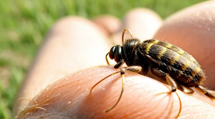
«Understanding Tick Bites»
«Initial Reaction to a Tick Bite»
«The Role of Tick Saliva»
Tick bites frequently produce a localized itching sensation, and the primary cause lies in the substances injected with the mouthparts. When a tick attaches, it releases a complex mixture of saliva that contains biologically active molecules designed to facilitate blood feeding.
The saliva is rich in anticoagulants, anti‑inflammatory proteins, proteases, and compounds that bind host histamine. These agents collectively modify the host’s immediate immune response, allowing the parasite to remain undetected for several days.
One effect of the saliva is the suppression of typical inflammatory signals, which paradoxically delays the recruitment of immune cells to the bite site. At the same time, certain salivary components provoke mast‑cell degranulation, leading to the release of histamine and other pruritogenic mediators. The simultaneous inhibition of inflammation and induction of histamine creates a delayed but noticeable itch.
Salivary factors most directly linked to pruritus include:
- Salivary cement proteins that anchor the mouthparts and irritate surrounding skin cells.
- Proteases that degrade extracellular matrix, exposing nerve endings.
- Histamine‑binding proteins that modulate host histamine levels, sometimes causing rebound release.
- Anti‑inflammatory peptides that dampen early cytokine signaling, extending the period before the immune system reacts.
Understanding the composition and actions of tick saliva clarifies why the bite area often becomes itchy after the initial attachment. The interplay of immune suppression and histamine activation produces the characteristic delayed pruritic response observed in most tick exposures.
«Immune Response Mechanisms»
A tick bite introduces saliva that contains anticoagulants, proteases, and immunomodulatory proteins. These substances disrupt the skin barrier and trigger the body’s immediate defensive reactions.
The innate immune system reacts within minutes. Mast cells in the dermis degranulate, releasing histamine, tryptase, and other mediators. Histamine binds to sensory nerve endings, producing the characteristic pruritus. Tryptase activates protease‑activated receptors, amplifying the itch signal.
Simultaneously, keratinocytes and resident dendritic cells release cytokines such as interleukin‑1β and tumor necrosis factor‑α. These cytokines recruit neutrophils and monocytes to the bite site, generating inflammation that sustains the sensation of itch.
A secondary, adaptive response may develop over days. Antigen‑presenting cells process tick salivary proteins and present them to T lymphocytes. Helper T cells secrete interleukin‑4 and interleukin‑13, promoting a Th2‑biased response. The resulting IgE production sensitizes mast cells, leading to heightened degranulation upon re‑exposure and prolonged pruritic episodes.
Key mechanisms contributing to itch after a tick bite:
- Mast cell degranulation → histamine release
- Protease‑activated receptor activation → neuronal sensitization
- Cytokine cascade (IL‑1β, TNF‑α, IL‑4, IL‑13) → inflammatory cell recruitment
- Th2‑driven IgE response → amplified mast cell activity
Collectively, these immune processes explain why the bite area frequently becomes itchy and why the sensation can persist or intensify after the initial exposure.
«Common Reasons for Itching Post-Bite»
«Allergic Reactions»
«Components of Tick Saliva Causing Allergy»
When a tick attaches, it injects saliva that contains several bioactive molecules capable of triggering an allergic response in the host’s skin. The immune system reacts to these proteins, releasing histamine and other mediators that produce the characteristic itching sensation at the bite site.
Key allergenic constituents of tick saliva include:
- Proteases (e.g., cysteine and serine proteases): degrade host proteins, expose hidden epitopes, and activate inflammatory pathways.
- Anticoagulants (e.g., apyrase, anticoagulant peptide): interfere with clotting, but also act as immunogenic proteins that sensitize skin cells.
- Immunomodulatory proteins (e.g., Salp15, Salp20): bind to host immune receptors, suppress certain defenses while provoking hypersensitivity in susceptible individuals.
- Lipocalins: transport small hydrophobic molecules; many function as allergen carriers that elicit IgE production.
- Histamine‑binding proteins: paradoxically modulate local histamine levels, leading to delayed or prolonged pruritus.
These components collectively disrupt normal skin homeostasis, promote vasodilation, and stimulate nerve endings. The resulting cascade of cytokine release and mast‑cell activation explains why the area around a tick bite often becomes itchy shortly after attachment.
«Individual Sensitivity Variations»
Tick bites frequently provoke a localized itch, but the intensity and timing of that sensation differ markedly among individuals. The variation stems from genetic, immunological, and environmental factors that shape each person’s cutaneous response to tick saliva.
Key determinants of personal sensitivity include:
- Genetic predisposition – Polymorphisms in genes encoding histamine receptors and cytokine pathways alter the magnitude of inflammatory signaling after a bite.
- Prior exposure – Repeated encounters with tick antigens can lead to either heightened reactivity (sensitization) or diminished response (tolerance), depending on the immune profile.
- Skin condition – Pre‑existing dermatoses, barrier defects, or chronic dryness increase susceptibility to pruritic reactions.
- Age and health status – Younger individuals and those with atopic tendencies generally report stronger itching, whereas immunocompromised patients may exhibit muted symptoms.
The underlying mechanism involves tick saliva, which contains anticoagulants, anesthetics, and proteins that modulate host immunity. In sensitive persons, these agents trigger mast cell degranulation, releasing histamine and other pruritogenic mediators. Less sensitive individuals experience limited mediator release, resulting in mild or absent itch.
Understanding these personal differences assists clinicians in predicting symptom severity, tailoring patient counseling, and selecting appropriate anti‑itch interventions.
«Inflammation and Skin Irritation»
«Mechanical Damage from Tick Attachment»
Ticks attach by inserting their barbed mouthparts, called the hypostome, into the epidermis and dermis. The physical penetration creates a puncture wound that disrupts keratinocytes and dermal collagen. This disruption damages cutaneous nerve endings, generating immediate sensory signals interpreted as itch. The wound also exposes underlying tissue to mechanical stress, prompting the release of endogenous pruritogenic mediators such as substance P and calcitonin‑gene‑related peptide from damaged nerves.
The mechanical injury contributes to itching through several mechanisms:
- Direct trauma to epidermal cells activates transient receptor potential (TRP) channels that convey itch signals.
- Disruption of the skin barrier allows rapid entry of environmental irritants, amplifying the pruritic response.
- Micro‑abrasions produced by the tick’s chelicerae create a focal area of inflammation, recruiting mast cells that degranulate and release histamine.
Consequently, the area surrounding a tick bite often itches shortly after attachment, even before the tick’s saliva introduces additional pharmacological agents. The itch intensity correlates with the depth of hypostome penetration and the extent of tissue disruption.
«Host Immune Cell Activity»
When a tick attaches to skin, host immune cells recognize foreign proteins in the saliva and in any transmitted pathogens. This recognition triggers a local inflammatory response that often manifests as pruritus.
- Neutrophils arrive within minutes, phagocytosing debris and releasing reactive oxygen species.
- Macrophages infiltrate later, presenting antigens to lymphocytes and secreting pro‑inflammatory cytokines.
- Dendritic cells capture tick antigens, migrate to draining lymph nodes, and activate adaptive immunity.
- Mast cells degranulate, liberating histamine and other mediators that directly stimulate sensory nerves.
- T‑helper cells, especially Th2 subsets, produce interleukin‑31, a potent itch‑inducing cytokine.
- Eosinophils accumulate in response to IL‑5, contributing additional pruritogenic substances.
The combined effect of histamine, leukotrienes, prostaglandins, and cytokines lowers the activation threshold of cutaneous C‑fibers. These fibers transmit itch signals to the spinal cord and brain, producing the characteristic sensation. The response peaks within several hours after the bite but can persist for days if tick saliva components continue to modulate immune activity or if secondary infection develops.
Understanding the specific cellular contributors clarifies why the bite site frequently itches and guides targeted interventions, such as antihistamines, corticosteroids, or cytokine blockers, to alleviate discomfort.
«Infections and Secondary Itching»
«Bacterial Skin Infections»
A tick bite often leaves a small puncture that can become a focus for bacterial colonisation. When skin‑resident bacteria such as Staphylococcus aureus or Streptococcus pyogenes invade the wound, they trigger an inflammatory response that commonly includes pruritus. The itch arises from cytokines and histamine released by immune cells reacting to bacterial toxins and tissue damage.
Typical features of a bacterial skin infection at a tick‑bite site include:
- Redness spreading beyond the original puncture
- Swelling and warmth
- Purulent discharge or crust formation
- Increased tenderness
- Persistent or worsening itch despite antihistamine use
The itch differs from the brief irritation caused by tick saliva, which usually subsides within hours. Bacterial inflammation sustains itch by maintaining mediator release and nerve sensitisation. Prompt cleaning of the bite, topical antiseptics, and, when signs of infection appear, systemic antibiotics reduce bacterial load and alleviate itching.
If the area remains itchy after 24–48 hours, especially with the signs listed above, bacterial involvement should be considered and evaluated by a healthcare professional.
«Tick-Borne Pathogens and Associated Symptoms»
Tick attachment often produces a localized pruritic response. The sensation results from mechanical disruption of skin, injection of salivary proteins, and subsequent release of histamine and other mediators by mast cells. In some individuals the reaction is mild; in others it may be pronounced, reflecting a hypersensitivity to tick saliva.
Pathogens transmitted by ticks generate additional cutaneous and systemic signs. Common agents and their typical manifestations include:
- Borrelia burgdorferi – erythema migrans (expanding, usually non‑pruritic rash), fever, arthralgia, fatigue.
- Rickettsia rickettsii – maculopapular rash often accompanied by intense itching, headache, high fever.
- Anaplasma phagocytophilum – fever, muscle aches, mild rash; itching is uncommon.
- Babesia microti – hemolytic anemia, chills, malaise; skin symptoms are rare.
- Powassan virus – encephalitis, meningitis; cutaneous irritation is not a primary feature.
When itching coincides with a spreading erythema or a papular rash, it may signal infection by rickettsial organisms rather than a simple mechanical irritation. Lyme disease typically presents with a non‑itchy target lesion, whereas Rocky Mountain spotted fever frequently produces a pruritic rash. Persistent or worsening itch after several days, especially with fever or systemic malaise, warrants medical evaluation to rule out pathogen‑induced dermatitis.
Early identification of pathogen‑related itch enables prompt antimicrobial therapy, reducing the risk of complications. Absence of systemic signs and rapid resolution of itching within 24–48 hours generally indicate a benign local reaction.
«Factors Influencing Itch Severity»
«Tick Species Differences»
Different tick species trigger varying skin reactions because their saliva contains distinct protein mixtures. Some proteins act as anticoagulants, others as immunomodulators, and the balance among them determines the intensity of local inflammation and pruritus.
- Ixodes scapularis (deer tick) – saliva rich in anti‑inflammatory compounds; initial bite often painless, itch may develop 24–48 hours later as the immune system recognizes foreign proteins.
- Dermacentor variabilis (American dog tick) – higher concentration of histamine‑like substances; itching can appear within a few hours, accompanied by redness and swelling.
- Amblyomma americanum (lone star tick) – secretes α‑galactosidase and other allergens; rapid onset of itch, sometimes severe, linked to allergic sensitization.
- Rhipicephalus sanguineus (brown dog tick) – limited salivary irritation; bite may remain unnoticed, itching typically mild or absent unless secondary infection occurs.
The duration and severity of itch depend on the host’s immune response, the duration of attachment, and whether the tick transmits pathogens that further stimulate inflammation. Species that inject potent vasodilators or histamine analogs provoke immediate pruritic sensations, while those with stronger immunosuppressive agents delay the reaction until the host’s defenses activate.
«Duration of Tick Attachment»
Ticks remain attached for a period that directly influences the host’s skin response. Most species attach for 24–48 hours before detaching if not removed, while some, such as Ixodes scapularis, may stay attached up to 72 hours or longer. The longer the attachment, the greater the probability that salivary proteins will provoke a localized inflammatory reaction, often perceived as itching.
Extended feeding increases exposure to tick saliva, which contains anticoagulants, anti‑inflammatory agents, and allergens. These substances disrupt normal skin homeostasis, trigger histamine release, and sensitize nerve endings. The host’s immune system may react within hours of attachment, producing a mild pruritic papule. If the tick remains for several days, cumulative allergen load can intensify the itch and may lead to a larger, erythematous wheal.
Typical attachment durations and associated pruritic outcomes:
- <12 hours – minimal saliva exposure; itch rarely reported.
- 12–24 hours – early inflammatory response; occasional mild itch.
- 24–48 hours – moderate saliva accumulation; itch common, often localized.
- >48 hours – high allergen load; pronounced itch, possible spreading erythema.
Prompt removal within the first 24 hours reduces saliva exposure, limits the inflammatory cascade, and markedly lowers the likelihood of itching.
«Location of the Bite»
Tick bites most often occur in regions where skin folds or hair is dense, because the parasite seeks a warm, protected site. Typical locations include the scalp, behind the ears, under the arms, the groin, the waistline, and the back of the knees. These areas differ in nerve density, skin thickness, and exposure to friction, all of which affect the perception of itch.
- Scalp and hair‑covered areas: Thin epidermis and abundant sensory nerves produce a sharp, early itch. Hair can conceal the attachment, delaying removal and prolonging irritation.
- Axillae and groin: Moist environment and frequent movement increase skin irritation, leading to persistent itching after the tick detaches.
- Waistline and belt line: Thin skin and constant friction from clothing amplify mechanical stimulation, intensifying the itch response.
- Leg creases (behind knees, ankles): Reduced airflow and sweat accumulation promote inflammation, sustaining itch for several days.
The intensity of itch correlates with the bite’s position because the local immune response varies with tissue type. Areas with higher concentrations of mast cells release more histamine when the tick’s saliva triggers inflammation, causing a more pronounced sensation. Additionally, regions that are less visible are often discovered later, allowing the tick to feed longer and inject more salivary proteins, which further amplifies the pruritic reaction.
«When to Seek Medical Attention»
«Persistent or Worsening Itching»
Persistent or worsening itching after a tick bite signals an ongoing skin response or a developing infection. The sensation typically begins within hours of attachment and may intensify over days. Several mechanisms explain this pattern:
- Local inflammatory reaction – Tick saliva contains proteins that suppress host immunity, provoking a delayed hypersensitivity response that manifests as pruritus.
- Allergic sensitization – Repeated exposure to tick antigens can trigger IgE‑mediated allergy, leading to persistent itch and swelling.
- Secondary bacterial infection – Scratching creates breaks in the epidermis, allowing Staphylococcus or Streptococcus species to colonize the bite site, which prolongs irritation.
- Tick‑borne pathogens – Early stages of Lyme disease, Rocky Mountain spotted fever, or other infections may produce erythema and itching before systemic symptoms appear.
- Tick paralysis toxin – Rarely, neurotoxic saliva induces localized pruritus that progresses to muscle weakness if untreated.
Clinical assessment should include: examination of the bite for erythema, central punctum, or necrosis; measurement of lesion size; evaluation for systemic signs such as fever, rash elsewhere, or joint pain. Laboratory testing may involve serology for Borrelia burgdorferi or PCR for other agents when indicated.
Management depends on the underlying cause. Immediate steps comprise gentle cleansing with antiseptic solution and application of a low‑potency corticosteroid cream to reduce inflammation. Oral antihistamines alleviate allergic itch, while topical antibiotics address confirmed bacterial involvement. If Lyme disease is suspected, a course of doxycycline (100 mg twice daily for 10–14 days) is recommended. Persistent or escalating symptoms beyond one week, or the emergence of fever, joint swelling, or neurological deficits, require prompt medical evaluation.
«Rash Development»
A tick bite introduces saliva containing anticoagulants and proteins that trigger a localized inflammatory response. The skin around the attachment site often becomes red, swollen, and itchy within minutes to hours. This early rash results from histamine release and irritation of nerve endings by the saliva’s irritant compounds.
If the tick remains attached for several days, the immune system may recognize foreign antigens and produce a delayed‑type hypersensitivity reaction. The reaction typically manifests as a larger, raised, erythematous area that intensifies in pruritus. The progression can be summarized as follows:
- Immediate phase (0–2 h): Redness and mild itching caused by mechanical irritation and saliva components.
- Early inflammatory phase (2–24 h): Increased swelling, warmth, and heightened itch due to histamine and cytokine release.
- Delayed hypersensitivity phase (24 h–72 h): Expanding erythema, possible papules or vesicles, and stronger itching as T‑cell–mediated immunity develops.
In some cases, a characteristic “bull’s‑eye” lesion appears days after the bite, indicating infection with Borrelia burgdorferi, the agent of Lyme disease. This annular rash often expands outward while the center clears, and it may be accompanied by persistent itching, though the itch can be less intense than in the earlier inflammatory stages.
The presence and severity of itching depend on:
- Saliva composition: Proteins that act as allergens provoke stronger pruritic responses.
- Duration of attachment: Longer feeding increases antigen exposure, amplifying immune activation.
- Host sensitivity: Individual variations in immune reactivity affect rash size and itch intensity.
Recognizing the pattern of rash development aids in distinguishing a simple irritant reaction from a tick‑borne infection that requires medical intervention. Prompt removal of the tick and observation of the skin’s response are essential steps in managing post‑bite symptoms.
«Systemic Symptoms of Illness»
A tick bite can produce a localized itching sensation caused by the saliva injected during feeding. In addition to this skin reaction, the bite may trigger systemic manifestations that indicate the spread of pathogens or an immune response.
Common systemic signs include:
- Fever or chills
- Headache
- Muscle or joint pain
- Generalized fatigue
- Nausea or vomiting
- Enlarged lymph nodes
- Rash that appears away from the bite site, often resembling a “bull’s‑eye” pattern
These symptoms arise when the tick transmits infectious agents such as Borrelia burgdorferi (Lyme disease), Rickettsia species (rocky‑mountain spotted fever), or Anaplasma phagocytophilum (anaplasmosis). The organisms enter the bloodstream, prompting the body’s inflammatory and immune pathways. Cytokine release produces fever, aches, and fatigue, while vascular inflammation can generate distant rashes. Lymphatic involvement leads to node enlargement.
When systemic signs develop, they signal that the infection has moved beyond the bite area. Prompt medical evaluation is required to confirm diagnosis and initiate antimicrobial therapy, reducing the risk of long‑term complications.
