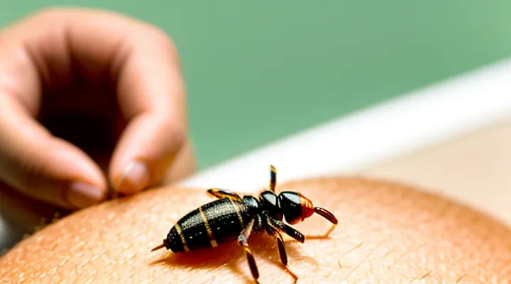
Understanding the Question
Distinguishing Between Types of Ticks
External Parasites
External parasites are organisms that live on the surface of a host and obtain nutrients from blood, tissue fluids, or skin debris. Among them, ticks belong to the order Ixodida and are primarily ectoparasites that attach to the epidermis of mammals, including humans.
Ticks normally embed their mouthparts into the superficial dermis, forming a feeding lesion that remains visible on the skin. In rare instances, the hypostome can penetrate deeper layers, creating a subdermal or subcutaneous nidus. This deeper placement results from prolonged attachment, aggressive feeding behavior, or host tissue reaction that drives the parasite inward.
Clinical manifestations of a subdermal tick include:
- Localized swelling or a firm nodule beneath the skin surface
- Minimal or absent surface puncture mark
- Possible erythema or mild pain at the site
- Absence of the typical engorged tick visible on the epidermis
Diagnosis relies on physical examination, ultrasonography, or magnetic resonance imaging to locate the embedded organism. Removal requires a sterile incision and careful extraction with forceps to avoid leaving mouthparts behind, which could provoke chronic inflammation.
Management steps:
- Confirm the presence of a tick within the subcutaneous tissue.
- Perform aseptic removal under local anesthesia.
- Clean the wound with antiseptic solution.
- Administer a prophylactic antibiotic if secondary bacterial infection is suspected.
- Monitor for signs of tick‑borne disease and initiate appropriate serologic testing if indicated.
Prevention focuses on avoiding tick exposure through protective clothing, repellents containing DEET or picaridin, and regular body inspections after outdoor activities. Prompt removal of attached ticks before they embed deeply reduces the risk of subdermal colonization and associated complications.
Mites vs. Ticks
Ticks belong to the order Ixodida, whereas mites are members of the order Acari. Both groups are arachnids, but ticks are generally larger (2–30 mm) and possess a specialized mouthpart called a capitulum adapted for prolonged blood feeding. Mites range from microscopic to a few millimetres and often have chelicerae suited for surface feeding or tissue scraping.
Key distinctions:
- Size: ticks > 2 mm; mites ≤ 2 mm.
- Mouthparts: ticks have a hypostome with barbs; mites possess simple chelicerae.
- Feeding duration: ticks remain attached for hours to days; mites feed for seconds to minutes.
- Life‑stage attachment: ticks embed across all stages (larva, nymph, adult); most mites do not penetrate deeply.
- Host specificity: many ticks are obligate blood feeders; many mites are opportunistic, feeding on skin, hair, or environmental debris.
Subcutaneous localization requires a parasite capable of penetrating the dermis and remaining viable. Ticks can insert their hypostome into the skin, sometimes migrating beneath the epidermis, leading to a palpable nodule. Mites lack the anatomical structures to achieve such deep insertion; they remain on the surface or within superficial follicles.
Documented cases of human subcutaneous infestation involve:
- Dermacentor spp. (American dog tick) – reported as a nodule after removal of a partially embedded tick.
- Ixodes ricinus (European castor‑bean tick) – occasional subdermal migration causing localized swelling.
Mite species such as Sarcoptes scabiei cause burrows within the stratum corneum but never reach the subcutaneous tissue. Consequently, the likelihood of a mite establishing a subcutaneous lesion is negligible compared with that of a tick.
True Subcutaneous Parasites in Humans
Human Parasitic Conditions
Scabies Mites
Scabies mites (Sarcoptes scabiei) are microscopic arthropods that inhabit the superficial layers of human skin. The female burrows into the stratum corneum, laying eggs within a tunnel that measures only a few millimetres deep. The organism never penetrates beyond the epidermis, and the host’s immune response is limited to the epidermal level, producing itching and a characteristic rash.
Ticks are arachnids that attach to the external surface of the skin, inserting their mouthparts into the dermis to obtain blood. While a tick’s hypostome can become lodged in the skin, the body of the tick remains external. Reports of “subcutaneous” ticks describe cases where the head is embedded, not a true intradermal location. Scabies mites differ fundamentally: they are not blood‑sucking parasites, they do not embed whole bodies beneath the skin, and their life cycle is confined to the epidermal surface.
Key distinctions:
- Habitat: scabies mites – epidermal burrows; ticks – surface attachment with shallow dermal penetration.
- Size: scabies mite ~0.3 mm; tick – several millimetres, visible to the naked eye.
- Feeding: mites consume skin cells and fluids; ticks ingest blood.
- Clinical presentation: scabies – intense nocturnal pruritus, linear burrows; tick – local erythema, possible attachment site pain.
Because scabies mites never reside beneath the epidermis, they cannot be considered a subcutaneous tick. The presence of a true subcutaneous tick in humans is exceedingly rare and, when reported, involves atypical embedding of the tick’s mouthparts rather than the entire organism. Treatment for scabies includes topical scabicides (e.g., permethrin 5 %) or oral ivermectin; tick removal requires careful extraction of the mouthparts and, if necessary, antimicrobial prophylaxis.
Other Skin-Dwelling Organisms
Humans host several ectoparasites that occupy the epidermis, hair follicles, or the superficial dermis. These organisms differ from subcutaneous ticks in depth of penetration, life cycle, and pathogenic potential.
-
Mites
• Sarcoptes scabiei – burrows within the stratum corneum, causing intense pruritus and papular eruptions.
• Demodex folliculorum and Demodex brevis – reside in hair follicles and sebaceous glands, often asymptomatic but may contribute to rosacea or blepharitis. -
Lice
• Pediculus humanus capitis – attaches to scalp hair, feeds on blood, produces nits adhered to shafts.
• Pediculus humanus corporis – inhabits clothing seams, migrates to skin to feed. -
Acari and larvae
• Chiggers (Trombiculidae) – larval stage penetrates epidermis, injects proteolytic enzymes, leading to erythematous wheals.
• Sand fleas (Tunga penetrans) – female embeds partially into skin, forming a nodular lesion with a central punctum. -
Leeches
• Aquatic leeches can attach to exposed skin, ingest blood, and create prolonged bleeding sites. -
Fungi
• Dermatophytes (Trichophyton, Microsporum, Epidermophyton) colonize keratinized layers, producing ring-shaped lesions and scaling.
Clinical presentation varies: itching, erythema, papules, nodules, or ulceration may indicate one of these organisms. Diagnosis relies on direct microscopic examination, skin scraping, or biopsy. Treatment protocols include topical acaricides (e.g., permethrin for scabies), oral ivermectin for severe infestations, pediculicidal shampoos for lice, and antifungal agents for dermatophyte infections. Removal of embedded larvae or fleas often requires surgical excision or careful extraction.
Recognition of these skin-dwelling parasites is essential when evaluating unexplained subcutaneous masses or persistent dermatitis, ensuring accurate differential diagnosis and appropriate therapeutic intervention.
The Mechanism of Tick Attachment
How Ticks Feed
Mouthpart Anatomy
Ticks attach to hosts using highly specialized mouthparts that enable penetration of skin layers and secure feeding. The apparatus consists of four primary structures:
- Palps (hypostomal palps): sensory appendages that locate suitable insertion sites and guide the feeding tube.
- Chelicerae: paired, blade‑like cutters that slice the epidermis and dermis, creating an entry channel.
- Hypostome: a barbed, tube‑shaped organ bearing numerous backward‑pointing teeth that anchor the tick within the host tissue.
- Scentor (or dorsal groove): a channel that houses saliva‑secreting glands, facilitating anticoagulant and immunomodulatory fluid delivery.
During attachment, the chelicerae separate the skin, allowing the hypostome to be driven into the dermal matrix. The barbs on the hypostome resist removal, while the palps maintain orientation. Saliva injected through the scentor suppresses host hemostasis and immune response, permitting prolonged blood ingestion.
If a tick penetrates beyond the superficial epidermis, the hypostome can lodge within the dermis or subdermal connective tissue. In such cases, the tick appears embedded beneath the skin surface, sometimes without a visible external body. This subcutaneous positioning results from deep insertion of the hypostome and may be facilitated by:
- Tick species with elongated hypostomes (e.g., Ixodes spp.).
- Prolonged attachment periods, allowing tissue remodeling around the barbs.
- Host skin thickness, which can accommodate deeper embedding without immediate expulsion.
Clinically, a subdermal tick may present as a localized nodule, swelling, or erythema. Removal requires careful extraction of the hypostome to avoid rupture and subsequent inflammation. Understanding the precise anatomy of tick mouthparts informs diagnostic suspicion and guides appropriate removal techniques.
Attachment Process
Ticks attach to a host by inserting their hypostome, a barbed feeding organ, into the skin. The process proceeds through distinct stages:
- Penetration: The tick’s forelegs locate a suitable site, then the hypostome pierces the epidermis and reaches the dermal layer.
- Anchoring: Barbs on the hypostome secure the tick, while salivary secretions contain proteins that inhibit coagulation and suppress the host’s immune response.
- Cement formation: After insertion, the tick secretes a cement-like substance that hardens around the mouthparts, creating a stable attachment that can persist for days.
- Engorgement: Blood is drawn through a canal in the hypostome; the tick expands while remaining fixed by the cement.
In rare cases, the hypostome may advance beyond the dermis into subcutaneous tissue, especially when the tick is forced to embed deeper by host movement or tissue inflammation. This deeper insertion can place the tick beneath the skin surface, making removal more difficult and increasing the risk of localized infection. The host’s inflammatory response may encapsulate the tick, forming a nodule that can be mistaken for other skin lesions. Early detection and proper extraction are essential to prevent complications.
Potential Misconceptions and Concerns
Symptoms of Tick Bites
Localized Reactions
A subcutaneous tick lodged beneath the skin elicits a confined inflammatory response that is often the first clinical clue. The reaction typically appears as a well‑defined, erythematous area surrounding the tick’s mouthparts. Swelling may accompany the redness, producing a palpable, tender nodule. Patients frequently report localized pruritus or a burning sensation that intensifies when the tick attempts to feed.
Common manifestations include:
- Erythema with clear margins, usually 1–2 cm in diameter.
- Edema that may fluctuate with the tick’s activity.
- Pain or tenderness on pressure.
- Pruritus that can persist after removal.
- A central punctum or ulceration where the tick’s hypostome penetrated the dermis.
The timeline of these signs varies. Initial erythema can develop within minutes of attachment; edema and pain often peak after several hours. If the tick remains embedded for days, a chronic granulomatous nodule may form, sometimes mimicking a cyst or skin tumor.
Differential diagnosis should consider:
- Bacterial cellulitis – distinguished by diffuse spreading, systemic fever, and rapid progression.
- Insect bite reaction – generally less localized, with multiple lesions.
- Foreign‑body granuloma – similar appearance but lacks the central punctum.
Prompt removal of the tick reduces the duration of the localized response. After extraction, applying a cold compress can alleviate swelling, while topical corticosteroids may diminish persistent inflammation. Monitoring for secondary infection or systemic tick‑borne disease remains essential.
Systemic Illnesses
A tick that embeds beneath the dermis can serve as a portal for pathogens that spread beyond the local bite site. Once the vector breaches the skin barrier, microorganisms may enter the bloodstream, initiating systemic disease processes that affect multiple organ systems.
Common systemic conditions linked to subdermal tick attachment include:
- Lyme disease – spirochete Borrelia burgdorferi disseminates to joints, heart, and nervous tissue; early signs involve erythema migrans, later stages may present arthritis, carditis, or neuroborreliosis.
- Rocky Mountain spotted fever – Rickettsia rickettsii causes vasculitis; fever, headache, and a characteristic rash appear after a febrile incubation.
- Ehrlichiosis and anaplasmosis – intracellular bacteria (Ehrlichia chaffeensis, Anaplasma phagocytophilum) produce leukopenia, thrombocytopenia, and hepatic dysfunction.
- Babesiosis – protozoan Babesia microti replicates within erythrocytes, leading to hemolytic anemia, hemoglobinuria, and possible organ failure in immunocompromised hosts.
- Tick‑borne encephalitis – flavivirus infection may cause meningitis, encephalitis, or meningoencephalitis, with neurological deficits persisting after acute illness.
- Tularemia – Francisella tularensis can cause ulceroglandular disease, pneumonic forms, or septicemia when disseminated systemically.
Diagnosis relies on serologic assays, polymerase chain reaction, or culture of blood and tissue specimens, often supplemented by imaging to assess organ involvement. Prompt antimicrobial therapy—doxycycline for most bacterial agents, specific antivirals or antiparasitics where indicated—reduces morbidity and prevents progression to severe systemic sequelae. Continuous monitoring of hematologic and hepatic parameters is essential during treatment to detect complications early.
When to Seek Medical Attention
A tick that has migrated beneath the skin can remain hidden while causing tissue damage, inflammation, and transmission of pathogens. Prompt professional evaluation prevents complications such as secondary infection, allergic reaction, or systemic illness.
Seek medical attention if any of the following occur:
- Persistent redness, swelling, or warmth surrounding the bite site for more than 24 hours.
- Increasing pain, throbbing, or a palpable lump that enlarges.
- Fever, chills, headache, muscle aches, or fatigue developing within two weeks of exposure.
- A rash resembling a bull’s‑eye (expanding erythema with central clearing) or any new skin lesions.
- Signs of an allergic response: hives, swelling of the face or throat, difficulty breathing.
- Evidence of tick‑borne disease in endemic areas (e.g., Lyme, Rocky Mountain spotted fever) regardless of symptom severity.
When a subdermal tick is suspected, a clinician should:
- Perform a thorough visual and tactile examination, possibly using dermoscopy or ultrasound.
- Remove the organism under sterile conditions, ensuring the head and mouthparts are extracted completely.
- Collect the tick for species identification and pathogen testing.
- Order appropriate laboratory studies (e.g., CBC, inflammatory markers, serology for Lyme disease) based on clinical presentation.
- Provide prophylactic antibiotics or antitoxin therapy if indicated by local guidelines.
Delaying care increases the risk of irreversible tissue injury and systemic infection. Immediate evaluation is essential for accurate diagnosis, safe removal, and timely treatment.
