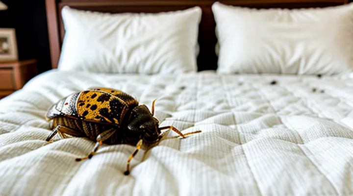
The Biology of Bed Bugs and Their Feeding Habits
Understanding Bed Bug Anatomy and Digestion
Bed bugs possess a flattened, oval body divided into three regions: head, thorax, and abdomen. The head bears piercing‑sucking mouthparts (the proboscis) equipped with stylets that penetrate skin to reach blood vessels. The thorax supports six legs and two short wings (vestigial in most species). The abdomen houses the digestive tract and reproductive organs, expanding considerably after a blood meal.
The digestive system begins with the foregut, a short tube that transports ingested blood to the midgut. In the midgut, proteolytic enzymes break down hemoglobin and plasma proteins, releasing nutrients that are absorbed into the hemolymph. Excess fluid and undigested components pass into the hindgut, where water is reabsorbed and waste is concentrated. The hindgut terminates at the anus, through which fecal material is expelled.
Excreted waste consists primarily of digested blood pigments, especially hemoglobin derivatives that darken upon exposure to air. As the fecal matter dries, oxidation intensifies the color, producing the characteristic black or dark brown spots found on mattresses, bedding, and furniture. Repeated defecation after consecutive feedings can create a linear trail of stains along the bed bug’s travel path.
Key anatomical and physiological factors that generate these stains:
- Proboscis‑mediated blood intake delivering large volumes of hemoglobin.
- Midgut enzymatic breakdown producing pigmented by‑products.
- Hindgut water reabsorption concentrating waste.
- Oxidative darkening of expelled feces upon contact with ambient oxygen.
The Feeding Process: How Bed Bugs Consume Blood
The Role of Anticoagulants and Anesthetics
Bed bugs inject a cocktail of salivary proteins when they pierce the skin. Two components—anticoagulants and anesthetics—directly influence the appearance of the dark marks that remain after feeding.
Anticoagulants prevent the blood from clotting at the bite site. By keeping the blood in a fluid state, they allow it to spread into surrounding tissue and to be drawn into the insect’s mouthparts. The excess fluid eventually leaks onto bedding or clothing, where it dries and oxidizes. Oxidized hemoglobin turns brown‑black, producing the characteristic stains.
Anesthetics suppress the host’s pain response. The bite goes unnoticed, so the insect can feed longer and ingest a larger volume of blood. Extended feeding increases the amount of liquid excreted onto the substrate, amplifying the size and darkness of the resulting spot.
Key points:
- Salivary anticoagulants (e.g., apyrases, nitrophorins) keep blood liquid, facilitating seepage onto fabrics.
- Anesthetic proteins (e.g., D7 family, salivary gland peptides) delay host detection, permitting prolonged feeding.
- The combination of larger blood volume and continuous fluid flow leads to more extensive deposition of hemoglobin.
- Drying and oxidation of hemoglobin produce a melanin‑like pigment that appears as a black or brown stain.
Understanding the biochemical actions of these salivary agents clarifies why the marks left by bed bugs are dark, persistent, and often mistaken for other types of stains.
The Composition and Formation of Black Spots
What Exactly Are These Black Spots?
Digested Blood and Fecal Matter
Bed bugs produce the dark marks commonly seen on mattresses and linens by depositing two types of residue: partially digested blood and fecal pellets. The blood that the insects ingest is broken down by enzymes, leaving a reddish‑brown fluid that oxidizes quickly after exposure to air, turning dark. This fluid can seep onto fabric, creating irregular stains that match the shape of the bug’s feeding site.
Fecal matter consists of concentrated waste that contains undigested hemoglobin. After the bug excretes the material, the hemoglobin oxidizes, resulting in a black or deep brown speck. The pellets are typically 1–3 mm in diameter and may appear as a line or cluster, depending on the insect’s movement.
Key characteristics of the stains:
- Color change from reddish to black as oxidation proceeds.
- Size correlates with the bug’s feeding frequency; frequent feeders leave larger, more numerous spots.
- Location aligns with the bug’s hiding places, often near seams, folds, or mattress edges.
Understanding that the marks are a combination of oxidized blood remnants and fecal deposits clarifies the source of the discoloration and aids in accurate identification of an infestation.
Distinguishing Bed Bug Feces from Other Stains
Bed bug excrement appears as small, dark specks that often resemble pepper grains. The spots are typically matte, ranging from deep brown to almost black, and they may be slightly raised when fresh. Over time, they fade to a lighter brown and become more powdery. These characteristics differ markedly from other household stains.
- Color and texture: Insect feces are uniformly dark and dry; mold stains show greenish or black fuzzy growth, while rust leaves a reddish‑brown wet smear.
- Typical locations: Bed bug droppings cluster on mattress seams, headboards, and nightstand surfaces near sleeping areas. Water damage stains spread across larger wall sections, and pet urine marks appear near feeding zones.
- Size and shape: Excrement measures 1–3 mm, often irregular but rounded; coffee spills create larger, irregular blotches, and ink leaks produce smooth, flowing lines.
- Reaction to moisture: A few drops of water dissolve bed bug spots, turning them into a faint brown solution; mold stains swell and become more pronounced, while rust stains darken.
- Microscopic evidence: Under magnification, bed bug feces reveal tiny, partially digested blood cells, a feature absent from other stains.
Identifying these markers enables precise determination of whether dark markings stem from Cimex lectularius activity or from unrelated sources such as mildew, corrosion, or pet waste. Accurate recognition is essential for targeted pest control measures and prevents unnecessary remediation of non‑infestation stains.
The Chemical and Biological Basis of Spot Formation
Hemoglobin Degradation and Pigment Production
Bed bug excrement appears as dark spots on bedding and furniture because the insects digest the hemoglobin they obtain from blood meals and convert it into pigmented waste. After a blood meal, proteolytic enzymes break down the globin chains, releasing free heme groups. The released heme is unstable in the gut environment and undergoes enzymatic oxidation. Heme oxygenase catalyzes the conversion of heme to biliverdin, which is subsequently reduced to bilirubin. Further oxidation of bilirubin produces insoluble, melanin‑like pigments that are dark brown to black.
- Ingestion of blood provides hemoglobin.
- Proteases cleave globin, freeing heme.
- Heme oxygenase transforms heme into biliverdin.
- Biliverdin is reduced to bilirubin.
- Oxidative polymerization of bilirubin yields dark pigments (hematin, melanin analogs).
- Pigments are excreted as solid particles that adhere to surfaces.
The resulting pigments are chemically resistant and bind tightly to fabric fibers, creating the characteristic stains that remain visible for days. Their formation occurs within hours after feeding, explaining the rapid appearance of the spots. Cleaning agents that do not disrupt the pigment–fiber bond often fail to remove the stains, requiring targeted oxidative or enzymatic treatments.
The Consistency and Appearance of the Stains
The stains produced by cimicids are primarily fecal deposits formed after the insect digests blood. Their texture ranges from a fine, powder‑like residue to a slightly tacky film when fresh. As the material dries, it becomes brittle and can be brushed off easily, unlike mold or mildew, which adheres more firmly.
Typical visual traits include:
- Dark brown to black coloration, often described as “ink‑spot” or “rusty” depending on the age of the deposit.
- Size varies from pinpoint specks (1–2 mm) to larger smears (up to 5 mm) when multiple excretions coalesce.
- Irregular edges, sometimes with a halo of lighter discoloration caused by blood oxidation.
- Location on seams, folds, or edges of mattresses, box springs, headboards, and nearby furniture.
- Occasionally accompanied by reddish stains where a crushed bug has released hemolymph.
The consistency and appearance together help differentiate these marks from other household blemishes. Fresh deposits feel slightly moist and may smudge under pressure; older spots are dry, flaky, and can be removed with a gentle brush or vacuum. Their distinct dark hue and location pattern are reliable indicators of an active infestation.
Identifying and Interpreting Black Spots
Locating Bed Bug Feces
Common Hiding Spots and Infestation Indicators
Bed bugs typically conceal themselves in locations that provide darkness, limited disturbance, and easy access to a host. Understanding these preferred refuges helps identify early signs of an infestation and explains the appearance of dark discolorations on bedding and furniture.
Common hiding spots include:
- Mattress seams, folds, and tags where the fabric creates tight crevices.
- Box springs and bed frames, especially wooden slats and metal joints.
- Upholstered furniture, such as cushions, seams, and under the fabric covering.
- Wall cracks, baseboards, and behind picture frames where temperature remains stable.
- Luggage racks, suitcases, and travel bags that have been placed near sleeping areas.
- Electrical outlets, switch plates, and wiring channels that offer concealed gaps.
Infestation indicators appear before visible insects are detected. The most reliable signs are:
- Small, dark‑brown to black specks on sheets, pillowcases, or mattress edges. These are fecal deposits that dry and turn black, often mistaken for dirt.
- Tiny, translucent or rust‑colored spots that result from crushed insects, leaving a reddish stain that darkens over time.
- Tiny, oval eggs measuring about 0.5 mm, often found in clusters near the same hiding sites.
- A sweet, musty odor that intensifies as the population grows.
- Live or dead bugs observed in the aforementioned refuges, especially after a thorough inspection with a flashlight.
Detecting these markers promptly allows targeted treatment, reducing the spread of stains and preventing further population expansion.
Patterns and Distribution of Stains
Bed bug excretions appear as small, dark‑brown to black specks that vary in size from a pinhead to a few millimeters. The spots are typically oval or round, with crisp edges when fresh and a slightly smudged appearance after exposure to air or cleaning attempts.
Distribution follows the insects’ preferred refuges. On mattresses, stains concentrate along seams, tags, and the edges of the box spring, where bugs hide during daylight hours. Pillows and mattress toppers may show linear streaks if bugs travel along fabric folds. Bed frames, headboards, and nightstands often exhibit clusters near cracks, joints, or upholstery seams, reflecting the proximity of harborages.
Furniture in the bedroom, such as upholstered chairs or sofas, can host scattered spots along cushions and armrests. Walls may display faint lines near baseboards or behind picture frames, indicating migration pathways. In severe infestations, stains spread outward in a radial pattern from a central nesting site, creating a gradient of density that diminishes with distance.
The progression of stains provides clues to infestation age. Fresh deposits are sharply defined and dark, while older ones fade to a reddish‑brown hue and may become powdery. Multiple layers of overlapping spots suggest repeated feeding events in the same location.
Understanding these patterns assists in pinpointing active harborages, evaluating infestation severity, and directing targeted treatment measures.
Differentiating Bed Bug Spots from Other Marks
Comparisons with Mold, Dirt, and Other Insect Droppings
Black stains that appear on sheets, mattress seams, or headboards are frequently identified as the excrement of Cimex lectularius. The droplets are typically 0.5–1 mm in diameter, deep brown to black, and maintain a distinct, almost glossy appearance after drying. Their distribution often follows the host’s resting positions, forming clusters or linear trails along seams and folds.
Mold colonies produce discoloration that can resemble dark spots, yet the underlying characteristics differ markedly. Mold growth displays fuzzy or powdery texture, expands outward in irregular patches, and frequently emits a musty odor. The coloration varies from greenish‑black to gray, depending on species, and the material is usually moist, allowing spores to spread in humid environments. In contrast, bed‑bug fecal spots remain dry, retain a defined shape, and lack the radiating hyphal structures typical of fungal colonies.
Dirt accumulation generates brown or black marks through contact with soil, dust, or fabric fibers. Such marks are generally irregular, lack the uniform size of insect excrement, and may contain visible particles of sand or lint. Dirt stains are not confined to the sleeping surface; they appear on any exposed area that contacts the floor or clothing, whereas bed‑bug spots concentrate near the insect’s feeding sites.
Other insects leave characteristic droppings that help distinguish them from bed‑bug excrement:
- Cockroach feces: small, cylindrical, dark brown to black, often found in clusters near food sources, with a crumbly consistency.
- Flea feces: tiny, dark specks resembling pepper grains, typically located on pet bedding or near animal hideouts, and often accompanied by a strong, unpleasant odor.
- Carpet beetle frass: fine, powdery, grayish‑white particles, usually associated with damaged fabrics rather than blood‑fed stains.
Key differentiators for bed‑bug stains include consistent droplet size, glossy texture, placement along seams or folds, and absence of fungal growth or granular debris. Recognizing these traits enables accurate identification and appropriate remediation.
Tools and Techniques for Confirmation
Confirming that the dark specks on sheets and mattress fabric originate from bed‑bug activity requires objective evidence rather than visual speculation. Reliable verification begins with a systematic visual survey, followed by targeted sampling and laboratory analysis.
- Hand lens or portable microscope (20–40× magnification) to examine spots for the characteristic hexagonal shape of fecal pellets and any residual cuticle fragments.
- Bright LED flashlight to illuminate concealed cracks and seams where insects hide, revealing fresh deposits.
- Sticky or glue‑board traps placed under the bed frame and along baseboards to capture active specimens for identification.
- Black‑light (UV) lamp to highlight fecal residues that fluoresce faintly, distinguishing them from other stains such as blood or food.
- Adhesive tape lifts taken from suspect spots, mounted on microscope slides for microscopic examination of particle morphology.
- DNA extraction kits applied to collected material, enabling polymerase chain reaction (PCR) confirmation of Cimex lectularius genetic markers.
Procedural steps reinforce accuracy:
- Record the location, size, and distribution pattern of each spot with a calibrated ruler and photographic documentation.
- Collect a representative sample from each cluster using a sterile swab or tape lift, placing it in a sealed container labeled with date and site.
- Submit samples to a certified entomology laboratory for microscopic and molecular analysis; request a detailed report specifying species confirmation and any associated pathogens.
- Correlate laboratory findings with field observations to differentiate bed‑bug stains from other sources, such as mold or household dust.
Employing these tools and techniques eliminates ambiguity, providing definitive proof of bed‑bug fecal deposits and informing effective remediation strategies.
