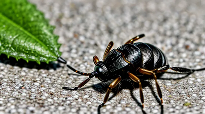
Ixodid Ticks: An Overview
What are Ixodid Ticks?
Morphology and Anatomy
Ixodid ticks, commonly called hard ticks, belong to the family Ixodidae and are distinguished by a rigid dorsal shield (scutum) that covers the entire back in males and a partial shield in females and nymphs. The anterior region, the capitulum, houses the mouthparts: chelicerae for cutting, a hypostome equipped with backward‑pointing barbs for anchorage, and the palps that guide the hypostome into the host’s skin. Six jointed legs emerge from the opisthosoma, each bearing sensory organs (Haller’s organs) that detect carbon dioxide, heat and movement, enabling precise host location.
The cuticle consists of a multilayered exoskeleton containing chitin, providing protection against dehydration and mechanical damage. Between the dorsal and ventral plates lies a flexible integument that expands dramatically during engorgement, allowing the tick to increase its body mass by up to 100‑fold. Internally, the foregut (pharynx and esophagus) connects to the midgut, where blood is stored and partially digested. Midgut cells secrete enzymes that break down hemoglobin, while the peritrophic matrix isolates the blood meal from the epithelium, reducing exposure to host immune factors.
Salivary glands occupy the anterior abdomen and release a complex cocktail of anticoagulants, immunomodulators and anti‑inflammatory proteins during feeding. These secretions prevent clot formation, suppress host immune responses and facilitate prolonged attachment, creating a conduit for pathogen transmission. The synganglion, a fused central nervous system located ventrally, coordinates muscle activity for limb movement and feeding behavior.
Reproductive structures differ between sexes. Males possess paired testes and accessory glands that produce spermatophores transferred to the female via the genital opening. Females contain a large, expandable ovary and a vitellarium that stores nutrients for egg development. After engorgement, the female detaches, lays thousands of eggs, and dies, completing the life cycle.
The combination of a robust exoskeleton, specialized mouthparts, expandable body cavity, and sophisticated salivary apparatus enables ixodid ticks to remain attached for days, ingest large blood volumes, and act as efficient vectors for bacteria, viruses and protozoa. Their anatomical adaptations directly underlie the health risks they pose to humans, livestock and wildlife.
Life Cycle Stages
Ixodid ticks, commonly known as hard ticks, develop through four distinct stages: egg, larva, nymph, and adult. Each stage requires a blood meal to progress to the next, creating opportunities for pathogen transmission.
-
Egg – Laid in clusters on the ground after the female detaches from the host. Development occurs in the environment; temperature and humidity dictate incubation time, typically ranging from weeks to months.
-
Larva – Emerges as a six‑legged organism. Seeks a small vertebrate, often a rodent or bird, for its first blood meal. After feeding, the larva detaches, digests the blood, and molts into a nymph.
-
Nymph – Possesses eight legs and resembles the adult in morphology but remains smaller. Requires a second host, frequently a medium‑sized mammal such as a dog or squirrel. Feeding duration may last several days; successful engorgement triggers molting into the adult stage.
-
Adult – Consists of male and female forms. Females require a final blood meal from a large host—commonly livestock, pets, or humans—to develop eggs. Males typically feed minimally, focusing on mating. After engorgement, females drop off the host to lay thousands of eggs, completing the cycle.
The interval between stages varies with species and climate. Rapid progression in warm, humid regions accelerates population growth, whereas cooler conditions prolong each phase. Pathogens, including bacteria, viruses, and protozoa, can be acquired during any blood meal and transmitted to subsequent hosts, rendering the life cycle a critical factor in disease epidemiology.
Habitat and Distribution
Ixodid ticks, commonly known as hard ticks, occupy a wide range of environments where they can locate suitable hosts. Their presence is concentrated in areas that provide adequate humidity, shelter, and access to vertebrate animals.
- Deciduous and mixed forests with leaf litter and understory vegetation
- Grasslands and meadows where grazing mammals graze
- Shrublands and scrubby habitats offering dense cover
- Agricultural fields, especially those bordering natural vegetation
- Urban parks and peri‑urban green spaces with wildlife activity
These habitats maintain the microclimatic conditions ticks require for survival, including moisture levels that prevent desiccation and temperature ranges that support their developmental cycles.
Geographically, ixodid ticks are distributed across temperate and subtropical zones on all inhabited continents. Their range extends from North America and Europe through parts of Asia, Africa, and South America. In each region, species composition reflects local climate and host availability, with some species confined to specific biogeographic zones while others exhibit broad, transcontinental distributions. Their spread is facilitated by migratory birds, livestock movement, and human travel, enabling colonization of new territories when environmental conditions are favorable.
Dangers Posed by Ixodid Ticks
Disease Transmission
Lyme Disease
Ixodid ticks, commonly known as hard ticks, belong to the family Ixodidae. They possess a hard dorsal shield (scutum) and feed for several days on the blood of mammals, birds, and reptiles. Their life cycle includes egg, larva, nymph, and adult stages, each requiring a blood meal to develop.
Lyme disease is a bacterial infection caused by Borrelia burgdorferi and transmitted primarily through the bite of infected nymphal or adult ixodid ticks. The pathogen resides in the tick’s midgut and migrates to the salivary glands during feeding, entering the host’s bloodstream.
The disease poses a health risk because it can progress from localized skin lesions to systemic involvement affecting joints, the nervous system, and the heart. Typical manifestations include:
- Erythema migrans (expanding rash)
- Arthritic pain, especially in large joints
- Neurological symptoms such as facial palsy and meningitis
- Cardiac abnormalities, notably atrioventricular block
Prompt removal of the attached tick and early antibiotic therapy reduce the likelihood of chronic complications.
Tick-borne Encephalitis
Ixodid ticks, commonly called hard ticks, belong to the family Ixodidae. Their mouthparts are enclosed in a scutum, allowing prolonged attachment to hosts while they ingest blood. This feeding habit facilitates the transfer of pathogens from animal reservoirs to humans.
Tick‑borne encephalitis (TBE) is a viral infection caused by members of the Flaviviridae family. The virus circulates in forested areas where ixodid ticks feed on small mammals, chiefly rodents, which serve as amplifying hosts. Human cases occur primarily in Europe and northern Asia, with incidence peaks in spring and early summer when nymphal ticks are most active.
After a bite, the incubation period lasts 7–14 days. The disease often follows a biphasic course: an initial febrile phase with headache, malaise, and myalgia, followed by a neurologic phase characterized by meningitis, encephalitis, or meningoencephalitis. Severe outcomes include long‑term cognitive deficits, paralysis, or fatality.
Laboratory confirmation relies on detection of specific IgM and IgG antibodies in serum or cerebrospinal fluid. Polymerase chain reaction can identify viral RNA during the early phase, while cerebrospinal fluid analysis typically reveals pleocytosis and elevated protein.
No antiviral therapy has proven efficacy; management is supportive, focusing on hydration, fever control, and monitoring of neurological status. In selected cases, corticosteroids are administered to reduce cerebral edema, though evidence remains limited.
Preventive strategies include:
- Vaccination with inactivated TBE vaccines for residents and travelers to endemic regions.
- Use of repellents containing DEET or picaridin on exposed skin.
- Wearing long sleeves and trousers, preferably treated with permethrin.
- Conducting thorough body checks after outdoor activities and removing attached ticks promptly with fine‑tipped tweezers.
These measures reduce the risk of infection transmitted by hard ticks and mitigate the public health impact of tick‑borne encephalitis.
Anaplasmosis and Ehrlichiosis
Ixodid ticks, commonly called hard ticks, belong to the family Ixodidae and serve as primary vectors for several intracellular bacterial infections in humans and animals. Their blood‑feeding behavior enables the transmission of pathogens directly into the host’s bloodstream, creating a rapid route for disease spread.
Anaplasmosis and ehrlichiosis are two tick‑borne illnesses caused by obligate intracellular bacteria of the genera Anaplasma and Ehrlichia. Both agents infect white‑blood cells, leading to systemic inflammation and, if untreated, potentially severe complications such as organ failure or death.
Typical clinical manifestations include:
- Fever and chills
- Headache and muscle pain
- Fatigue and malaise
- Laboratory abnormalities: thrombocytopenia, leukopenia, elevated liver enzymes
Prompt diagnosis relies on serologic testing, polymerase chain reaction, or microscopic identification of morulae in peripheral blood. Effective treatment consists of doxycycline administered for a minimum of 10 days; early therapy reduces morbidity and prevents progression.
The danger associated with hard ticks stems from their capacity to acquire, maintain, and transmit these bacteria across life stages, allowing sustained infection cycles in endemic regions. Control measures focus on personal protection, habitat management, and regular inspection of hosts to limit exposure and interrupt pathogen transmission.
Other Tick-borne Pathogens
Ixodid ticks transmit a wide spectrum of microorganisms beyond the classic agents of Lyme disease and Rocky Mountain spotted fever. These additional pathogens contribute significantly to the public‑health burden of tick‑borne illnesses.
- Anaplasma phagocytophilum – obligate intracellular bacterium causing human granulocytic anaplasmosis; induces fever, leukopenia, and thrombocytopenia.
- Ehrlichia muris‑like agent – emerging ehrlichial species linked to febrile illness with myalgia and elevated liver enzymes.
- Babesia microti and related Babesia spp. – intra‑erythrocytic protozoa responsible for babesiosis; may trigger hemolytic anemia, especially in immunocompromised patients.
- Coxiella burnetii – agent of Q fever; tick‑borne transmission documented in several regions, leading to atypical pneumonia and hepatitis.
- Tick‑borne encephalitis virus (TBEV) – flavivirus endemic to Europe and Asia; causes meningitis, encephalitis, or meningoencephalitis with potential long‑term neurological deficits.
- Powassan virus – North‑American flavivirus; neuroinvasive disease with high mortality and severe sequelae.
- Bourbon virus – novel orthomyxovirus identified in the United States; associated with febrile illness and thrombocytopenia.
- Rickettsia slovaca, Rickettsia raoultii – spotted fever group rickettsiae producing a mild scalp eschar and regional lymphadenopathy (tick‑borne lymphadenopathy).
These agents share common ecological characteristics: they reside in the midgut or salivary glands of hard ticks, persist through molting stages, and are delivered to vertebrate hosts during blood feeding. Co‑infection is frequent; a single tick may harbor multiple pathogens, complicating diagnosis and treatment. Prompt recognition of these diseases relies on laboratory testing specific to each organism, as clinical presentations often overlap. Effective control measures focus on reducing tick exposure, early removal of attached ticks, and surveillance of pathogen prevalence in tick populations.
Symptoms of Tick-borne Illnesses
Ixodid ticks—hard-bodied arachnids that attach to mammals, birds, and reptiles—are vectors for several pathogenic microorganisms. Their bite can introduce bacteria, viruses, or protozoa that produce distinct clinical manifestations.
Typical early signs after a tick bite include:
- Localized erythema at the attachment site, often expanding in diameter
- Mild to moderate pain or itching around the lesion
- Low‑grade fever developing within 2–10 days
Systemic symptoms vary according to the specific pathogen:
-
Lyme disease (Borrelia burgdorferi)
-
Rocky Mountain spotted fever (Rickettsia rickettsii)
- Sudden high fever, chills
- Severe headache, photophobia
- Maculopapular rash beginning on wrists and ankles, spreading centrally
- Nausea, vomiting, abdominal pain
-
Anaplasmosis (Anaplasma phagocytophilum)
- Fever, chills, muscle aches
- Nausea, vomiting, diarrhea
- Elevated liver enzymes, leukopenia, thrombocytopenia
-
Ehrlichiosis (Ehrlichia chaffeensis)
- Fever, headache, malaise
- Rash on trunk in ~30 % of cases
- Laboratory evidence of low platelet count and abnormal liver function
-
Babesiosis (Babesia microti)
- Hemolytic anemia, jaundice
- Dark urine, fatigue, fever
- Splenomegaly, especially in severe infections
-
Tularemia (Francisella tularensis)
Prompt recognition of these patterns enables early antimicrobial therapy, reducing the risk of severe complications such as neurological impairment, organ failure, or persistent infection. Monitoring for rash evolution, fever trajectory, and laboratory abnormalities is essential after any known or suspected tick exposure.
Prevention and Protection
Personal Protective Measures
Ixodid ticks, commonly called hard ticks, transmit bacteria, viruses, and protozoa that cause illnesses such as Lyme disease, anaplasmosis, and babesiosis. Their bites often go unnoticed, allowing pathogens to enter the bloodstream within hours. Effective personal protection reduces exposure and limits disease transmission.
Protective actions include:
- Wear light‑colored, tightly woven clothing that covers the body; tuck shirts into trousers and secure pant legs with elastic cuffs.
- Apply repellents containing 20‑30 % DEET, picaridin, IR3535, or oil of lemon eucalyptus to exposed skin and clothing, reapplying according to product instructions.
- Perform thorough tick checks at least every two hours in tick‑infested areas; remove attached ticks promptly with fine‑point tweezers, grasping close to the skin and pulling straight upward.
- Shower or bathe within 30 minutes after outdoor activity; bathing increases the chance of finding and removing unattached ticks.
- Avoid high‑grass, brush, and leaf litter where ticks quest for hosts; stay on cleared paths whenever possible.
- Use permethrin‑treated clothing and gear for extended exposure; treat items once and wash separately from untreated fabrics.
Consistent use of these measures significantly lowers the probability of tick attachment and subsequent infection.
Tick Removal Techniques
Ticks of the hard‑shell family attach firmly to skin and can transmit pathogens within hours. Prompt, proper removal reduces infection risk and prevents prolonged feeding that enhances disease transmission.
Effective removal follows a precise sequence. First, use fine‑pointed tweezers or a specialized tick‑removal tool. Grasp the tick as close to the skin as possible, avoiding compression of the abdomen. Second, apply steady, upward pressure without twisting; pull straight out until the mouthparts detach. Third, disinfect the bite area with an antiseptic solution. Fourth, place the tick in a sealed container with alcohol for identification if needed. Finally, monitor the site for signs of erythema, swelling, or fever and seek medical advice if symptoms develop.
Avoid common errors: squeezing the body, using heat or chemicals to force detachment, and leaving mouthparts embedded. These actions increase the likelihood of pathogen entry and inflammatory reactions.
When removal is impossible with tweezers, a calibrated tick‑removal hook can be employed, ensuring the same straight upward motion. In pediatric or sensitive cases, topical anesthetic may improve patient comfort without interfering with the extraction process.
Environmental Control
Ixodid ticks, commonly referred to as hard ticks, transmit bacterial, viral, and protozoan pathogens that cause diseases such as Lyme borreliosis, anaplasmosis, and babesiosis. Their life cycle—egg, larva, nymph, adult—requires blood meals from vertebrate hosts, allowing pathogens to enter human and animal populations. Environmental control reduces tick abundance, limits host‑tick contact, and consequently lowers disease incidence.
Effective environmental strategies include:
- Habitat modification: clear low vegetation, trim grass to 2–3 cm, remove leaf litter and brush where ticks quest for hosts.
- Host management: treat domestic animals with acaricides, implement wildlife feeding restrictions, and use deer‑exclusion fencing where feasible.
- Chemical interventions: apply approved acaricide sprays or granules to high‑risk zones, rotate active ingredients to prevent resistance.
- Biological agents: introduce entomopathogenic fungi (e.g., Metarhizium anisopliae) or nematodes that infect ticks, monitor efficacy regularly.
- Landscape planning: create buffer zones of non‑host plants (e.g., lavender, rosemary) that deter tick attachment, incorporate hard‑scape elements such as stone pathways to reduce suitable microclimates.
Monitoring protocols involve systematic drag sampling, tick density mapping, and pathogen testing of captured specimens. Data guide targeted interventions and assess long‑term impact on tick populations. Integrating these measures within public health and veterinary programs establishes a coordinated approach to mitigate the health threats posed by ixodid ticks.
Medical Considerations and Treatment
Ixodid, or hard, ticks act as vectors for bacterial, viral, and protozoan pathogens that cause illnesses such as Lyme disease, Rocky Mountain spotted fever, anaplasmosis, babesiosis, and tick‑borne encephalitis. Bite sites often present with a localized erythema, sometimes developing into a characteristic expanding rash (e.g., erythema migrans) or a petechial eruption. Systemic symptoms may include fever, headache, myalgia, and, in severe cases, organ dysfunction or neurologic deficits. Prompt recognition of these patterns is essential for accurate diagnosis and timely therapy.
Diagnostic work‑up relies on clinical assessment supported by laboratory tests: serologic assays for Borrelia burgdorferi, PCR for Rickettsia spp., and blood smears for Babesia parasites. When serology is unavailable or early infection is suspected, empiric antimicrobial treatment based on epidemiologic exposure is recommended.
Treatment protocol:
- Immediate removal of the tick with fine‑pointed tweezers, grasping close to the skin and pulling straight upward; avoid crushing the body to prevent pathogen release.
- For confirmed or strongly suspected Lyme disease, prescribe doxycycline 100 mg twice daily for 10–21 days; alternative regimens include amoxicillin or cefuroxime for patients unable to tolerate doxycycline.
- Rocky Mountain spotted fever requires doxycycline 100 mg twice daily for at least 7 days, continued until the patient is afebrile for 48 hours.
- Anaplasmosis and ehrlichiosis are also treated with doxycycline, 100 mg twice daily for 10–14 days.
- Babesiosis demands a combination of atovaquone plus azithromycin for 7–10 days; severe cases may need clindamycin plus quinine.
- Supportive care includes antipyretics, hydration, and monitoring for organ involvement; severe neurologic or cardiac complications warrant specialist referral.
Prevention measures—regular body checks after outdoor exposure, use of permethrin‑treated clothing, and application of EPA‑approved repellents—reduce the likelihood of attachment and subsequent infection, thereby decreasing the burden of tick‑borne disease.
