Immediate Actions After Finding a Tick
Assessing the Situation
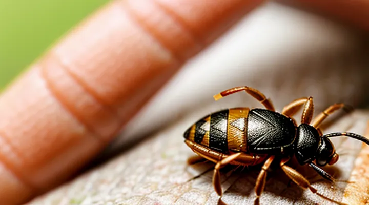
Identifying the Tick
When a tick attaches, accurate identification guides appropriate management.
Key visual characteristics include overall size, body shape, coloration, and the presence of distinct markings. Size ranges from a few millimeters in larvae to over a centimeter in engorged adults. Body shape varies between oval, elongated, or rounded forms, often reflecting the species. Coloration may shift from light brown in unfed stages to dark brown or gray after feeding. Distinct markings, such as white or silver scutal patterns, assist in differentiating species.
Life‑stage identification follows a predictable progression. Larvae possess six legs, lack visible eyes, and appear as tiny, translucent specks. Nymphs retain six legs, display a slightly larger body, and may exhibit faint markings. Adults bear eight legs, prominent eyes, and more defined coloration. Recognizing the stage informs potential disease risk and removal technique.
Species clues arise from geographic distribution, preferred hosts, and characteristic patterns. For example, ticks found in temperate regions often belong to the genus « Ixodes », while those in grasslands may be « Dermacentor ». Host preference—such as attachment to rodents, deer, or domestic animals—narrows identification. Specific patterns, like the “ornate” scutal design of « Dermacentor variabilis », provide decisive evidence.
Effective identification employs simple tools. A magnifying lens or handheld microscope reveals fine details. Reference materials, including regional tick identification keys, reputable websites, and photographic databases, enable comparison. When uncertainty persists, consulting a medical professional or an entomology laboratory ensures precise species determination.
Determining the Duration of Attachment
Assessing how long a tick has remained attached is a prerequisite for evaluating the risk of pathogen transmission.
Visual inspection provides the most immediate estimate. An unfed tick measures a few millimetres, whereas a partially or fully engorged specimen can exceed 10 mm. Size correlates with feeding duration:
- < 4 mm – likely attached for less than 24 hours.
- 4‑8 mm – suggests 2‑3 days of feeding.
-
8 mm – indicates 4 days or more, often approaching full engorgement.
Species‑specific data refine the estimate. For example, Ixodes scapularis typically requires 36‑48 hours to become noticeably engorged, while Dermacentor variabilis may reach comparable size after 48‑72 hours. Recognizing the species therefore narrows the time window.
Additional indicators support the assessment:
- Presence of a swollen, reddened area around the attachment site implies prolonged feeding.
- Detection of a clear “halo” of skin discoloration often accompanies ticks that have been attached for several days.
- Observation of a visible mouthpart or “capitulum” still embedded suggests incomplete removal and possibly extended attachment.
Recording the removal date and, when possible, the approximate bite date creates a documented timeline. Cross‑referencing this information with known incubation periods of tick‑borne pathogens enables targeted prophylactic decisions.
In practice, combine size measurement, species identification, and local tissue reaction to derive a reliable estimate of attachment duration. This systematic approach underpins appropriate medical management.
Essential Tools for Tick Removal
Fine-Tipped Tweezers
Fine‑tipped tweezers provide the precision necessary for safe extraction of an attached tick. Their narrow jaws grip the tick close to the skin without compressing the body, which reduces the risk of pathogen release.
To remove a tick with fine‑tipped tweezers, follow these steps:
- Grasp the tick as close to the skin surface as possible, holding the head or mouthparts.
- Apply steady, downward pressure to pull the tick straight out, avoiding twisting or jerking motions.
- Inspect the extracted tick to confirm that the entire mouthparts have been removed; retained fragments may cause local irritation.
- Disinfect the bite area with an antiseptic solution.
- Clean the tweezers with alcohol or another appropriate disinfectant before storage.
Using fine‑tipped tweezers eliminates the need for excessive force, minimizes tissue damage, and aligns with recommended medical guidelines for tick removal. The tool’s design ensures a controlled grip, facilitating complete extraction while preserving the surrounding skin integrity.
Other Recommended Equipment
When a tick adheres to the skin, precise instruments reduce the risk of pathogen transmission and tissue damage.
- Fine‑point tweezers or straight‑edge forceps, sterilized before use.
- Dedicated tick‑removal device, often shaped like a curved hook, marked «tick remover».
- Disposable nitrile gloves to prevent direct contact with the arthropod.
- Antiseptic solution (e.g., 70 % isopropyl alcohol) for post‑removal skin cleansing.
- Small magnifying glass to verify complete removal of mouthparts.
- Sealable biohazard container for safe disposal of the extracted tick.
Apply the tweezers or remover to grasp the tick as close to the skin as possible, pull upward with steady pressure, and avoid twisting. After extraction, cleanse the site with antiseptic, inspect for remaining parts, and discard the tick in the sealed container.
Step-by-Step Tick Removal Process
Grasping the Tick Correctly
When removing a tick, secure grip is essential to prevent mouthparts from remaining embedded. Use fine‑pointed tweezers or a specialized tick‑removal tool. Position the instrument as close to the skin as possible, grasp the tick’s head or mouthparts, not the body, to avoid crushing the abdomen.
- Pinch the tick’s mouthparts firmly.
- Apply steady, gentle upward pressure.
- Avoid twisting or jerking motions.
- Continue pulling until the entire tick separates from the skin.
After extraction, cleanse the bite area with antiseptic solution and monitor for signs of infection. Dispose of the tick by submerging it in alcohol or sealing it in a plastic bag before discarding.
Pulling the Tick Out
When a tick is attached to the skin, immediate removal reduces the risk of disease transmission. The removal process should be swift, clean, and precise.
- Use fine‑point tweezers or a specialized tick‑removal tool; avoid blunt instruments.
- Grasp the tick as close to the skin’s surface as possible, holding the mouthparts, not the body.
- Apply steady, downward pressure to pull the tick straight out without twisting or jerking.
- After extraction, place the tick in a sealed container for identification if needed.
- Disinfect the bite area with an antiseptic solution and wash hands thoroughly.
If any part of the tick remains embedded, repeat the removal steps with fresh tweezers. Persistent remnants may require medical evaluation. Monitoring the site for signs of infection, such as redness or swelling, is advisable for several days following removal.
Avoiding Common Mistakes
When a tick becomes lodged beneath the skin, prompt and correct removal reduces the risk of infection. Errors during extraction often compromise the outcome.
Common mistakes to avoid:
- «Squeezing the tick’s body» – pressure may force infectious fluids into the wound.
- «Using hot objects, chemicals, or petroleum products to detach the tick» – these methods can irritate the mouthparts, causing them to break off.
- «Pulling the tick with fingers or tweezers without a steady grip» – uneven force can leave mouthparts embedded.
- «Delaying removal for several hours or days» – prolonged attachment increases pathogen transmission probability.
- «Applying excessive force after the tick’s head has detached» – can cause additional tissue damage.
Correct practice involves grasping the tick as close to the skin as possible with fine‑point tweezers, applying steady, upward pressure, and cleaning the area with antiseptic after extraction. Monitoring the site for several weeks ensures early detection of any adverse reaction.
Post-Removal Care and Monitoring
Cleaning the Bite Area
Antiseptic Application
When a tick has been removed from the skin, immediate antiseptic treatment reduces the risk of secondary infection. The area should be cleaned promptly after extraction, before the skin dries or any residue remains.
- Apply a broad‑spectrum antiseptic, such as povidone‑iodine or chlorhexidine, directly to the bite site.
- Use a sterile gauze pad to spread the solution evenly, ensuring contact with the entire wound margin.
- Allow the antiseptic to remain in place for at least one minute; do not rinse off prematurely.
If a patient exhibits sensitivity to iodine‑based products, substitute with an alcohol‑based antiseptic containing at least 70 % ethanol. For individuals with known allergies to chlorhexidine, a hydrogen peroxide solution (3 %) may be employed, recognizing its lower residual activity.
After the initial application, cover the area with a clean, non‑adhesive dressing to protect against contamination. Re‑apply the antiseptic once daily, or more frequently if the wound shows signs of moisture or debris accumulation. Monitor for erythema, increasing pain, or purulent discharge, which may indicate infection requiring medical evaluation.
Proper antiseptic use, combined with careful removal technique, constitutes a critical component of effective tick‑bite management.
Hand Hygiene
Proper hand hygiene is a fundamental preventive measure when a tick adheres to the skin. Clean hands minimize the transfer of pathogens from the tick’s mouthparts to the surrounding tissue and reduce the risk of secondary infection after removal.
Before handling the tick, wash hands thoroughly with soap and water for at least 20 seconds. Rinse completely, dry with a disposable paper towel, and consider wearing disposable gloves if available. Alcohol‑based hand rubs may be used when soap and water are not immediately accessible, but they do not replace washing in this context.
After the tick is extracted, repeat hand washing with soap and water. Apply an antiseptic solution such as povidone‑iodine or chlorhexidine to the fingertips, then dry with a clean disposable towel. Dispose of gloves and any contaminated materials in a sealed bag before discarding.
Key steps for hand hygiene during tick removal:
- Wash hands with soap and water ≥ 20 seconds before contact.
- Dry hands with a single‑use paper towel.
- Wear disposable gloves if possible.
- After removal, repeat washing and apply antiseptic.
- Discard gloves and contaminated items in a sealed container.
Adhering to these practices supports effective tick management and protects against infection.
Monitoring for Symptoms
Localized Reactions
When a tick remains attached to the skin, the immediate area often exhibits a localized reaction. The reaction typically manifests as erythema, swelling, or a small papule surrounding the mouthparts. In some cases, a central puncture wound may be visible, sometimes accompanied by a clear or serous fluid exudate.
Common characteristics of the localized response include:
- Redness extending 2–5 mm from the attachment site
- Mild to moderate edema that may persist for several days
- Pruritus or tenderness upon palpation
- Occasional formation of a small vesicle or pustule
Management focuses on prompt removal of the tick and care of the affected skin. The following steps are recommended:
- Grasp the tick as close to the skin surface as possible with fine‑point tweezers.
- Apply steady, upward traction without twisting to avoid breaking the mouthparts.
- Disinfect the bite area with an antiseptic solution such as povidone‑iodine or chlorhexidine.
- Observe the site for 24–48 hours; if erythema expands beyond the initial margin, or if necrosis develops, seek medical evaluation.
Persistent redness, increasing pain, or the appearance of a bull’s‑eye lesion may indicate an early infection and warrants professional assessment. Documentation of the reaction’s progression assists clinicians in distinguishing benign inflammation from vector‑borne disease manifestations.
Systemic Illnesses
When a tick embeds in the skin, prompt removal reduces the risk of systemic infections. Certain pathogens can disseminate beyond the bite site, producing illness that affects multiple organ systems. Recognizing these conditions guides timely medical intervention.
Common systemic illnesses transmitted by ticks include:
- Lyme disease – characterized by erythema migrans, fever, fatigue, and joint pain; may progress to neurological or cardiac involvement if untreated.
- Anaplasmosis – presents with fever, headache, muscle aches, and leukopenia; can evolve to severe respiratory distress.
- Ehrlichiosis – causes fever, thrombocytopenia, and hepatic dysfunction; may lead to multiorgan failure.
- Rocky Mountain spotted fever – marked by rash, high fever, and vascular injury; rapid progression can result in renal or neurologic failure.
- Babesiosis – produces hemolytic anemia, hemoglobinuria, and splenomegaly; severe cases may cause acute respiratory distress syndrome.
- Tick‑borne encephalitis – leads to meningitis, encephalitis, and long‑term neurological deficits.
- Southern tick‑associated rash illness – manifests as a maculopapular rash, fever, and arthralgia; may evolve into chronic joint disease.
Key clinical indicators of systemic spread include:
- Fever exceeding 38 °C persisting beyond 24 hours after removal.
- New rash distant from the bite site, especially with central clearing or petechiae.
- Neurological symptoms such as facial palsy, meningismus, or altered mental status.
- Cardiovascular signs including palpitations, chest discomfort, or conduction abnormalities.
- Laboratory abnormalities: elevated liver enzymes, low platelet count, or abnormal renal function.
If any of these signs appear, seek medical evaluation promptly. Laboratory testing should target specific tick‑borne pathogens, and empirical antimicrobial therapy may be initiated based on regional prevalence and clinical presentation. Early treatment reduces the likelihood of chronic complications and supports full recovery.
When to Seek Medical Attention
Incomplete Tick Removal
When a tick is only partially extracted, the remaining mouthparts can remain embedded in the skin and may release pathogens. Immediate action reduces the risk of infection and minimizes tissue irritation.
- Clean the area with antiseptic solution before further manipulation.
- Use fine‑point tweezers to grasp the tick as close to the skin as possible, avoiding squeezing the body.
- Apply steady, upward pressure to pull the mouthparts out in one motion; do not twist or jerk.
- If the mouthparts break off, do not dig them out with a needle or scalpel; such attempts increase tissue damage.
- Disinfect the site again after removal and monitor for redness, swelling, or fever.
Seek professional medical care if any of the following occur: visible fragments remain after repeated attempts, the bite area becomes inflamed, or systemic symptoms develop within weeks. Documentation of the bite date and location assists healthcare providers in assessing potential disease exposure.
Signs of Infection
After a tick is removed, the skin should be inspected regularly for any indication of infection. Prompt identification of adverse changes reduces the risk of complications.
Typical signs of infection include:
- Redness extending beyond the bite site
- Swelling or warmth surrounding the area
- Pain that intensifies rather than fades
- Pus or other discharge
- Fever, chills, or unexplained fatigue
If any of these symptoms develop, immediate medical evaluation is required. Treatment may involve topical antiseptics, oral antibiotics, or further wound care, depending on severity. Continuous observation for at least two weeks ensures that delayed reactions, such as Lyme disease, are not overlooked.
Symptoms of Tick-Borne Diseases
When a tick attaches to the skin, pathogens may be introduced, producing a range of clinical manifestations. Early recognition of these signs enables prompt treatment and reduces the likelihood of complications.
Common tick‑borne infections and their characteristic symptoms include:
- Lyme disease: expanding erythema migrans rash, fever, fatigue, headache, neck stiffness, arthralgia, and occasional facial palsy.
- Rocky Mountain spotted fever: abrupt fever, severe headache, rash that begins on wrists and ankles and spreads centrally, nausea, and vomiting.
- Ehrlichiosis and anaplasmosis: fever, chills, muscle aches, leukopenia, thrombocytopenia, and elevated liver enzymes.
- Babesiosis: hemolytic anemia, jaundice, dark urine, fever, and chills, often accompanied by fatigue.
- Tularemia: ulceroglandular form with a skin ulcer at the bite site, painful swollen lymph nodes, fever, and chills.
- Powassan virus infection: encephalitis, meningitis, fever, weakness, and loss of coordination.
Incubation periods vary from a few days to several weeks, depending on the pathogen. Symptoms may overlap, making laboratory confirmation essential for accurate diagnosis. Persistent or worsening signs—particularly fever, rash, or neurological deficits—warrant immediate medical evaluation.
Healthcare providers assess exposure history, perform physical examination, and order appropriate serologic or molecular tests. Early antimicrobial therapy, especially for bacterial infections such as Lyme disease and rickettsial illnesses, improves outcomes and prevents long‑term sequelae.
Preventing Future Tick Bites
Personal Protective Measures
Personal protective measures reduce the likelihood of tick attachment and limit health risks associated with embedded arthropods. Wearing long sleeves, long trousers, and closed shoes creates a physical barrier. Light‑colored clothing facilitates visual detection of ticks before they attach.
Effective deterrents include applying EPA‑registered repellents containing DEET, picaridin, or IR3535 to exposed skin and treating clothing with permethrin. Repellents must be reapplied according to manufacturer guidelines, especially after swimming or heavy sweating.
Routine inspection of the body after outdoor activity is essential. Conduct systematic checks by:
- Examining scalp, behind ears, armpits, and groin.
- Running fingers over clothing seams and skin folds.
- Using a mirror or a partner for hard‑to‑see areas.
If a tick is discovered attached, immediate removal should follow standard procedures, after which protective actions continue. Disinfect the bite site with an iodine‑based solution or alcohol swab. Dispose of the tick by placing it in a sealed container with ethanol or by flushing it down the toilet; avoid crushing the body to prevent pathogen release.
Post‑removal monitoring involves recording the date of the bite, the tick’s developmental stage, and any emerging symptoms. Consulting a healthcare professional is advised if fever, rash, or joint pain develop within weeks. Maintaining personal protection habits for the duration of the tick season sustains reduced exposure and supports early detection.
Environmental Controls
When an engorged arachnid attaches to the dermis, prompt extraction is essential; however, controlling the surrounding habitat limits subsequent incidents.
- Maintain short grass and clear leaf litter in yards to eliminate humid microclimates favored by ticks.
- Apply registered acaricides to perimeter zones, focusing on shaded, wooded edges where tick populations concentrate.
- Install physical barriers, such as fine-mesh fencing, to restrict wildlife movement into residential areas.
- Treat domestic animals with veterinarian‑approved tick preventatives to reduce host availability.
- Use landscape mulch that dries quickly, avoiding damp organic material that supports tick development.
After removal, decontaminate clothing and footwear with hot water or a tumble‑dry cycle, then store items in sealed containers. Regularly inspect and clean outdoor equipment, especially hunting gear and gardening tools, to prevent inadvertent transport of ticks into the home environment.
Implementing these measures creates an environment hostile to tick survival, thereby decreasing the likelihood of future attachments and supporting effective personal protection.
