Immediate Actions After Tick Removal
Assessing the Situation
Identifying Remaining Parts
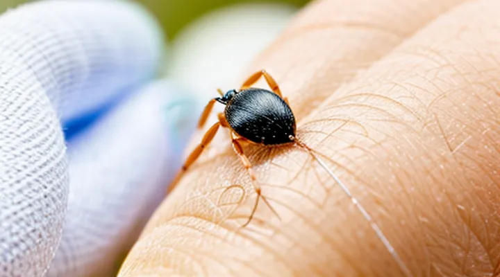
When a tick is removed, the first priority is to confirm that no mouthparts or legs remain embedded. Visual inspection should begin immediately after extraction. Use a well‑lit area and, if possible, a magnifying glass or a smartphone camera with zoom to examine the bite site closely. Look for any dark, pointed fragments protruding from the skin or a small, raised bump that may indicate retained parts.
If the area is difficult to see, gently stretch the surrounding skin with clean fingers or a sterile instrument to expose hidden fragments. A sterile needle or fine‑point tweezers can be used to lift the skin without pushing deeper. Do not dig aggressively; excessive pressure can embed the fragment further.
Key signs that something remains include:
- A visible piece of the tick’s mouthparts or legs.
- Persistent redness, swelling, or a puncture that does not close.
- Ongoing itching or pain at the site after the initial removal.
When any of these indicators are present, take the following steps:
- Disinfect the area with an antiseptic solution.
- Attempt to grasp the visible fragment with fine‑point tweezers; pull straight upward with steady, even pressure.
- If the fragment is not easily reachable or removal causes discomfort, cease attempts.
- Seek professional medical assistance promptly; a clinician can use specialized tools and may prescribe antibiotics if infection risk is high.
After removal, monitor the bite site for several days. Document any changes such as increasing redness, swelling, or fever, and report them to a healthcare provider without delay. This systematic approach ensures that no tick remnants remain to cause complications.
Signs of Complete Removal
After extracting a tick, verify that the removal is complete. Visible confirmation is the first indicator: the entire body, including mouthparts, should be absent from the skin. If no fragment remains attached to the puncture site, the removal is likely successful.
Additional signs include:
- The skin around the bite is smooth, without protruding or embedded material.
- Absence of lingering pain or a sharp, localized sensation that could suggest a hidden part.
- No swelling, redness, or inflammation that expands beyond the immediate area of the bite.
- The wound edges close naturally within a few minutes, indicating that the tissue was not torn by retained parts.
Observe the site for 24–48 hours. If the area remains stable—no new redness, warmth, or discharge—the removal can be considered complete. Should any of these symptoms develop, seek medical evaluation, as they may signal a retained fragment or secondary infection.
Initial Self-Care Steps
Cleaning the Area
When a tick is only partially extracted, the surrounding skin must be decontaminated promptly to prevent infection and reduce the risk of pathogen transmission.
First, wash your hands with soap and water. Then cleanse the bite site with an antiseptic solution such as povidone‑iodine or chlorhexidine. Apply the antiseptic for at least 30 seconds, ensuring the entire area around the remnant mouthparts is covered. Rinse with sterile saline if the product instructions advise.
After disinfection, dry the skin with a sterile gauze pad. Avoid rubbing, which could embed remaining fragments deeper.
If the wound shows signs of inflammation—redness extending beyond a few centimeters, swelling, heat, or pus—seek medical evaluation promptly.
Maintain cleanliness by:
- Re‑applying antiseptic twice daily for the next 24‑48 hours.
- Monitoring the site for changes in size, color, or sensation.
- Keeping the area covered with a breathable, sterile dressing if irritation occurs.
Document the incident, including the date of tick exposure, removal method, and any symptoms, to aid healthcare providers in assessing potential tick‑borne diseases.
Applying Antiseptics
When a fragment of a tick remains embedded, immediate wound care reduces infection risk. First, wash the area with mild soap and running water to remove debris. Pat dry with a clean gauze.
Apply a broad‑spectrum antiseptic directly to the site. Suitable options include:
- 70 % isopropyl alcohol, applied with a sterile swab and left to evaporate.
- Povidone‑iodine solution, applied in a thin layer and allowed to dry.
- Chlorhexidine gluconate, applied with a cotton tip and left on the skin.
After antiseptic application, cover the wound with a sterile adhesive bandage to maintain a clean environment. Re‑apply the antiseptic once daily or whenever the bandage is changed.
Observe the area for signs of infection—redness spreading beyond the margin, increasing pain, swelling, pus, or fever. If any of these symptoms develop, seek medical evaluation promptly.
Seeking Professional Medical Attention
When to Consult a Doctor
Visible Tick Fragments
When a tick is only partially detached, the remaining mouthparts may be visible on the skin. Their presence can increase the risk of pathogen transmission, so prompt action is required.
First, examine the area under good lighting. Visible fragments usually appear as a small, dark, pin‑point or linear shape embedded in the epidermis. If the fragment is clearly exposed, attempt removal without delaying.
Removal procedure
- Disinfect a pair of fine‑pointed tweezers with alcohol.
- Grip the fragment as close to the skin surface as possible, avoiding squeezing surrounding tissue.
- Pull upward with steady, even pressure; do not twist or jerk, which can cause deeper embedment.
- After extraction, clean the site with an antiseptic solution (e.g., povidone‑iodine or chlorhexidine).
Aftercare
- Observe the wound for 24‑48 hours. If redness, swelling, or a rash develops, seek medical evaluation.
- Document the date of removal and any symptoms; this information aids clinicians in assessing infection risk.
- If removal fails or the fragment cannot be seen clearly, consult a healthcare professional for possible surgical extraction.
Prevention of future incidents
- Use tick‑preventive clothing and repellents when in endemic areas.
- Perform full‑body tick checks after outdoor exposure, paying special attention to hidden sites such as scalp, groin, and armpits.
By following these steps, visible tick fragments can be safely eliminated, reducing the likelihood of disease transmission.
Symptoms of Infection
If a fragment of a tick stays embedded, monitor the site for signs of infection. Early detection prevents complications and guides timely medical intervention.
Typical indicators include:
- Redness expanding beyond the immediate bite area
- Swelling or palpable warmth around the puncture
- Persistent throbbing or sharp pain at the location
- Pus or other discharge from the wound
- Fever, chills, or unexplained fatigue within days of the bite
- Headache, muscle aches, or joint pain, especially if accompanied by a rash
These symptoms may suggest bacterial infection, such as cellulitis, or transmission of tick‑borne pathogens like Lyme disease. If any of the listed signs appear, seek professional medical evaluation promptly. Treatment may involve antibiotics, wound cleaning, or further removal of residual tick material under sterile conditions. Continuous observation for at least two weeks after removal is advisable, as some infections develop gradually.
History of Tick-Borne Illnesses
The earliest recorded tick‑borne illness appears in the 5th‑century Chinese medical text Zhubing yuanhou lun, which describes a fever associated with tick exposure. European physicians noted similar symptoms in the 16th century, but systematic study began only in the late 19th century when scientists isolated Rickettsia rickettsii, the agent of Rocky Mountain spotted fever. This discovery established the link between arthropod vectors and systemic infection.
In 1975, a cluster of arthritis cases in Lyme, Connecticut, led to the identification of Borrelia burgdorferi as the cause of Lyme disease. The subsequent development of serologic testing and antibiotic protocols reduced morbidity and highlighted the need for prompt removal of the tick, especially when any part remains embedded.
Key milestones in the understanding of tick‑borne diseases include:
- 1889 – Discovery of Rickettsia species by Howard Ricketts.
- 1910 – First description of ehrlichiosis in cattle, later linked to human infection.
- 1975 – Isolation of Borrelia burgdorferi and definition of Lyme disease.
- 1990s – Recognition of emerging pathogens such as Babesia and Anaplasma phagocytophilum.
- 2000s – Genome sequencing of multiple tick‑borne agents, enabling targeted therapies.
Historical evidence shows that incomplete removal of a tick increases the risk of pathogen transmission, because salivary glands may remain attached to the host tissue. Contemporary guidelines, rooted in this legacy, advise careful extraction with fine‑point tweezers, ensuring that the mouthparts are fully withdrawn. If a fragment persists, the wound should be cleansed with antiseptic, and medical evaluation sought to assess the need for prophylactic antibiotics or monitoring for early signs of infection.
What to Expect at the Doctor's Office
Examination and Removal
When only a portion of a tick remains embedded, immediate visual assessment is essential. Locate the residual mouthparts by inspecting the bite site under good lighting, possibly using a magnifying glass. Confirm that the skin around the fragment is clean and free of excessive blood or pus, which could indicate infection.
Removal should follow a sterile, controlled technique:
- Disinfect the area with an alcohol swab or antiseptic solution.
- Grasp the visible part of the tick fragment with fine-tipped tweezers, keeping the forceps as close to the skin as possible.
- Apply steady, gentle pressure to pull the fragment straight out, avoiding twisting or squeezing, which can cause additional tissue damage.
- If the fragment is difficult to grasp, a sterile needle can be used to lift the tip, then extract with tweezers.
- After removal, cleanse the wound again with antiseptic and cover with a clean bandage if needed.
Post‑removal care includes monitoring the site for signs of infection—redness, swelling, warmth, or discharge—and for systemic symptoms such as fever or rash. Seek medical attention promptly if any of these develop, or if the fragment cannot be removed completely. Professional evaluation may involve a minor surgical procedure or prescription of antibiotics to prevent complications.
Prescribing Medications
If a tick fragment stays embedded, remove the remaining part promptly with fine‑point tweezers, grasping as close to the skin as possible, and pull upward with steady pressure. Clean the site with antiseptic solution and observe for signs of infection.
Prescribe medication based on risk assessment for tick‑borne diseases and local guidelines:
- Prophylactic doxycycline: 200 mg orally, single dose, if removal occurred within 72 hours of attachment and the tick was attached for ≥36 hours in an area where Lyme disease is common.
- Extended doxycycline course: 100 mg orally twice daily for 10–14 days when early localized infection (e.g., erythema migrans) is suspected.
- Amoxicillin: 500 mg orally three times daily for 10 days as an alternative in children, pregnant patients, or those with doxycycline contraindications.
- Tetanus booster: administer if immunization status is uncertain or the wound is dirty and the last booster was >10 years ago.
Monitor the patient for fever, rash, arthralgia, or neurologic symptoms. If such manifestations develop, initiate targeted therapy for the specific pathogen (e.g., ceftriaxone for suspected neuroborreliosis). Document the encounter, including the tick removal method, medication prescribed, dosage, and follow‑up plan.
Monitoring and Follow-up
If a tick fragment remains in the skin, immediate observation is essential. Inspect the site daily for changes and record any developments.
Key indicators that require medical attention include:
- Expanding redness or a bullseye pattern
- Persistent itching, burning, or pain
- Swelling beyond the immediate area
- Fever, chills, or flu‑like symptoms
- Joint pain or stiffness
Schedule a follow‑up appointment with a healthcare professional within 48‑72 hours. During the visit, the clinician will assess the wound, consider laboratory testing for tick‑borne pathogens, and decide whether prophylactic antibiotics are warranted.
If the initial evaluation shows no complications, continue self‑monitoring for at least four weeks. Document temperature readings, new skin lesions, and any systemic symptoms. Contact a medical provider promptly if any of the above signs emerge at any point during this period.
Potential Complications and Prevention
Understanding Tick-Borne Diseases
Common Illnesses
A fragment of a tick left in the skin can introduce pathogens that cause common tick‑borne illnesses such as Lyme disease, anaplasmosis, babesiosis, and Rocky Mountain spotted fever. Prompt removal of the remaining part reduces the risk of infection and minimizes tissue irritation.
If a tick fragment stays embedded, follow these actions:
- Clean the area with antiseptic solution.
- Use fine‑pointed tweezers to grasp the visible portion as close to the skin as possible.
- Pull upward with steady pressure, avoiding twisting or squeezing the body.
- After extraction, disinfect the site again and apply a sterile bandage.
- Record the date of the bite and the removal attempt.
Monitor the wound for signs of infection: redness spreading beyond the bite site, swelling, warmth, or pus. Observe the body for systemic symptoms within the next weeks, including fever, headache, fatigue, muscle aches, joint pain, or a rash resembling a bull’s‑eye. Any of these signs warrants immediate medical evaluation.
Seek professional care if removal is incomplete, if the bite area becomes painful or inflamed, or if systemic symptoms develop. A clinician may prescribe antibiotics to prevent or treat disease transmission and can order laboratory tests to confirm infection. Early intervention improves outcomes for the illnesses commonly associated with tick exposure.
Symptoms to Watch For
If a tick mouthpart is left in the skin, monitor the site and overall health for signs that may indicate infection or disease transmission. Immediate observation helps determine whether medical intervention is required.
Key symptoms to watch for include:
- Redness or swelling that spreads beyond the immediate area of the bite.
- A rash resembling a target or bull’s‑eye pattern, often associated with Lyme disease.
- Fever, chills, or unexplained fatigue.
- Joint pain or stiffness, particularly in the knees or elbows.
- Headache, neck stiffness, or facial palsy.
- Nausea, vomiting, or abdominal pain.
- Neurological changes such as tingling, numbness, or difficulty concentrating.
Any of these manifestations appearing within weeks of the incident warrants prompt medical evaluation. Early treatment reduces the risk of complications and supports faster recovery.
Preventing Future Tick Bites
Protective Measures
If a tick fragment remains lodged in the skin, immediate protective actions reduce the risk of infection.
- Disinfect the area with an antiseptic solution such as povidone‑iodine or alcohol before any manipulation.
- Use fine‑point tweezers or a sterile needle to grasp the exposed part of the fragment as close to the skin surface as possible.
- Pull upward with steady, even pressure; avoid twisting or squeezing the fragment to prevent further tissue damage.
- After removal, clean the site again with antiseptic and apply a sterile dressing.
- Observe the wound for signs of redness, swelling, fever, or a rash over the next 2–3 weeks.
- If any of these symptoms appear, seek medical evaluation promptly; a clinician may prescribe a short course of doxycycline or another appropriate antibiotic.
- Record the date of the bite and the removal procedure; this information assists healthcare providers in assessing potential tick‑borne diseases.
- Ensure tetanus immunization is up to date, especially if the skin is broken or the removal was traumatic.
These steps constitute the core protective measures after a tick fragment is left in the body. Prompt, clean removal and vigilant monitoring are essential to prevent complications.
Tick Checks
Tick checks involve systematic examination of the skin after outdoor exposure to locate and remove attached ticks before they embed deeply. The process starts with a visual inspection of the entire body, paying special attention to hair‑covered areas, armpits, groin, and behind the knees. Use a fine‑toothed comb or a mirror to enhance visibility.
If a tick is found, grasp it as close to the skin as possible with fine‑point tweezers and pull upward with steady, even pressure. Removing the whole organism reduces the risk of pathogen transmission. When a portion of the mouthparts or abdomen remains lodged, immediate action is required to prevent infection.
- Clean the site with antiseptic solution.
- Apply a sterile, flat‑head instrument (e.g., a needle or the tip of a scalpel) to gently lift the residual fragment.
- Use tweezers to grasp the exposed part and extract it in the same upward motion used for whole‑tick removal.
- Disinfect the wound again after extraction.
- Preserve the removed fragment in a sealed container with alcohol for possible laboratory identification.
- Monitor the bite area for redness, swelling, or fever over the next several weeks; seek medical evaluation if symptoms develop.
Prompt, thorough removal and proper wound care are essential components of effective tick checks and minimize the likelihood of disease transmission.
