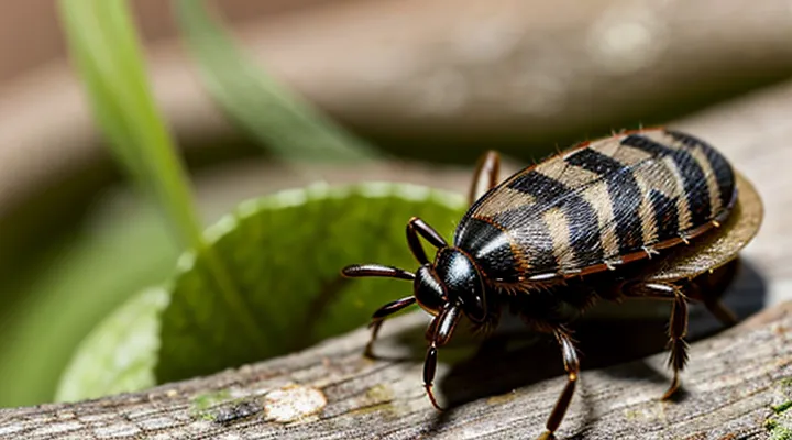
Understanding Tick-borne Diseases
The Threat of Infected Ticks
Ticks that carry pathogens pose a measurable public‑health risk. Studies across North America and Europe show that 10‑30 % of adult Ixodes scapularis and Ixodes ricinus specimens test positive for Borrelia burgdorferi, the agent of Lyme disease. Similar surveys report 5‑15 % infection rates for Anaplasma phagocytophilum and 1‑3 % for the tick‑borne encephalitis virus. These figures vary with region, host density, and seasonal activity.
Key aspects of the threat include:
- Disease spectrum – Lyme disease, anaplasmosis, babesiosis, and tick‑borne encephalitis are the most common outcomes of a bite from an infected tick.
- Geographic hotspots – Northeastern United States, Upper Midwest, and Central Europe exhibit the highest prevalence of infected vectors.
- Seasonal peaks – Nymphal activity in late spring and early summer raises exposure risk because nymphs are small and often go unnoticed.
- Host interaction – High deer and rodent populations sustain tick life cycles and increase pathogen circulation.
Preventive measures reduce the probability of transmission. Regular landscape management, use of EPA‑registered repellents containing DEET or picaridin, and prompt removal of attached ticks within 24 hours lower infection odds. Post‑exposure monitoring for fever, rash, or flu‑like symptoms enables early diagnosis and treatment, which limits complications.
Overall, the likelihood that any given tick carries a disease‑causing organism ranges from a few percent to nearly one‑third, depending on local ecology and tick stage. Recognizing these rates informs risk assessment and guides effective control strategies.
Factors Influencing Infection Rates
Geographic Location and Endemic Areas
Geographic location determines the prevalence of tick‑borne pathogens because vector species and host reservoirs vary across regions. In areas where a pathogen is endemic, a higher proportion of ticks carry the organism, raising the probability of transmission to humans or animals.
- Temperate zones of North America and Europe host Ixodes scapularis and Ixodes ricinus, vectors for Borrelia burgdorferi; infection rates in these ticks often exceed 20 % in established foci.
- Subtropical regions of the United States, such as the Gulf Coast, support Amblyomma americanum, a vector for Ehrlichia chaffeensis; local infection rates typically range from 5 % to 15 %.
- Eastern Asia, particularly parts of China and Japan, contains Haemaphysalis longicornis populations infected with severe fever with thrombocytopenia syndrome virus; prevalence can reach 10 % in hotspot districts.
- High‑altitude or arid environments usually sustain fewer competent vectors, resulting in infection rates below 1 % for most pathogens.
Endemic zones emerge where competent hosts—small mammals, birds, or deer—maintain the pathogen life cycle. Human exposure intensifies in habitats overlapping residential areas, recreational trails, and agricultural fields. Seasonal activity peaks, often in spring and early summer, further concentrate infected ticks, amplifying risk during these periods.
Consequently, assessing infection probability requires precise knowledge of the region’s vector species, documented pathogen prevalence, and the presence of established transmission cycles. Data from local surveillance programs and published prevalence studies provide the most reliable estimates for any given location.
Tick Species and Life Stage
Tick species differ markedly in their capacity to acquire and transmit pathogens, which directly influences the probability that an individual tick carries an infection. In North America, the most studied vectors include:
- Ixodes scapularis (black‑legged tick) – primary carrier of Borrelia burgdorferi; infection rates often exceed 30 % in nymphs from endemic regions.
- Amblyomma americanum (lone star tick) – associated with Ehrlichia chaffeensis and Francisella tularensis; nymphal infection prevalence typically ranges from 5 % to 15 %.
- Dermacentor variabilis (American dog tick) – vector of Rickettsia rickettsii; adult infection rates usually fall between 2 % and 10 %.
- Ixodes pacificus (western black‑legged tick) – transmits B. burgdorferi on the West Coast; nymphal infection frequencies often lie between 10 % and 20 %.
Life stage strongly modulates infection likelihood. After hatching, larvae are generally uninfected because they acquire pathogens only during the first blood meal. Consequently, larval infection prevalence is near zero across species. Nymphs, having fed once, exhibit the highest infection rates, reflecting the cumulative exposure of the previous stage. Adult ticks, which have fed twice, retain or increase prevalence, but species‑specific patterns differ: some adults show lower rates than nymphs due to mortality of heavily infected individuals, while others maintain or exceed nymphal levels.
The interaction of species‑specific competence and developmental stage creates a variable risk landscape. For any given tick, the chance of harboring a pathogen can be estimated by multiplying the species‑associated baseline prevalence by the stage‑specific adjustment factor. For example, a Ixodes scapularis nymph in a high‑incidence area may have a >30 % probability of infection, whereas an Amblyomma americanum larva would be expected to be virtually uninfected. Understanding these distinctions is essential for accurate risk assessment and targeted public‑health interventions.
Probability of Encountering an Infected Tick
Data Collection and Surveillance
Methods for Tick Testing
Accurate assessment of tick infection prevalence relies on laboratory testing of individual specimens. Reliable data enable calculation of the probability that a tick carries a pathogen and inform public‑health decisions.
- Polymerase chain reaction (PCR) – amplifies pathogen‑specific DNA fragments; provides rapid, highly sensitive detection of bacteria, viruses, and protozoa.
- Quantitative PCR (qPCR) – measures DNA copy number; yields infection load, useful for distinguishing low‑level carriers.
- Reverse transcription PCR (RT‑PCR) – targets RNA viruses; converts RNA to cDNA before amplification.
- Enzyme‑linked immunosorbent assay (ELISA) – detects antibodies or antigens in tick homogenates; suitable for large sample sets.
- Immunofluorescence assay (IFA) – visualizes pathogen antigens with fluorescent antibodies; confirms microscopy findings.
- Next‑generation sequencing (NGS) – sequences all genetic material; identifies known and novel pathogens simultaneously.
- Culture methods – isolates viable organisms on selective media; confirms infectivity but requires extended incubation.
Specimens must be collected with sterile tools, stored at –20 °C or in RNA‑preserving buffer, and processed within recommended timeframes to prevent DNA degradation. Homogenization protocols standardize tissue disruption, ensuring consistent nucleic‑acid yields across samples.
Result interpretation follows binary or quantitative criteria defined by assay validation. Positive detections are tallied, and infection prevalence is calculated as the ratio of positive ticks to total tested, expressed as a percentage or probability. Confidence intervals derived from binomial statistics provide precision estimates for the reported prevalence.
Reporting and Public Health Initiatives
Accurate estimation of tick infection prevalence depends on systematic surveillance and transparent data exchange. Health agencies collect specimens from wildlife, domestic animals, and human cases, then test for pathogens such as Borrelia, Anaplasma, and Rickettsia. Laboratory results are entered into centralized databases, providing a real‑time picture of infection rates across regions.
Mandatory reporting frameworks require clinicians and veterinarians to submit confirmed tick‑borne disease diagnoses within defined timeframes. Complementary passive surveillance captures incidental findings, while sentinel programs target high‑risk habitats for routine tick sampling. Data from these sources are aggregated, standardized, and released to researchers and policymakers through open‑access portals.
Public health initiatives translate surveillance outputs into actionable measures. Core activities include:
- Targeted public education campaigns that explain tick avoidance, proper removal, and symptom recognition.
- Environmental management programs that reduce tick habitats through vegetation control and wildlife management.
- Geographic risk mapping that overlays infection prevalence with human activity patterns, guiding resource allocation.
- Interagency collaborations that align veterinary, wildlife, and human health sectors under a One Health approach.
By linking robust reporting mechanisms with coordinated intervention strategies, authorities generate reliable infection probability estimates and mitigate the impact of tick‑borne diseases on communities.
Regional Variations in Tick Infection Rates
Lyme Disease Prevalence
Lyme disease prevalence reflects the proportion of ticks that carry the bacterium Borrelia burgdorferi and the resulting incidence in human populations. Surveillance data from the United States indicate that approximately 20–30 % of adult black‑legged ticks (Ixodes scapularis) and 10–15 % of nymphs are infected in high‑risk regions such as the Northeast, Mid‑Atlantic, and Upper Midwest. In contrast, infection rates fall below 5 % in the Pacific Northwest and are rarely detected in the Southwest.
Key determinants of tick infection prevalence include:
- Host diversity: abundance of competent reservoir species (e.g., white‑footed mice) raises bacterial carriage.
- Habitat characteristics: deciduous forests with dense understory provide optimal microclimates for tick development.
- Seasonal dynamics: nymphal activity peaks in late spring, coinciding with the highest human exposure risk.
- Climate trends: warmer temperatures expand tick ranges northward and elevate infection percentages in newly colonized areas.
Human Lyme disease case reports correspond closely with these tick infection patterns. The Centers for Disease Control and Prevention recorded roughly 35 000 confirmed cases annually, though modeling estimates suggest actual infections may exceed 300 000 per year, indicating under‑reporting. Age‑specific incidence peaks among children aged 5–15 and adults aged 45–55, aligning with typical outdoor activity periods.
Efforts to quantify the probability of encountering an infected tick rely on regional surveillance reports, tick‑testing programs, and ecological modeling. Accurate risk assessment requires integrating local infection rates, tick density estimates, and human behavior data to guide public‑health interventions and personal protective measures.
Other Tick-borne Pathogens
Ticks transmit a diverse array of microorganisms beyond the agents most commonly associated with Lyme disease. These additional pathogens include bacteria, viruses, and protozoa that can cause severe clinical syndromes in humans and animals.
Key bacterial agents carried by ticks are Anaplasma phagocytophilum (human granulocytic anaplasmosis), Ehrlichia chaffeensis (human monocytic ehrlichiosis), and Rickettsia rickettsii (Rocky Mountain spotted fever). Viral infections transmitted by ticks encompass Powassan virus, which can lead to encephalitis, and tick-borne encephalitis virus, prevalent in parts of Europe and Asia. Protozoan parasites such as Babesia microti cause babesiosis, a malaria‑like illness that may be life‑threatening in immunocompromised individuals.
Geographic distribution of these pathogens varies:
- Anaplasma and Ehrlichia: United States, Europe, Asia.
- Rickettsia rickettsii: Eastern and central United States, Mexico.
- Powassan virus: Northeastern United States and Canada.
- Tick‑borne encephalitis virus: Central and Eastern Europe, Russia, parts of East Asia.
- Babesia microti: Northeastern United States, parts of Europe, China.
Prevalence rates reported in field studies typically range from 1 % to 10 % of questing ticks, depending on region, tick species, and sampling methodology. Surveillance data indicate that co‑infection with multiple agents occurs in up to 5 % of examined ticks, increasing diagnostic complexity and potential disease severity. Accurate risk assessment therefore requires consideration of the full spectrum of tick‑borne pathogens, not solely the most widely recognized agents.
Implications for Public Health
Personal Protective Measures
Prevention Strategies
Effective prevention of tick-borne disease begins with reducing exposure to infected arthropods. Personal measures include wearing long sleeves and trousers, tucking clothing into socks, and applying repellents containing DEET, picaridin, or permethrin. After outdoor activity, conduct a thorough body inspection, focusing on hidden areas such as the scalp, groin, and behind the knees; remove attached ticks promptly with fine-tipped tweezers, grasping close to the skin and pulling steadily.
Environmental control reduces tick density in residential zones. Maintain lawns at a low height, remove leaf litter, and create a barrier of wood chips or gravel between wooded areas and play spaces. Treat perimeters with acaricides approved for residential use, following label instructions to avoid overuse. Encourage wildlife management practices that limit host populations, such as deer fencing or controlled feeding.
Community-level interventions amplify individual efforts. Support public health campaigns that provide educational materials on tick identification and removal techniques. Participate in local tick surveillance programs that map infection rates, enabling targeted pesticide applications and habitat modifications. Advocate for policies that fund research on vaccine development and novel control technologies.
When planning travel to endemic regions, consult travel clinics for prophylactic advice and consider pre‑exposure vaccination where available. Carry a tick removal kit and a portable repellent, and schedule regular skin checks during extended stays in high‑risk habitats.
Early Detection and Removal
Early detection reduces the probability of pathogen transmission because the longer a tick remains attached, the greater the chance that infectious agents migrate into the host’s bloodstream. Most species begin to transmit after 24–48 hours of attachment; removal before this window limits exposure dramatically.
Effective detection relies on systematic skin examinations. Conduct full-body checks after outdoor activities, focusing on hidden areas such as the scalp, behind the ears, armpits, groin, and behind the knees. Use a bright flashlight and a fine-toothed comb to expose small ticks that may be difficult to see with the naked eye.
When a tick is found, remove it promptly using fine-tipped tweezers or a specialized tick‑removal device. Follow these steps:
- Grasp the tick as close to the skin surface as possible, avoiding squeezing the body.
- Pull upward with steady, even pressure; do not twist or jerk.
- Disinfect the bite site with an antiseptic after removal.
- Preserve the tick in a sealed container (e.g., a zip‑lock bag) for possible laboratory testing if symptoms develop.
Documentation of the removal date, location on the body, and tick size aids clinicians in assessing infection risk. Prompt action combined with thorough inspection substantially lowers the likelihood that a bite results in disease transmission.
When to Seek Medical Attention
Symptoms of Tick-borne Illnesses
Tick exposure carries a measurable risk of acquiring pathogens; early identification of disease manifestations reduces complications. Recognizing the clinical picture is essential for prompt treatment and for estimating the likelihood that a bite transmitted an infection.
Common tick‑borne illnesses present with overlapping and disease‑specific signs:
- Lyme disease – expanding erythema migrans rash, fever, chills, headache, fatigue, joint pain, occasionally facial nerve palsy.
- Anaplasmosis – sudden fever, severe headache, muscle aches, nausea, low white‑blood‑cell count, elevated liver enzymes.
- Babesiosis – hemolytic anemia, high fever, chills, sweats, dark urine, possible splenomegaly.
- Rocky Mountain spotted fever – fever, headache, nausea, rash beginning on wrists and ankles then spreading centrally, potential confusion or seizures.
- Ehrlichiosis – fever, muscle pain, malaise, rash (less common), low platelet count, liver dysfunction.
- Tularemia – ulcerating skin lesion at bite site, swollen lymph nodes, fever, chills, respiratory symptoms if inhaled.
- Powassan virus infection – fever, headache, vomiting, encephalitis, long‑term neurological deficits.
Symptoms typically emerge within days to weeks after a bite, depending on the pathogen. Persistent or worsening signs warrant laboratory testing and immediate medical evaluation. Accurate symptom recognition directly informs risk assessment of tick‑borne infection.
Diagnosis and Treatment
Ticks transmit pathogens such as Borrelia, Anaplasma, Ehrlichia, and tick‑borne encephalitis virus. Determining whether a removed tick carries these agents requires laboratory analysis, while managing a bite relies on clinical evaluation and prompt antimicrobial therapy.
Laboratory diagnosis of tick infection includes:
- Polymerase chain reaction (PCR) on tick tissue to detect bacterial or viral DNA.
- Enzyme‑linked immunosorbent assay (ELISA) or immunofluorescence assay (IFA) on tick extracts for specific antigens.
- Culture of spirochetes or rickettsiae from the tick, reserved for specialized laboratories.
- Quantitative real‑time PCR to estimate pathogen load, useful for epidemiological risk assessment.
Clinical diagnosis after a bite focuses on:
- Detailed exposure history (geographic region, tick species, attachment duration).
- Physical examination for erythema migrans, fever, headache, or neurologic signs.
- Laboratory tests on the patient: serology for IgM/IgG antibodies, PCR of blood or cerebrospinal fluid when indicated, complete blood count to detect leukopenia or thrombocytopenia.
Treatment protocols depend on the identified or suspected pathogen:
- Doxycycline 100 mg orally twice daily for 10–14 days is first‑line for most bacterial tick‑borne diseases, including Lyme disease, anaplasmosis, and ehrlichiosis.
- Ceftriaxone 2 g intravenously once daily for 14–21 days is recommended for neurologic Lyme manifestations or severe infections.
- Amoxicillin 500 mg orally three times daily for 14–21 days serves as an alternative in doxycycline‑intolerant patients with early Lyme disease.
- Antiviral therapy (e.g., ribavirin) is not standard for tick‑borne encephalitis; supportive care and vaccination remain primary preventive measures.
Follow‑up includes repeat serology at 4–6 weeks to confirm seroconversion, assessment of symptom resolution, and monitoring for post‑treatment Lyme disease syndrome. Early identification of an infected tick and appropriate antimicrobial initiation markedly reduce the likelihood of chronic complications.
