Understanding the Initial Tick Bite Mark
Immediate Appearance of a Tick Bite
Size and Shape of the Initial Mark
The initial lesion produced by a feeding tick is typically a small, well‑defined area of erythema. Diameter ranges from 2 mm to 5 mm in most cases; larger species may generate marks up to 8 mm. The shape is usually circular or slightly oval, reflecting the attachment site of the mouthparts. A central punctum, sometimes visible as a tiny red dot, marks the exact insertion point. Occasionally, the margin appears slightly raised or raised in a halo pattern, but the core remains flat. The uniformity of color and the sharp border distinguish the early tick bite from other insect bites, which often present with diffuse or irregular edges.
Coloration at the Bite Site
A tick bite usually produces a localized change in skin color that can be observed within minutes to hours. The most common presentation is a small, reddish papule centered on the attachment point. The surrounding area often shows a diffuse erythema that may expand to a few centimeters in diameter. In some cases, the central punctum appears darker, ranging from brown to black, reflecting the tick’s mouthparts and engorged blood.
When the bite is associated with infection, such as Lyme disease, the coloration can develop a concentric pattern. An inner red zone is surrounded by a clear or slightly pink ring, which in turn is encircled by a broader outer erythema, creating the classic “bull’s‑eye” appearance. This pattern typically emerges days to weeks after the initial bite.
Color changes evolve over time. Early inflammation may fade, leaving a faint pink or ivory scar. Persistent redness beyond two weeks warrants medical evaluation, as it may indicate ongoing infection or an allergic response. In rare cases, a pale or blanching area appears due to local vasoconstriction, especially in individuals with strong immune reactions.
Typical coloration patterns at a tick bite site:
- Small, bright red papule at the attachment point.
- Diffuse erythema extending 1–3 cm from the papule.
- Darkened central punctum (brown‑black).
- Bull’s‑eye pattern (central red, clear ring, outer erythema) for Lyme‑related lesions.
- Fading pink or ivory discoloration during healing.
- Pale, blanching zone in hypersensitive reactions.
Presence of the Tick Itself
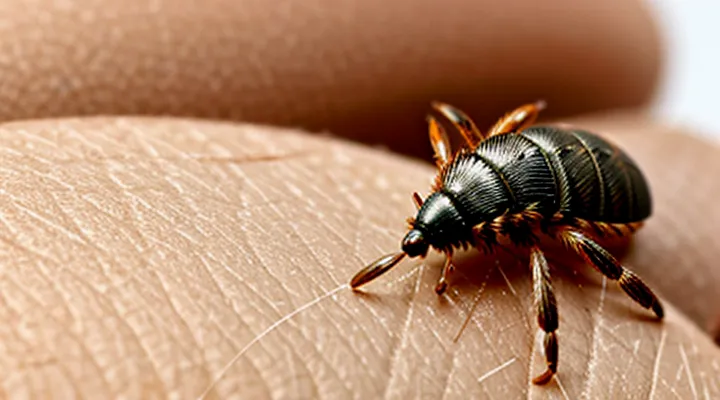
The tick itself may remain attached to the skin after the bite, providing the most reliable visual clue of a recent feeding event. The insect appears as a small, rounded or oval body, typically 2‑5 mm in length, though engorged females can reach up to 10 mm. Its color ranges from light brown to dark reddish‑brown, often darkening as it fills with blood. The abdomen is noticeably expanded, creating a dome‑shaped silhouette that contrasts with the flatter, lighter‑colored anterior part (the capitulum) where the mouthparts emerge.
Key visual features of an attached tick include:
- A defined outline distinct from surrounding skin, often visible as a raised bump.
- A central black or gray mouthpart (hypostome) protruding from the skin surface.
- A clear demarcation between the engorged abdomen and the thinner anterior region.
- Possible movement or twitching when the tick is disturbed.
If the tick detaches, a small puncture wound may remain, but the presence of the engorged arthropod itself confirms the bite and allows for immediate removal and identification.
Common Symptoms Associated with an Uncomplicated Bite
Itching and Irritation
A tick bite typically leaves a small, erythematous papule at the attachment site. The lesion often measures 2–5 mm in diameter and may be surrounded by a faint halo of redness. Itching is the most common sensory complaint; the pruritus can appear within minutes to several hours after the bite and may persist for days. Irritation frequently manifests as a mild to moderate burning sensation, especially when the skin is stretched or exposed to heat.
- Localized itching, sometimes intense enough to provoke scratching.
- Tingling or burning feeling around the bite.
- Swelling of the surrounding tissue, producing a raised, palpable bump.
- Occasionally, a central punctum or a tiny scar where the tick’s mouthparts detached.
The intensity of itching and irritation varies with individual sensitivity, tick species, and duration of attachment. In most cases, the symptoms subside within 3–7 days without intervention. Persistent or worsening pruritus, spreading erythema, or the development of a target‑shaped lesion may indicate secondary infection or an allergic response and warrants medical evaluation.
Localized Swelling
Localized swelling is the most common visible reaction at the site of a tick attachment. The tissue expands within a few hours to a day after the bite, forming a raised, firm area that may be slightly tender to pressure. The edges of the swelling are usually well‑defined, and the skin over it often appears normal in color or exhibits a faint pink hue. In some cases, the swelling may be accompanied by a central punctum, the tiny opening left by the tick’s mouthparts.
Typical features of the reaction include:
- Diameter ranging from a few millimeters to several centimeters, depending on the host’s immune response and the duration of attachment.
- Consistent firmness; the area feels solid rather than fluid‑filled.
- Absence of extensive erythema or spreading redness beyond the immediate perimeter.
- Persistence for several days, gradually diminishing as the tick is removed and the inflammatory process resolves.
When the swelling fails to subside within a week, enlarges, or is accompanied by fever, headache, or a rash, medical evaluation is warranted to rule out tick‑borne infections such as Lyme disease or Rocky Mountain spotted fever. Immediate removal of the tick with fine tweezers, followed by cleaning the site, reduces the likelihood of secondary complications and promotes faster resolution of the localized edema.
Mild Redness
Mild redness is the most common early manifestation of a tick bite on the skin. The area appears as a faint pink‑to‑light red discoloration, usually confined to a diameter of 0.5–2 cm. The coloration is uniform, without the central clearing or target pattern that characterizes some later reactions. The lesion may be slightly raised or flat, and it typically develops within minutes to a few hours after the tick detaches.
Key features of mild redness after a tick attachment:
- Color: pale pink to light red, matching the surrounding skin tone but visibly distinct.
- Size: small, generally not exceeding 2 cm across.
- Edge: smooth, without irregular borders or concentric rings.
- Sensation: may be barely perceptible; itching or tenderness is minimal.
- Duration: persists for 1–3 days, gradually fading without intervention.
The appearance remains stable unless secondary infection or an allergic response occurs. Progression to a larger erythema, a bull’s‑eye lesion, or the development of a rash elsewhere suggests a need for clinical evaluation. Monitoring the site for changes in size, color intensity, or the onset of pain helps determine whether further medical assessment is required.
Recognizing Concerning Marks and Symptoms
Rash Patterns Associated with Tick-Borne Illnesses
Erythema Migrans «Bullseye» Rash
Erythema migrans, commonly referred to as the “bullseye” rash, is the earliest cutaneous manifestation of Lyme disease following a tick attachment. The lesion typically emerges within 3–30 days after the bite and expands outward from the site of attachment. Its classic appearance consists of a central erythematous area surrounded by a peripheral ring of paler skin, creating a concentric pattern. The overall diameter ranges from a few millimetres to more than 30 cm; enlargement proceeds at an average rate of 2–3 mm per hour.
Key visual characteristics include:
- Irregular, well‑demarcated borders that may be smooth or slightly raised.
- Uniform redness in the central zone, often accompanied by mild swelling.
- A lighter, sometimes vesicular, annular rim that may appear slightly raised.
- Absence of purpura, ulceration, or necrosis in the initial phase.
- Possible accompanying symptoms such as mild itching or warmth, but typically no pain.
Variability is common; some patients display a solid red macule without a distinct ring, while others present multiple overlapping lesions. Lesions on the trunk, thighs, or arms are more readily noticed than those in concealed areas, such as the groin or scalp.
Clinical relevance:
- Presence of erythema migrans confirms early Lyme infection and warrants prompt antibiotic therapy, usually doxycycline or amoxicillin, to prevent dissemination.
- Differential diagnoses include cellulitis, allergic reactions, and other arthropod‑borne rashes; however, the concentric “target” configuration and recent tick exposure strongly favor erythema migrans.
- Absence of the rash does not exclude infection; serologic testing and clinical assessment remain essential.
Early identification of the bullseye rash facilitates timely treatment and reduces the risk of systemic complications such as arthritis, neurologic involvement, or cardiac conduction abnormalities.
Other Atypical Rash Presentations
A tick bite often produces a cutaneous lesion that differs from the classic expanding red ring. Clinicians must recognize less common patterns to avoid delayed diagnosis.
- Uniform macular erythema – flat, evenly red area without central clearing; may measure 2–5 cm and persist for several weeks.
- Papular eruption – raised, firm bumps that may coalesce into a plaque; occasionally accompanied by mild itching.
- Vesicular lesions – small fluid‑filled blisters that appear within days of attachment; can rupture, leaving shallow erosions.
- Urticarial hives – transient, raised welts that migrate across the skin; often mistaken for allergic reactions.
- Necrotic focus – dark, indurated center with surrounding erythema; suggests severe local inflammation or secondary infection.
- Multiple discrete lesions – several small erythematous spots scattered around the bite site; may indicate disseminated early infection.
Atypical rashes can develop within 3–30 days after exposure, may remain static in size, and are not always painful. Absence of a target shape does not exclude tick‑borne illness. When any of these manifestations appear after a known or suspected bite, prompt serologic testing and empirical therapy are warranted.
Signs of Infection at the Bite Site
Increased Redness and Warmth
After a tick attaches, the surrounding skin often becomes visibly redder than the adjacent tissue. The erythema may appear as a uniform pink‑to‑crimson halo or as an irregularly shaped patch that expands gradually over several hours. The affected area feels noticeably warmer to the touch compared with surrounding skin, indicating increased blood flow and local inflammation. This combination of heightened color intensity and temperature is a typical early sign of the body’s response to the bite.
- Color: pink, red, or deep crimson, sometimes with a central pale spot where the tick mouthparts are embedded.
- Temperature: 1–2 °C higher than surrounding skin, detectable by gentle palpation.
- Onset: develops within 30 minutes to a few hours after attachment.
- Duration: persists for 24–48 hours unless secondary infection or an allergic reaction develops, in which case redness may spread and warmth intensify.
Pus or Drainage
A tick bite may develop a localized collection of purulent material that appears as a small, raised nodule or ulcerated area. The exudate is usually creamy‑white to yellow, sometimes tinged with blood, and may ooze spontaneously or when the skin is compressed. The surrounding skin often shows erythema extending a few millimeters beyond the drainage point.
The formation of pus typically begins within 24–48 hours after the bite if a secondary bacterial infection occurs. Early lesions may be clear or serous; as neutrophils infiltrate, the fluid becomes thicker and more opaque. The discharge can persist for several days, gradually diminishing as the immune response resolves the infection.
Key visual cues that differentiate infectious drainage from a simple tick bite mark include:
- Presence of a visible plug or crust that can be expressed.
- Swelling that enlarges rather than contracts over time.
- Redness that spreads outward, forming a halo.
- Warmth and tenderness on palpation.
- Foul odor emanating from the site.
Persistent or worsening drainage, increasing pain, fever, or the appearance of a bull’s‑eye rash suggest systemic involvement and warrant prompt medical evaluation. Early antimicrobial therapy can prevent complications such as Lyme disease or other tick‑borne infections.
Pain and Tenderness
A tick bite frequently produces little or no immediate pain; the puncture is often described as a faint prick that may go unnoticed. When discomfort occurs, it is typically mild and localized to the site of attachment.
Tenderness develops as the skin around the bite becomes inflamed. The area may feel sore to pressure, especially when the tick is removed or the skin is rubbed. Tenderness usually peaks within the first 24–48 hours and diminishes as the bite heals, unless secondary infection or an allergic reaction develops.
Key clinical observations related to pain and tenderness:
- Absence of sharp or throbbing pain; most bites are painless or produce only a subtle ache.
- Mild to moderate tenderness that intensifies with palpation.
- Persistence of tenderness beyond a few days, accompanied by redness, swelling, or pus, suggests bacterial infection or a tick‑borne disease and warrants medical evaluation.
- Sudden increase in pain or spreading tenderness may indicate an allergic response or cellulitis.
Monitoring the intensity and duration of tenderness helps differentiate a normal healing reaction from complications that require prompt treatment.
Systemic Symptoms Indicating Potential Illness
Fever and Chills
A tick attachment typically leaves a small, round, erythematous lesion at the feeding site. The center may appear slightly raised or depressed, and the surrounding skin often shows a pale halo that contrasts with the redness. This pattern, sometimes called a “target” or “bull’s‑eye” lesion, is not exclusive to any single disease but serves as a visual cue for clinicians.
Fever and chills frequently accompany tick‑borne infections, emerging days to weeks after the bite. Their clinical significance includes:
- Sudden rise in body temperature, often exceeding 38 °C (100.4 °F).
- Accompanying rigors or shivering episodes that may occur in cycles.
- Persistence for several days if untreated, potentially indicating progression to systemic illness such as Lyme disease, Rocky Mountain spotted fever, or ehrlichiosis.
The presence of a target‑shaped skin mark together with unexplained pyrexia and chills should prompt immediate medical evaluation. Laboratory testing for specific pathogens and early antimicrobial therapy can reduce complications and shorten the febrile period.
Body Aches and Fatigue
A tick bite typically leaves a small, erythematous puncture or a raised, red macule at the attachment site. The lesion may enlarge, develop a central clearing, or become a target‑shaped rash within days. While the cutaneous sign is the most obvious clue, systemic manifestations often accompany the skin change.
Body aches and fatigue are common early complaints after a tick attachment. Muscular soreness usually appears 2–7 days post‑bite and may involve the entire skeleton or be localized to the neck, shoulders, and back. Fatigue often presents as a persistent lack of energy that does not improve with rest and may be accompanied by low‑grade fever. These symptoms are frequently reported in the initial phase of tick‑borne infections such as Lyme disease, anaplasmosis, and babesiosis.
The presence of generalized myalgia and exhaustion signals possible dissemination of the pathogen beyond the bite site. Persistent or worsening discomfort after the skin lesion has resolved warrants medical evaluation, as early treatment reduces the risk of chronic complications.
- Physical examination: assess the bite mark, check for expanding erythema, evaluate joint tenderness, and measure temperature.
- Laboratory testing: order serologic assays for Borrelia burgdorferi, PCR for Anaplasma phagocytophilum, and complete blood count to detect leukopenia or thrombocytopenia.
- Follow‑up: schedule reassessment within 1–2 weeks to monitor symptom progression and treatment response.
Management includes prompt antibiotic therapy when a tick‑borne infection is confirmed or strongly suspected, typically doxycycline for adults and children over eight years. Analgesics such as acetaminophen or ibuprofen alleviate muscle pain, while adequate hydration and sleep support recovery from fatigue. Patients should be instructed to report any new neurological signs, joint swelling, or persistent fever, as these may indicate disease advancement.
Headache and Nausea
A tick bite typically leaves a localized skin change that may appear as a small, raised papule or flat macule. The lesion often measures 2–5 mm in diameter, with a reddish hue that can expand to form a concentric ring of erythema surrounding a paler center, creating a target‑like pattern. In some cases the border is irregular, and the surrounding area may show slight swelling or a faint halo. The mark usually persists for several days to weeks, gradually fading without scarring unless secondary infection occurs.
Headache and nausea frequently accompany the early systemic response to tick exposure.
- Headache: sudden onset, moderate to severe intensity, may be throbbing or pressure‑like; often coincides with fever or fatigue.
- Nausea: sensation of unease in the stomach, may progress to vomiting; commonly reported alongside dizziness or malaise.
Both symptoms can emerge within 24–72 hours after the bite and may persist for several days. Their presence warrants prompt medical evaluation to exclude tick‑borne infections such as Lyme disease, Rocky Mountain spotted fever, or ehrlichiosis. Early treatment reduces the risk of complications and accelerates recovery.
