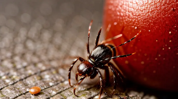
Immediate Post-Bite Appearance
Initial Reaction
Redness and Swelling
After a tick attaches, the skin around the bite typically becomes red and swollen. The redness appears as a well‑defined, pink to crimson halo that may expand outward over several hours. Swelling is often soft, raised, and may feel warm to the touch. In many cases, the erythema measures 2–5 cm in diameter, but larger zones can develop if the bite triggers an allergic reaction or secondary infection.
Key characteristics to observe:
- Onset: Redness usually emerges within 12–24 hours; swelling may follow shortly after.
- Color progression: Initial pink shade can deepen to a darker hue if inflammation intensifies.
- Texture: The area feels tender; edema may cause the skin to appear stretched.
- Duration: Mild erythema and edema often resolve within 3–5 days without intervention; persistent or worsening signs warrant medical evaluation.
When redness spreads rapidly, forms a bullseye pattern, or is accompanied by fever, headache, or joint pain, the presentation may indicate a vector‑borne infection such as Lyme disease. Prompt removal of the tick and consultation with a healthcare professional are recommended in these situations.
Small Bump or Pimple-like Mark
A small, raised lesion often appears at the site where a tick has attached. The bump typically measures 2–5 mm in diameter, is firm to the touch, and may resemble a pimple. Its surface is usually smooth, though occasional central puncture marks the tick’s mouthparts. Early coloration ranges from pink to reddish‑brown; as inflammation progresses, the lesion can become darker or develop a surrounding halo of erythema.
Key visual and clinical features:
- Size: 2–5 mm, occasionally up to 1 cm if swelling increases.
- Shape: round, dome‑shaped, sometimes with a tiny central point.
- Color: pink, red, or brown; may darken with time.
- Texture: firm, non‑fluctuant; no pus unless secondary infection occurs.
- Evolution: appears within hours to a day after attachment, may persist 3–7 days, then fades unless an infection or disease (e.g., Lyme disease) develops.
If the bump enlarges rapidly, becomes painful, develops ulceration, or is accompanied by fever, joint pain, or a spreading rash, medical evaluation is recommended.
Sensation and Symptoms
Itchiness
Itchiness often accompanies the skin changes that follow a tick attachment. The sensation typically begins within a few hours after the bite and may persist for several days.
- The area around the bite becomes mildly pruritic, sometimes intensifying when the skin is warm or moist.
- Scratching can aggravate the surrounding erythema, leading to a larger, irregularly shaped redness that may spread outward from the original puncture site.
- In some individuals, the itch is accompanied by a raised, raised halo that contrasts with the central punctum, creating a target‑like appearance.
- Persistent or worsening itchiness, especially if combined with swelling, fever, or a rash extending beyond the bite, warrants medical evaluation for possible infection or tick‑borne disease.
Understanding the pattern of itch and its correlation with the evolving mark helps differentiate a simple tick bite reaction from complications that require treatment.
Mild Pain or Discomfort
A tick bite often leaves a small, circular erythema that may be accompanied by a faint, localized ache. The discomfort is usually described as a mild pressure or tingling sensation at the site of attachment. It does not typically impair movement or cause systemic symptoms.
Typical characteristics of mild pain or discomfort include:
- Slight tenderness when the area is touched
- A faint, persistent throbbing that fades within a few hours
- Minimal swelling that does not extend beyond the immediate perimeter of the bite
The intensity of these sensations generally diminishes within 24–48 hours as the skin begins to heal. Persistent or worsening pain, expanding redness, or the development of a bullseye pattern may indicate an infection and warrants medical evaluation.
Later Stage Marks and Potential Complications
Non-Infected Bite Marks
Fading of Redness
After a tick attachment, the skin around the bite often shows a localized erythema that may be bright red or pink. Within the first 24–48 hours the redness typically reaches its peak intensity, then begins to diminish. The fading process proceeds gradually, with the color shifting from vivid red to a lighter pink or pale hue before disappearing completely. The rate of disappearance varies according to individual skin tone, the depth of the tick’s mouthparts, and the presence of any secondary inflammation.
Factors influencing the speed of color loss include:
- Depth of attachment: superficial bites resolve faster than deeper lesions.
- Host immune response: robust inflammatory activity can prolong redness.
- Skin characteristics: darker skin tones may retain a subtle discoloration longer.
- Secondary infection: bacterial involvement can maintain or darken the area, delaying fading.
If the redness persists beyond two weeks, expands, becomes intensely painful, or is accompanied by a bullseye pattern, fever, or flu‑like symptoms, medical evaluation is warranted to rule out tick‑borne diseases such as Lyme disease or Rocky Mountain spotted fever.
Disappearance of Swelling
After a tick attaches, the bite site often swells within the first 24–48 hours. In most cases the edema resolves spontaneously as the inflammatory response subsides. The reduction usually follows a predictable pattern:
- Day 1–2: Peak swelling; redness may be pronounced.
- Day 3–5: Gradual decrease in volume; the area may feel less tender.
- Day 6–10: Most visible swelling disappears; faint discoloration or a small punctate mark may remain.
- Beyond day 10: Complete resolution in uncomplicated cases; any lingering discoloration fades over several weeks.
Factors that accelerate the decline include early removal of the tick, proper wound cleaning, and avoidance of secondary infection. Topical anti‑inflammatory agents or oral antihistamines can shorten the duration of edema but are not required for normal healing.
Persistent or worsening swelling after the first week, especially if accompanied by fever, rash, or joint pain, signals possible infection or tick‑borne disease. In such instances, medical evaluation is warranted to rule out conditions such as Lyme disease or cellulitis.
Signs of Infection
Increasing Redness and Warmth
After a tick attachment, the skin around the bite often turns red and feels warm to the touch. The redness typically spreads outward from the puncture site, forming a halo that may become more pronounced over several hours. Heat generated by the local inflammatory response can be detected even before the color change becomes obvious.
- Initial stage: faint pink discoloration, slight warmth.
- Early progression: deepening erythema, noticeable increase in temperature.
- Advanced stage: bright crimson ring, pronounced heat, possible swelling.
If redness expands rapidly, temperature rises sharply, or the area becomes painful, these signs may indicate infection or an early allergic reaction. Prompt medical evaluation is recommended when the lesion exceeds a few centimeters, is accompanied by fever, or shows signs of necrosis. Early intervention reduces the risk of complications such as cellulitis or tick‑borne disease transmission.
Pus or Drainage
Pus or drainage emerging from a tick bite site signals a secondary infection rather than the normal inflammatory response. The exudate typically appears as yellow‑white, creamy material that may be mixed with blood‑tinged fluid. It often accumulates in a small pocket beneath the skin, creating a raised, fluctuating nodule that can be pressed to release the contents.
Key characteristics of purulent discharge include:
- Consistency: thick, viscous, and may form a thin film over the wound.
- Color: ranging from clear yellow to greenish‑brown if bacterial enzymes are present.
- Odor: faintly foul, especially when anaerobic bacteria are involved.
- Timing: usually develops 3–7 days after the bite, following an initial erythematous or papular stage.
The presence of drainage warrants prompt medical evaluation because it may indicate:
- Bacterial colonization such as Staphylococcus aureus or Streptococcus species.
- Progression to cellulitis, abscess formation, or systemic infection.
- Potential need for antimicrobial therapy and, in some cases, incision and drainage.
Monitoring the lesion for changes in size, pain intensity, or spread of redness can help differentiate uncomplicated inflammation from a true infectious process. Early intervention reduces the risk of complications and promotes faster resolution of the wound.
Spreading Rash
A spreading rash following a tick attachment typically begins as a small, reddish spot at the bite site and enlarges outward in a circular or oval pattern. The lesion often reaches 5 cm or more in diameter within days, displaying a clear central area surrounded by a paler halo. Texture remains smooth; the skin is not raised, vesicular, or ulcerated. The rash may be accompanied by mild itching or tenderness, but systemic symptoms such as fever or fatigue can also develop.
Key characteristics:
- Diameter expands gradually, often exceeding 5 cm.
- Borders are well defined, with a uniform red coloration.
- Central clearing creates a target‑like appearance.
- No pus, crust, or necrosis.
- May persist for several weeks if untreated.
Recognition of these features enables prompt clinical evaluation and appropriate antimicrobial therapy.
Disease-Specific Rashes
Lyme Disease: «Bull's-Eye» Rash (Erythema Migrans)
After a tick attachment, the earliest cutaneous sign of Lyme disease is erythema migrans, frequently termed a “bull’s‑eye” rash. The lesion typically begins as a small, expanding erythematous macule or papule at the bite site and enlarges over days to weeks.
Key characteristics include:
- Diameter ranging from 5 mm to more than 30 cm.
- Central clearing that creates a lighter zone surrounded by a red, raised border.
- Asymmetrical shape; the concentric pattern is not required for diagnosis.
- Possible accompanying symptoms such as mild itching, warmth, or tenderness.
Variations may present as:
- Uniformly red plaques without central pallor.
- Multiple lesions appearing simultaneously on different body areas.
- Lesions that fade or become less distinct as they enlarge.
The rash appears within 3–30 days after exposure. Presence of erythema migrans warrants prompt antimicrobial therapy to prevent dissemination to joints, heart, and nervous system. Absence of the classic bull’s‑eye pattern does not exclude infection; clinical judgment should consider exposure history and symptomatology. Immediate medical evaluation is advised when any expanding erythematous lesion follows a tick bite.
Other Tick-Borne Illnesses: Varied Rashes
Tick‑borne infections often produce skin eruptions that differ markedly from the classic erythema migrans of Lyme disease.
Rocky Mountain spotted fever generates a rapid‑onset rash that begins on the wrists and ankles, spreading centrally to the trunk. Lesions appear as small, erythematous macules that may become petechial, sometimes forming a confluent, blanching pattern.
Ehrlichiosis and anaplasmosis frequently cause a non‑specific, faint maculopapular rash, most often on the trunk. In severe cases, petechiae may develop on the palms and soles, indicating thrombocytopenia.
Rickettsial infections such as Mediterranean spotted fever produce an inoculation eschar—a dark, necrotic papule at the bite site—surrounded by a peripheral erythematous halo. The surrounding rash typically consists of discrete, pink macules that may coalesce.
Tularemia may present with a painless ulcer or papule at the attachment point, occasionally accompanied by a surrounding erythema that can evolve into a necrotic ulcer.
Babesiosis rarely causes cutaneous manifestations; however, hemolytic anemia can lead to jaundice and occasional petechial spots due to platelet consumption.
Key distinguishing features include:
- Distribution: wrists/ankles (RMSF) vs. trunk (Ehrlichiosis/Anaplasmosis).
- Lesion type: maculopapular, petechial, eschar, ulcerative.
- Onset timing: RMSF rash appears within 2–5 days; erythema migrans emerges after 3–30 days.
Recognizing these patterns enables prompt differentiation among tick‑borne diseases and guides appropriate antimicrobial therapy.
Allergic Reactions
Hives or Widespread Rash
Hives that develop after a tick attachment appear as raised, erythematous wheals that may merge into larger plaques. The lesions are typically pruritic, vary from a few millimeters to several centimeters in diameter, and can be scattered across the trunk, limbs, or face. Their borders are often ill‑defined, and the surface may be smooth or slightly edematous.
A widespread rash may manifest as diffuse erythema with multiple confluent areas of inflammation. The coloration ranges from pink to deep red, sometimes accompanied by a faint, mottled pattern. The rash often spreads rapidly, involving extensive skin regions within hours to a few days after the bite. Accompanying sensations include burning, itching, or a mild stinging feeling.
Typical timeline:
- Onset: 12 – 48 hours post‑exposure.
- Peak intensity: 24 – 72 hours.
- Resolution: 5 – 10 days with or without treatment; persistence beyond two weeks warrants medical evaluation.
Key clinical clues that differentiate tick‑related urticaria from other dermatoses:
- Sudden appearance after known tick exposure.
- Absence of vesicles or necrotic centers.
- Lack of systemic symptoms such as fever or joint pain, unless associated with a specific tick‑borne infection.
Management approach:
- Antihistamines (second‑generation preferred) to control pruritus.
- Topical corticosteroids for localized inflammation.
- Oral corticosteroids in severe, extensive cases.
- Monitoring for secondary signs of infection (e.g., fever, lymphadenopathy) and initiating appropriate antimicrobial therapy if indicated.
Severe Swelling
Severe swelling following a tick attachment presents as a rapid, pronounced enlargement of the skin around the bite site. The affected area often exceeds several centimeters in diameter, appears raised, and may feel firm to the touch. Skin coloration ranges from pink to deep red, sometimes with a mottled or purplish hue indicating underlying inflammation.
The swelling typically develops within hours to a day after the bite and can expand despite initial attempts at cold compresses or antihistamines. Accompanying features may include:
- Tightness or stretching of the skin surface
- Warmth localized to the swollen region
- Pain or tenderness that intensifies with movement of nearby joints
- Possible development of a central erythematous or necrotic spot, known as an eschar, surrounded by the edematous tissue
When swelling progresses beyond the immediate vicinity of the bite, involves multiple joints, or is associated with fever, headache, or malaise, immediate medical evaluation is warranted. Prompt treatment can prevent complications such as tick‑borne infections or severe allergic reactions.
