The Appearance of an Embedded Tick
Size and Shape Variations
Ticks that have penetrated the skin display a broad spectrum of dimensions. Unfed larvae measure roughly 1–2 mm in length, while nymphs range from 2–4 mm. Adult females, after several days of blood intake, can expand to 6–10 mm or more. Males remain smaller, typically not exceeding 5 mm, even when fully fed.
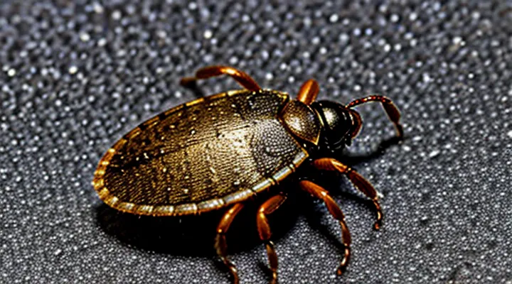
Shape transitions accompany the feeding process. Early attachment presents a flattened, oval body with visible dorsal scutum. As engorgement progresses, the abdomen elongates and swells, giving the tick a more rounded, balloon‑like silhouette. Mouthparts, including the hypostome, remain visible at the anterior end throughout all stages.
Key visual indicators:
- Length: 1 mm (larva) to >10 mm (engorged female).
- Width: proportional to length; engorged specimens may appear twice as wide as unfed forms.
- Body profile: flat and oval initially; progressively rounded and distended during feeding.
- Color: pale to reddish‑brown when unfed; deepening to dark brown or black as blood fills the gut.
Species differences affect size limits. For example, the American dog tick (Dermacentor variabilis) rarely exceeds 8 mm when engorged, whereas the Lone Star tick (Amblyomma americanum) can reach 12 mm. Recognizing these size and shape variations assists in accurate identification and timely removal.
Color Changes Due to Feeding
A tick attached to the skin appears as a small, rounded object that may be difficult to see against the surrounding tissue. Immediately after attachment, the body is typically pale or light brown, reflecting the unfed state of the arthropod.
During the feeding period, the tick’s abdomen expands dramatically as it ingests blood. This expansion produces a series of observable color changes:
- Early feeding (0–12 hours): abdomen remains light brown; legs and mouthparts are visible; surface may show a faint grayish hue.
- Mid feeding (12–48 hours): abdomen begins to swell; color deepens to a darker brown or reddish‑brown; edges become more pronounced.
- Late feeding (48–72 hours): abdomen reaches maximum size, often several times the original length; color turns dark brown to almost black; surface becomes glossy and may appear slightly translucent.
«The tick’s body expands and darkens as it fills with blood», a characteristic that aids in distinguishing an engorged specimen from other skin lesions. Recognizing these color stages assists healthcare providers in confirming the presence of a feeding tick and in determining the appropriate removal timing.
Visible Body Parts
An embedded tick appears as a small, often darkened protrusion on the skin surface. The abdomen swells as the parasite fills with blood, increasing in size from a few millimeters to several centimeters. The surrounding skin may show a tiny puncture point where the mouthparts have entered.
Visible structures include:
- Capitulum (mouthparts) that remain anchored beneath the skin, typically invisible externally but responsible for the central puncture.
- Scutum, a hard shield covering the anterior portion of the tick’s body, often visible as a lighter‑colored patch on the engorged abdomen.
- Legs, usually folded against the body and not readily seen once the tick is fully attached.
- Engorged abdomen, the most conspicuous feature, presenting a rounded, balloon‑like shape that may be reddish, brown, or black depending on the species and feeding stage.
- The attachment site, a small, sometimes reddened or inflamed area surrounding the puncture.
Identification relies on observing the enlarged abdomen and the presence of the scutum. The puncture point marks the location of the capitulum, indicating that the tick is still attached and must be removed carefully to avoid leaving mouthparts in the skin.
Distinguishing Ticks from Other Skin Blemishes
Moles and Freckles
Ticks that have penetrated the epidermis appear as compact, dark masses. The anterior mouthparts often create a tiny central puncture, and the body may be slightly raised above the surrounding skin. Color ranges from deep brown to black, sometimes with a sheen that reflects the engorged abdomen.
Moles, or «nevus», present as uniformly pigmented lesions. They may be flat or raised, with smooth or slightly textured surfaces. Borders are regular, and the color stays consistent throughout the lesion, typically ranging from light brown to dark brown or black.
Freckles, known as «ephelides», are small, flat spots of increased melanin. Their size does not exceed a few millimetres, and the colour varies from light tan to dark brown. Distribution is often widespread, especially on sun‑exposed areas, and the lesions lack any raised component.
Key visual distinctions:
- Elevation: Tick – raised; mole – flat or mildly raised; freckle – flat.
- Border definition: Tick – irregular, may show a central point; mole – regular; freckle – diffuse.
- Color uniformity: Tick – dark with possible sheen; mole – consistent pigment; freckle – uniform but lighter than a tick.
- Size range: Tick – up to several millimetres, may enlarge with feeding; mole – variable, often larger than a freckle; freckle – consistently small.
Recognizing these characteristics enables accurate identification of an embedded tick versus benign pigmented skin markings.
Scabs and Dirt
When a tick penetrates the epidermis, the surrounding tissue often reacts by forming a thin, dry crust. This scab typically appears as a pale, slightly raised ring that encircles the mouthparts. The crust may be partially translucent, allowing the tick’s body to be seen through the outer layer.
Dirt commonly adheres to the scab’s surface, especially if the bite occurs outdoors. Soil particles create a speckled pattern that can obscure the tick’s outline but also serve as an indicator that the lesion is recent.
Key visual characteristics:
- Thin, dry crust surrounding a central dark spot (the tick’s body)
- Slight elevation of the crust above surrounding skin
- Presence of fine, gray‑brown specks embedded in or around the crust
- Absence of fluid‑filled blister; the area remains firm
Recognition of these features assists in differentiating an embedded tick from other skin lesions such as simple abrasions or insect bites. Prompt removal of the tick, followed by cleaning of the scab and surrounding dirt, reduces the risk of infection and disease transmission.
Other Insect Bites
A tick that has penetrated the skin usually appears as a tiny, dark, oval object partially visible through the epidermis. The body may be engorged, creating a raised, firm nodule. A central puncture point often remains visible, sometimes surrounded by a faint, red halo. Swelling may extend a few millimeters from the site, and the surrounding skin can feel warm to the touch.
Other arthropod bites present distinct visual patterns:
- Mosquito: small, pink, raised wheal that fades within 24 hours; often accompanied by intense itching.
- Flea: clusters of tiny, red papules with a central punctum; lesions may appear in a line or “breakfast‑scrambled” pattern.
- Bedbug: irregular, raised, red welts with a dark‑colored spot at the center; lesions frequently occur in a linear or zig‑zag arrangement.
- Spider: punctate wound surrounded by a larger area of erythema; some species produce necrotic lesions with a yellow‑white core.
- Ant: localized swelling and a sharp, white‑colored sting mark; pain is immediate and may be accompanied by a burning sensation.
Key differentiators include size, shape, distribution, and the presence of a central puncture. Ticks remain attached for several days, whereas most other insects detach after a single bite. Persistent attachment, a visible engorged body, and a stable nodule suggest a tick rather than a bite from another arthropod.
Recognizing Symptoms of a Tick Bite
Redness and Swelling
When a tick penetrates the epidermis, the immediate cutaneous response typically presents as a localized area of redness surrounded by a small, raised swelling. The erythema often appears as a uniform pink to reddish halo that matches the size of the tick’s mouthparts, while the edema forms a palpable bump that may be slightly tender to pressure.
Key characteristics of the reaction include:
- Uniform coloration without sharp demarcation, indicating a vascular response rather than a necrotic lesion.
- Swelling confined to the perimeter of the bite, rarely extending beyond a few millimeters.
- Persistence for several days; gradual fading signals normal healing, whereas expansion or discoloration may suggest secondary infection.
Recognition of these signs enables prompt differentiation between a simple tick attachment and complications such as cellulitis or tick‑borne disease, facilitating appropriate medical assessment.
Itching and Irritation
When a tick secures its mouthparts beneath the epidermis, the surrounding tissue often reacts with pronounced itching. The sensation typically begins within minutes to a few hours after attachment and may intensify as the tick continues to feed.
Irritation manifests as a localized erythema that surrounds the entry point. The skin may appear slightly raised, forming a papule or a small wheal. In many cases, a faint halo of redness expands outward, indicating an inflammatory response.
Common visual indicators of the reaction include:
- A central puncture mark, sometimes visible as a tiny dark dot where the tick’s hypostome entered the skin.
- Redness that is brighter than the surrounding area, often circular or oval in shape.
- Swelling that may produce a raised bump, occasionally accompanied by a thin fluid‑filled blister.
- Persistent scratching marks, revealing excoriations that develop from repeated irritation.
If itching persists beyond 24 hours or the erythema expands rapidly, the possibility of an allergic or infectious complication increases, warranting medical assessment.
Pain or Discomfort
A tick that has penetrated the skin creates a focal point of irritation. The mouthparts anchor deep in the epidermis, producing a small, often pale, raised bump surrounded by a halo of reddened tissue. The surrounding area may be moist or slightly swollen, reflecting the tick’s secretions that prevent clotting.
Typical sensations associated with the attachment include:
- Sharp, localized sting at the moment of insertion.
- Persistent itching that intensifies as the tick feeds.
- Tingling or burning around the bite site, especially after several hours.
- Dull ache that may spread to adjacent muscles or joints if inflammation progresses.
The intensity of pain or discomfort varies with the tick’s species, feeding duration, and the host’s skin sensitivity. Absence of immediate pain does not guarantee that the tick has not begun feeding; many individuals experience only mild irritation while the parasite remains attached. Prompt removal, followed by cleansing of the area, reduces the risk of secondary infection and minimizes prolonged discomfort.
What to Do if You Find an Embedded Tick
Safe Removal Techniques
When a tick has penetrated the epidermis, prompt removal reduces the risk of pathogen transmission. The parasite’s mouthparts embed shallowly; the body remains visible as a small, raised nodule. Immediate action focuses on extracting the tick whole, without crushing the abdomen.
- Grasp the tick as close to the skin as possible with fine‑point tweezers.
- Apply steady, upward pressure; pull straight out in a continuous motion.
- Disinfect the bite site with an antiseptic solution such as povidone‑iodine.
- Store the removed specimen in a sealed container for potential laboratory analysis, labeling with date and location.
- Monitor the area for several days; seek medical advice if redness, swelling, or fever develop.
Avoid methods that involve heat, chemicals, or squeezing the tick’s body, as these can force infected fluids into the host. Use only tools designed for precision gripping; disposable tweezers eliminate cross‑contamination. After removal, wash hands thoroughly with soap and water.
Tools for Tick Removal
An engorged tick embedded in the skin appears as a dark, swollen body attached firmly to the epidermis, with a visible capitulum (mouthparts) extending into the tissue. The surrounding area may show a small puncture mark and, occasionally, a faint halo of irritation.
Effective removal relies on tools that grasp the tick close to the skin without crushing the abdomen. Recommended instruments include:
- Fine‑tipped, straight‑pointed tweezers — allows precise grip on the tick’s head.
- Tick removal hook (also called a tick key) — slides beneath the mouthparts for clean extraction.
- Small, flat‑bladed forceps — useful for ticks attached in hard‑to‑reach locations.
- Disposable gloves — prevents direct contact with potential pathogens.
- Antiseptic solution or alcohol wipes — cleans the bite site before and after removal.
- Sealable container with alcohol — stores the extracted tick for identification or testing.
When using tweezers or forceps, position the tips as close to the skin as possible, grasp the tick’s head, and apply steady, downward pressure while pulling straight upward. The hook is inserted beneath the mouthparts, then lifted gently to release the attachment. After extraction, disinfect the bite area, discard gloves, and wash hands thoroughly. If the tick’s body ruptures, clean the wound and monitor for signs of infection.
Post-Removal Care
After extracting a tick from the skin, immediate attention reduces irritation and infection risk. Clean the bite site with mild soap and water, then apply an antiseptic such as povidone‑iodine or chlorhexidine. Pat the area dry with a clean gauze pad.
Monitor the wound for signs of inflammation. If redness, swelling, or warmth expands beyond the immediate perimeter, seek medical evaluation. A short course of topical antibiotic ointment may be applied to prevent bacterial colonisation, especially if the skin appears broken.
Maintain the area in a dry, protected state for 24–48 hours. Change dressings only if they become wet or soiled. Avoid scratching or applying excessive pressure, which can introduce pathogens. Record the date of removal and any subsequent symptoms; this information assists health‑care providers in assessing potential tick‑borne diseases.
When to Seek Medical Attention
Incomplete Tick Removal
A tick that has not been fully extracted remains anchored by its mouthparts, which appear as a small, dark point protruding from the skin. The surrounding area may show a slight depression where the body sits, often resembling a tiny, brownish lump. The tick’s head can be visible as a pin‑point or a thin, black line extending into the epidermis. Surrounding skin may be mildly erythematous, but inflammation is usually minimal unless infection develops.
Incomplete removal leaves the hypostome embedded, creating a portal for pathogen transmission. Risks include prolonged attachment, increased chance of bacterial colonisation, and potential development of Lyme disease or other tick‑borne illnesses. The retained mouthparts may cause local irritation, itching, or secondary infection if not addressed promptly.
Proper management of a partially removed tick involves:
- Cleaning the site with antiseptic solution.
- Applying a fine‑pointed sterile tweezers to grasp the tick’s head as close to the skin as possible.
- Pulling upward with steady, even pressure without twisting.
- Inspecting the extracted specimen to confirm that the mouthparts are intact.
- If any portion remains, seeking medical evaluation for surgical removal or antibiotic prophylaxis.
Monitoring after removal is essential. Observe the bite area for expanding redness, fever, fatigue, or joint pain. Any such symptoms warrant immediate medical consultation.
Symptoms of Tick-Borne Illnesses
An embedded tick presents as a small, darkened, engorged nodule often resembling a tiny black speck or a raised papule. The mouthparts may be visible as a tiny point protruding from the skin surface, especially if the tick’s body has expanded after feeding. The surrounding area can appear slightly erythematous but may remain otherwise unremarkable, making early recognition dependent on careful visual inspection.
Symptoms of illnesses transmitted by ticks typically emerge days to weeks after attachment. Common clinical manifestations include:
- Fever or chills
- Headache, often described as severe or throbbing
- Fatigue and malaise
- Muscle or joint aches, sometimes progressing to migratory arthralgia
- Rash, frequently presenting as a red, expanding lesion with central clearing (often termed a “bull’s‑eye” pattern)
- Nausea, vomiting, or abdominal pain
Less frequent but serious signs involve neurological involvement such as facial palsy, meningitis‑like symptoms, or cognitive disturbances, as well as cardiac manifestations like heart block. Prompt identification of these symptoms and correlation with a recent tick bite are essential for early diagnosis and treatment of tick‑borne diseases.
Allergic Reactions
An embedded tick appears as a small, oval body attached to the skin surface, often 2–5 mm in diameter when unfed and up to 10 mm after engorgement. The dorsal shield is dark brown to gray, while the ventral side may show a lighter hue. Mouthparts, including the hypostome, protrude into the epidermis, forming a pinpoint entry point that may be difficult to see without magnification. Engorged specimens become noticeably raised, sometimes resembling a tiny blister.
Allergic reactions to a tick bite manifest in two principal patterns:
- Localized erythema and edema around the attachment site, typically appearing within hours and peaking within 24 hours.
- Systemic hypersensitivity, presenting as urticaria, pruritus, or, in severe cases, anaphylaxis characterized by urticaria, angioedema, respiratory distress, and hypotension.
Immediate signs of a severe allergic response include rapid swelling of the face or neck, difficulty breathing, and a sudden drop in blood pressure. Mild reactions may resolve spontaneously, but persistent or worsening symptoms warrant medical evaluation.
If a tick remains attached for more than 24 hours, removal with fine‑point tweezers is recommended, grasping the mouthparts close to the skin and pulling upward with steady pressure. After extraction, observe the bite area for increasing redness, swelling, or systemic signs. Consultation with a healthcare professional is advised when symptoms progress beyond localized irritation, especially if respiratory or cardiovascular involvement occurs.
