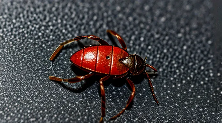
Initial Observation: Distinguishing a Fed Tick
Size and Shape Changes
A tick that has taken a blood meal expands dramatically. The body elongates and the dorsal shield swells, turning a previously flat organism into a markedly convex, balloon‑like shape. The abdomen, which houses the ingested blood, dominates the visual profile, while the legs remain relatively short and clustered near the anterior margin.
Typical dimensions illustrate the change:
- Unfed adult females: 3–5 mm long, 2 mm wide.
- Fully engorged females: 10–12 mm long, 8–10 mm wide.
- Unfed nymphs: 1.5–2 mm long, 0.5 mm wide.
- Fully engorged nymphs: 4–5 mm long, 2–3 mm wide.
The transformation is not merely linear growth; the tick’s silhouette becomes rounded, with the dorsal surface achieving a near‑spherical contour. The ventral side also expands, creating a noticeable bulge that distinguishes a fed tick from its unfed counterpart.
Species differences affect the magnitude of enlargement. Ixodes ricinus, for example, may double its length, whereas Dermacentor variabilis can increase volume by a factor of ten. Nonetheless, all blood‑filled ticks share the characteristic shift from a thin, oval profile to a thick, rounded form that signals successful feeding.
Color Variations
A tick that has completed a blood meal expands dramatically and its coloration shifts from the typical brown or reddish‑brown of an unfed specimen to a range of hues that reflect the ingested blood and the tick’s species.
Common color presentations include:
- Light tan or creamy‑white when the tick is freshly engorged and the blood has not yet darkened.
- Deep reddish‑brown or mahogany, especially in species that feed on mammals with high hemoglobin content.
- Dark brown to almost black in hard‑tick species where the cuticle thickens and the blood oxidizes.
- Mixed mottled patterns where the abdomen shows a translucent pinkish hue while the dorsal shield remains darker.
Variations arise from factors such as the host’s blood composition, the duration of feeding, and the tick’s developmental stage. Recognizing these color changes aids accurate identification and informs appropriate removal or control measures.
Anatomical Modifications After Feeding
Engorged Abdomen: The Primary Indicator
An engorged abdomen is the most reliable visual cue that a tick has recently fed. After a blood meal the body expands dramatically, often increasing the overall length by two to three times. The abdomen becomes balloon‑like, rounded, and visibly distended from the normally narrow, tapered shape of an unfed specimen. Color shifts from a light brown or gray to a deep, glossy red or purplish hue, reflecting the presence of host blood beneath the cuticle. Surface texture may appear smoother, as the stretched cuticle reduces the visible segmentation seen on a flat, unfed tick.
- Length: 4–6 mm for larvae, up to 12 mm for adult females.
- Width: abdomen width exceeds half of the total body width.
- Shape: rounded, convex, lacking the typical “hourglass” silhouette.
- Color: dark red, mahogany, or deep brown; translucency may reveal blood flow.
- Movement: slower, often remaining attached to the host for several days.
These characteristics differentiate a fed tick from one that is questing or recently detached. Recognizing the engorged abdomen enables prompt removal and reduces the risk of pathogen transmission.
Leg and Head Proportions in Relation to the Body
A blood‑engorged tick expands dramatically, its abdomen swelling to several times the size of an unfed specimen. The cuticle stretches, giving the body a rounded, balloon‑like silhouette that dominates the overall profile.
The legs retain their original length while the body enlarges, creating a stark contrast. Each of the eight legs appears relatively short, emerging from the anterior margin at shallow angles. The leg segments remain proportionally constant, but the distance between the attachment points and the expanded abdomen increases, making the legs look stubby compared to the massive torso.
The head region, comprised of the capitulum (mouthparts) and the anterior shield, does not enlarge with the abdomen. The capitulum stays compact, positioned near the front of the swollen body. Its size remains comparable to that of an unfed tick, resulting in a pronounced disparity between the tiny head and the voluminous rear.
- Leg length: unchanged, appears short relative to engorged abdomen.
- Leg angle: shallow, directed forward and outward.
- Head size: constant, situated at the anterior edge, markedly smaller than the expanded body.
- Body shape: rounded, dominant, obscuring the proportional balance of legs and head.
These morphological changes provide a reliable visual cue for identifying a tick that has recently fed on blood.
Why Ticks Change So Dramatically
Blood Meal: A Nutrient-Rich Resource
A blood‑engorged tick expands dramatically; the body can increase three to five times its unfed size. The dorsal shield (scutum) remains relatively unchanged, while the abdomen swells into a balloon‑like shape, often turning a pale gray or bluish hue due to the volume of ingested blood. The tick’s legs may appear shorter relative to the enlarged body, and the cuticle becomes more translucent, revealing the internal meal.
The blood meal supplies a concentrated source of nutrients essential for development and reproduction. Key components include:
- Hemoglobin‑derived iron, supporting egg formation.
- High‑quality proteins for tissue growth.
- Lipids providing energy reserves.
- Carbohydrates for immediate metabolic needs.
- Vitamins and trace elements that facilitate enzymatic processes.
During the post‑feeding period, the tick digests the blood, converting these nutrients into reserves that sustain molting, egg production, and survival through adverse conditions. The physical transformation and nutrient composition together define the tick’s capacity to complete its life cycle.
Preparation for Reproduction and Survival
A tick that has recently taken a blood meal expands dramatically; its body swells to several times its unfed size, often reaching a diameter of 5–10 mm in females. The cuticle becomes translucent, revealing a pale, creamy interior where blood pools, while the dorsal surface may appear reddish or brownish due to the hemoglobin content. Legs remain proportionally short, and the mouthparts are visible as a dark, curved structure near the anterior margin. The abdomen dominates the silhouette, giving the organism a rounded, balloon‑like profile.
These morphological changes serve two critical functions:
- Egg production: The enlarged abdomen stores sufficient nutrients to support the development of hundreds to thousands of eggs, directly linking engorgement to reproductive output.
- Energy reserves: The accumulated blood provides a high‑energy substrate for metabolism during the prolonged off‑host period, enhancing survival through harsh environmental conditions.
After detachment, the tick descends into a protected microhabitat where it digests the meal, synthesizes vitellogenin, and initiates oviposition. The visible engorged state therefore signals both imminent fecundity and a temporary physiological resilience that ensures the species’ continuation.
Implications for Human and Animal Health
Increased Disease Transmission Risk
A blood‑engorged tick expands dramatically, often reaching the size of a small grape. The abdomen swells, turning a deep gray‑brown or bluish hue, while the mouthparts remain anchored in the host’s skin. The cuticle stretches, making the tick appear soft and translucent in places.
The engorged state directly elevates the probability of pathogen transmission. When the tick’s body fills with blood, the following factors increase risk:
- Higher pathogen load: the tick ingests more infected blood, amplifying the number of microorganisms it can release.
- Prolonged attachment: larger size usually means the tick has remained attached for several days, providing a longer window for disease agents to migrate.
- Enhanced salivary secretions: expanded glands produce more saliva, the primary vehicle for bacteria, viruses, and protozoa.
- Greater skin disruption: the enlarged mouthparts cause a larger wound, facilitating easier entry of pathogens into the host’s bloodstream.
These physiological changes make an engorged tick a more efficient vector than an unfed counterpart, underscoring the importance of prompt removal and regular inspection after exposure.
Removal Challenges of Engorged Ticks
Engorged ticks present a set of practical obstacles that differ markedly from those associated with unfed specimens. Their bodies can swell to several times the original size, often assuming a balloon‑like silhouette that obscures the exact location of the mouthparts. The expansion stretches the hypostome, increasing the risk that manual traction will detach the head while leaving fragments embedded in the skin. Such remnants can become infection portals and may retain pathogen reservoirs.
Key challenges in extraction include:
- Visibility of attachment points – the engorged abdomen masks the chelicerae, making precise placement of removal tools difficult.
- Increased attachment strength – the hypostome’s barbed structure penetrates deeper as the tick expands, requiring steady, controlled force rather than jerking motions.
- Risk of tearing – excessive pulling can snap the mouthparts, leaving them behind and necessitating additional medical intervention.
- Potential for pathogen transmission – prolonged handling raises the chance of saliva or hemolymph exposure, especially if the tick’s cuticle ruptures.
Effective removal protocols recommend:
- Fine‑point tweezers or a specialized tick removal device – grasp the tick as close to the skin as possible, securing the head without squeezing the abdomen.
- Steady, upward traction – apply consistent pressure parallel to the skin surface; avoid twisting or rapid movements.
- Immediate post‑removal inspection – verify that the entire mouthpart is present; if any fragment remains, seek professional care.
- Disinfection of the bite site – cleanse with an antiseptic solution to reduce secondary infection risk.
Understanding the altered morphology of a blood‑filled tick clarifies why conventional pinching or crushing methods prove ineffective and highlights the necessity of precise, controlled techniques for safe extraction.
Recognizing Different Stages of Engorgement
Partially Fed vs. Fully Engorged
A tick that has begun to ingest blood displays a noticeable change in size and coloration, but the degree of transformation varies with feeding stage.
In the partially fed stage, the body expands to roughly one‑third of its maximum size. The dorsal surface remains relatively flat, with a light brown to reddish hue that does not yet cover the entire abdomen. Legs retain a normal proportion to the body, and the mouthparts are visible but not fully extended.
In the fully engorged stage, the tick’s body swells to its greatest volume, often increasing tenfold in weight. The abdomen becomes balloon‑like, uniformly dark red or deep purple, and the cuticle stretches tightly over the internal blood load. Legs appear short relative to the swollen body, and the tick may appear immobile as it prepares to detach.
Key visual differences
- Size: 1/3 vs. up to 10× original length
- Shape: flat‑to‑slightly convex vs. round, balloon‑shaped
- Color: light brown/reddish vs. dark red/purple
- Leg proportion: normal vs. shortened appearance
Recognition of these traits enables accurate identification of feeding status, which is essential for assessing disease transmission risk.
Differences Among Tick Species
Engorged ticks vary noticeably among species, a fact that aids identification during field surveys or veterinary examinations.
- Ixodes ricinus (castor bean tick): abdomen expands to a rounded, dome‑shaped mass; coloration shifts from reddish‑brown to a uniform dark brown; legs remain relatively short, giving a compact silhouette.
- Dermacentor variabilis (American dog tick): body swells into a markedly oval, balloon‑like form; dorsal surface retains a pale, creamy hue with distinctive white‑spotted patterns; legs elongate proportionally, creating a more “stretched” appearance.
- Amblyomma americanum (lone star tick): engorgement produces a markedly elongated, spindle‑shaped abdomen; dorsal surface appears bright red to orange; scutum remains visible as a contrasting dark patch near the head, unlike the fully concealed scutum of Ixodes.
- Rhipicephalus sanguineus (brown dog tick): abdomen expands into a cylindrical, brick‑red column; overall body darkens to a uniform brown; scutum is small and hidden, and the tick maintains a relatively flat profile despite swelling.
These morphological cues—shape of the abdomen, color shift, leg proportion, and visibility of the scutum—constitute reliable markers for distinguishing blood‑filled ticks across the most common genera.
