«General Appearance and Characteristics»
«Color and Shape»
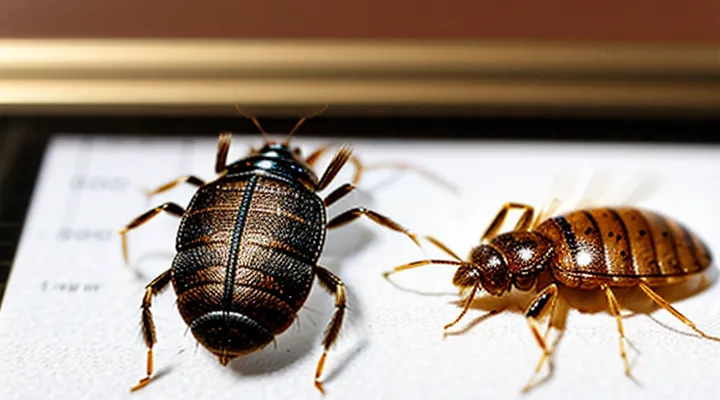
Bedbugs are small, oval‑shaped insects measuring 4–5 mm in length when unfed and expanding to about 6–7 mm after a blood meal. Their bodies are dorsoventrally flattened, facilitating movement through fabric seams and mattress crevices. The exoskeleton bears a smooth, glossy surface without visible segmentation.
Coloration varies with feeding status and age:
- Unfed adults: reddish‑brown or mahogany hue, often described as “brown‑red.”
- Engorged adults: deep rust or brick‑red, sometimes appearing almost black due to the expanded abdomen filled with blood.
- Nymphal stages: lighter tan or amber tones, progressively darkening with each molt.
The head is short and concealed beneath the pronotum, which forms a distinct, shield‑like plate extending laterally. Antennae consist of six slender segments, and the legs are slender, ending in clawed tarsi that grip fabric fibers. The overall silhouette resembles a tiny, flattened seed or a small apple seed, with a blunt anterior and a tapered posterior.
«Size and Dimensions»
Bedbugs (Cimex lectularius) measure between 4.5 mm and 5.5 mm in length when fully fed, shrinking to 3 mm after digestion. Their width ranges from 2 mm to 3 mm, giving an oval profile that is flatter on the dorsal surface and slightly convex ventrally. Adult females are typically 0.5 mm longer than males, reflecting the additional space required for egg development.
Nymphal stages progress through five instars; each molt adds approximately 0.5 mm to overall length. The smallest instar measures about 1.5 mm, while the final instar reaches 4 mm before the adult molt.
Key dimensions:
- Length (adult, unfed): 3 mm – 4 mm
- Length (adult, fed): 4.5 mm – 5.5 mm
- Width (adult): 2 mm – 3 mm
- Female length increase: ≈0.5 mm over male
- Instar growth increment: ≈0.5 mm per molt
These measurements allow reliable visual identification and differentiate bedbugs from similarly sized arthropods such as carpet beetles or flea larvae.
«Body Segmentation»
Bedbugs exhibit a distinct body segmentation that directly influences their recognizable shape and measurable size. The insect’s anatomy is divided into three primary regions: head, thorax, and abdomen, each separated by visible sutures that create a flattened, oval silhouette. The head houses compound eyes and antennae, the thorax bears three pairs of legs and a pair of wings reduced to vestigial structures, and the abdomen consists of five visible dorsal plates (tergites) that expand after feeding.
Key dimensions correlate with this segmentation. Adult specimens typically measure 4–5 mm in length and 2.5–3 mm in width at the widest point of the abdomen. Nymphal stages retain the same segmented layout but are proportionally smaller, ranging from 1.5 mm (first instar) to 4 mm (fifth instar). The segmented abdomen enlarges noticeably after a blood meal, increasing overall thickness by up to 2 mm.
- Head: small, triangular, equipped with a pair of antennae.
- Thorax: central, bearing three leg pairs; wing pads absent.
- Abdomen: five dorsal tergites, expandable, dominating visual profile.
«Distinguishing Bed Bugs from Other Pests»
«Comparison with Fleas»
Bedbugs and fleas are often confused because both are small, blood‑feeding insects, yet they differ markedly in appearance and anatomy.
The adult bedbug measures 4–5 mm in length, roughly the size of an apple seed. Its body is flat, oval, and reddish‑brown after feeding, with a smooth dorsal surface and a distinct, elongated head that points forward. Six legs emerge from the thorax, each ending in short, claw‑like tarsi. Antennae are short, composed of four segments, and the abdomen expands dramatically when engorged, taking on a swollen, balloon‑like shape.
In contrast, an adult flea is 1.5–3 mm long, considerably smaller and more laterally compressed. Its body is dark brown to black, with a hard exoskeleton that gives a glossy sheen. The head is concealed beneath a large, rounded pronotum, and the legs are long relative to body size, adapted for powerful jumps; each leg ends in a tiny, hooked claw. Antennae are very short and hidden beneath the head capsule. The abdomen remains relatively uniform in size, even after a blood meal.
Key comparative points:
- Length: Bedbug ≈ 4–5 mm; Flea ≈ 1.5–3 mm.
- Body shape: Bedbug – flat, oval, expands when fed; Flea – laterally flattened, rigid.
- Color after feeding: Bedbug – reddish‑brown, visibly swollen; Flea – retains dark brown/black hue.
- Leg structure: Bedbug – short, walking legs; Flea – elongated, jumping legs with strong claws.
- Antennae: Both short, but bedbug’s are four‑segmented and visible; flea’s are concealed.
These morphological distinctions enable reliable identification without photographic reference.
«Comparison with Ticks»
Bedbugs are small, flattened insects measuring 4–5 mm in length and 2–3 mm in width when unfed; they expand to about 7 mm after a blood meal. Their bodies are oval, reddish‑brown, and lack visible segmentation. Ticks, by contrast, range from 2 mm in the larval stage to 12 mm or more in adult females, depending on species. Their bodies are round to oval, covered with a hard scutum in many species, and display distinct segmental plates.
Key visual differences:
- Body shape: Bedbugs have a smooth, streamlined silhouette; ticks possess a more robust, shield‑like dorsal surface.
- Color: Bedbugs are uniformly reddish‑brown; ticks vary from brown to gray, often with mottled patterns.
- Surface texture: Bedbugs’ exoskeleton is glossy and soft; ticks’ cuticle is rough, with tiny hairs or spines.
- Leg count: Bedbugs have six legs typical of insects; ticks have eight legs, a characteristic of arachnids.
- Mouthparts: Bedbugs display a concealed beak‑like proboscis; ticks exhibit visible chelicerae and a feeding tube extending from the front.
Size comparison:
- Unfed adult bedbug: ~4.5 mm long.
- Fed adult bedbug: up to ~7 mm long.
- Larval tick: ~0.5–1 mm.
- Adult female tick: 10–12 mm, sometimes larger in engorged state.
These distinctions enable reliable identification when examining specimens under magnification or in photographs.
«Comparison with Small Beetles»
Bed bugs are oval, dorsoventrally flattened insects measuring 4–5 mm in length when unfed and expanding to about 7 mm after a blood meal. Their coloration ranges from reddish‑brown to deep mahogany, and the abdomen displays a distinctive pale, crescent‑shaped marking on the dorsal surface. Antennae consist of four short segments, and the legs are slender with tarsal claws adapted for clinging to fabric. Wings are absent; locomotion relies on rapid crawling.
Small beetles, such as the common ladybird or carpet beetle, typically measure 3–6 mm in length. Their bodies are hard‑shelled (elytra) and often exhibit bright, patterned colors or matte brown tones. Antennae are longer, usually composed of 11 segments, and the legs terminate in spined or toothed tarsi. Most beetles possess fully developed forewings (elytra) covering membranous hindwings, even in species that rarely fly.
Key morphological differences
- Body covering: Bed bugs have soft, leathery exoskeletons; beetles possess rigid elytra.
- Coloration: Bed bugs display uniform reddish tones; beetles often show contrasting patterns or metallic hues.
- Antennae length: Bed bug antennae are short and stubby; beetle antennae are elongated and segmented.
- Wings: Bed bugs lack wings; beetles have hardened forewings covering functional hindwings.
- Shape: Bed bugs are oval and flattened; beetles are typically more rounded with a pronounced dorsal shield.
- Size range: Both groups overlap in length, but fed bed bugs can exceed the maximum size of many small beetles.
«Life Stages and Their Appearance»
«Eggs»
Bedbug eggs are tiny, oval‑shaped capsules that measure approximately 0.5 mm in length and 0.3 mm in width. Their shells are translucent white, often appearing slightly cloudy when freshly laid, and become more opaque as embryonic development progresses. Eggs are typically deposited in clusters of 5‑10, known as “egg batches,” which are glued to fabric fibers, mattress seams, or crevices using a thin, adhesive secretion.
Key visual characteristics:
- Oval form with smooth, rounded ends.
- Color shifts from clear to milky white over a 7‑10‑day incubation period.
- Surface may show faint ridges caused by the adhesive matrix.
Dimensional details:
- Length: 0.4 mm – 0.6 mm
- Width: 0.25 mm – 0.35 mm
- Thickness: roughly 0.2 mm
- Cluster size: 5‑12 eggs per batch
Photographic identification relies on macro imaging at 40‑100× magnification, which reveals the egg’s smooth curvature and subtle sheen. Under such magnification, the adhesive rim surrounding each egg becomes visible, aiding differentiation from debris or fungal spores.
«Nymphs (Instars)»
Bedbug nymphs are immature stages that differ markedly from adults in coloration, size, and wing‑pad development. Immediately after hatching, the first‑instar nymph measures approximately 1.2 mm in length, exhibits a translucent, light‑yellow body, and lacks visible abdominal markings. As the insect molts through successive instars, its body elongates, pigmentation deepens, and the characteristic “walnut‑shaped” silhouette becomes apparent.
Key morphological changes per instar:
- First instar (1 – 2 mm): translucent, no visible dorsal spots, no wing pads.
- Second instar (2 – 3 mm): pale brown, faint dorsal markings appear, short wing pads visible near the thorax.
- Third instar (3 – 4 mm): medium brown, distinct dark spots on the dorsal surface, wing pads extend further but remain undeveloped.
- Fourth instar (4 – 5 mm): darker brown, clear pattern of three reddish‑brown spots on the abdomen, wing pads approach the length of the abdomen.
- Fifth instar (5 – 6 mm): nearly adult coloration, pronounced dorsal spots, wing pads almost reach the abdomen’s end, ready for the final molt to adulthood.
Photographic documentation typically shows each instar against a neutral background, emphasizing the progressive darkening of the cuticle and the elongation of the wing pads. Close‑up images reveal the lack of fully formed wings in all nymphal stages, distinguishing them from mature bedbugs whose wings are fully developed but remain non‑functional for flight.
«Adult Bed Bugs»
Adult bed bugs are small, oval‑shaped insects that flatten when not feeding and swell after a blood meal. Their bodies are dorsoventrally flattened, allowing them to hide in narrow crevices. The exoskeleton exhibits a matte, reddish‑brown hue; newly emerged adults appear lighter, almost tan, and darken to a deep mahogany after feeding. Six legs emerge from the thorax, each ending in a tiny claw that aids in climbing fabrics and furniture.
Key visual characteristics:
- Length: 4.5 mm ± 0.5 mm (≈ 0.18 in)
- Width: 2.5 mm ± 0.3 mm (≈ 0.10 in)
- Body shape: flat, elongated oval
- Color: light tan to dark reddish‑brown, depending on feeding status
- Antennae: four segmented, slender, brown
- Mouthparts: piercing‑sucking proboscis concealed beneath the head when not feeding
The dorsal surface bears tiny, pale markings that form a faint “M” or “W” pattern on the hemelytra, visible only under magnification. Adult males and females are morphologically similar; females are slightly larger and possess a more rounded abdomen when gravid. Photographs typically capture the insect from a top‑down perspective, highlighting the flat profile and the characteristic coloration gradient. Close‑up images reveal the segmented antennae and the delicate forelegs used for grasping fabric fibers.
«Identifying Bed Bugs in Different States»
«Fed vs. Unfed Appearance»
Bedbugs exhibit distinct visual changes after a blood meal. Recognizing these differences aids accurate identification and effective monitoring.
-
Unfed individuals:
- Length ≈ 4–5 mm; width ≈ 2–3 mm.
- Flat, oval silhouette.
- Light brown to tan coloration; dorsal surface matte, lacking shine.
- Antennae and legs appear slender, with clear segmentation.
-
Fed individuals:
- Length expands to ≈ 6–7 mm; width may reach ≈ 3–4 mm.
- Body swells, becoming more rounded and convex.
- Color deepens to reddish‑brown or burgundy, reflecting blood ingestion.
- Abdomen appears glossy; edges of the exoskeleton become less defined.
- Legs and antennae remain proportionally unchanged, but may appear shorter relative to the enlarged abdomen.
Photographic documentation typically shows unfed bugs on a white background, emphasizing the flat profile, while fed specimens are captured after 24–48 hours, highlighting the engorged, bulbous abdomen. The transition from flat, pale insects to swollen, darkened ones occurs within hours of feeding and persists until digestion concludes.
«After Molting»
Bedbugs undergo five developmental stages, each ending with a molt. After the final molt, the adult measures 4.5–5.5 mm in length and 1.5–2.5 mm in width, appearing as a flat, oval body with a reddish‑brown hue. The dorsal surface is smooth, lacking noticeable segmentation, while the ventral side shows a lighter, creamy‑colored abdomen.
Key visual changes after each molt:
- First instar (≈1 mm): translucent, almost invisible; legs appear proportionally long.
- Second instar (≈1.5 mm): faint brown tint develops; body becomes more oval.
- Third instar (≈2 mm): color deepens to brown; wing pads remain absent.
- Fourth instar (≈3 mm): abdomen enlarges; eyes become more defined.
- Adult (4.5–5.5 mm): fully pigmented; antennae and legs exhibit darker tips; scent glands become visible at the abdomen’s posterior edge.
Photographic documentation typically shows the adult’s dorsal view with a clear contrast between the dark thorax and lighter abdomen, while lateral images reveal the characteristic flattened profile that enables the insect to hide in narrow crevices. High‑resolution macro photos capture the minute hairs on the legs and the subtle curvature of the head capsule, features that distinguish a post‑molting adult from earlier stages.
«Signs of Infestation Beyond the Bugs Themselves»
«Fecal Spots»
Fecal spots are a primary visual indicator of bedbug activity. They appear as small, dark‑colored stains on bedding, mattresses, and surrounding furniture. The spots are composed of digested blood and excrement, giving them a pepper‑like texture.
Typical dimensions range from 0.5 mm to 2 mm in diameter. Fresh deposits are glossy and deep black; older spots dry to a brownish hue and may become powdery. The color contrast is most noticeable against light fabrics, where the spots form irregular clusters or linear trails.
Key identification features:
- Size: 0.5–2 mm, roughly the width of a pencil lead.
- Shape: rounded to slightly elongated, often with a jagged edge.
- Color progression: fresh (jet black) → aged (brown to reddish‑brown).
- Distribution: concentrated near sleeping areas, especially along seams, folds, and creases.
- Texture: moist and shiny when newly deposited; powdery after desiccation.
Detecting fecal spots enables early confirmation of infestation without requiring live specimens. Their presence, combined with visual evidence of adult or nymphal bedbugs, provides a reliable basis for treatment decisions.
«Shed Skins»
Shed skins, or exuviae, are the remnants left behind when a bedbug molts. They retain the insect’s distinctive flattened oval shape and the characteristic dorsal pattern of dark brown to reddish‑brown coloration. The exoskeleton is semi‑transparent, allowing the underlying body outline to be discerned.
Key visual traits of bedbug exuviae:
- Length: 4–5 mm, slightly shorter than a fully grown adult.
- Width: 2–3 mm at the widest point.
- Surface: smooth, with faint ridges where the thorax meets the abdomen.
- Color: uniform dark brown, fading to lighter tones toward the edges as the cuticle dries.
Exuviae are commonly discovered in seams of mattresses, behind headboards, and within cracks near baseboards. Their presence confirms an established infestation because molting occurs only after the insect has fed and begun reproduction.
When comparing shed skins to live specimens, note that the former lacks legs and antennae, but the overall silhouette and size remain reliable for identification. Photographic documentation of exuviae should capture the dorsal view at a scale bar to verify dimensions.
«Bite Marks»
Bite marks serve as a practical indicator when confirming a bedbug infestation. The lesions typically appear as small, red, raised welts that are 1–3 mm in diameter. They often develop in clusters or linear patterns, reflecting the insect’s feeding behavior of probing multiple sites during a single session.
- Size: 1–3 mm, matching the length of a bedbug’s proboscis.
- Shape: Oval or slightly irregular, with a central puncture point.
- Distribution: Groups of three to five bites arranged in a line or staggered formation.
- Timing: Appear within 24–48 hours after feeding; may persist for several days.
- Sensation: Itchiness or mild pain, varying with individual sensitivity.
Distinguishing bedbug bites from those of mosquitoes, fleas, or mites relies on pattern and location. Mosquito bites are usually isolated and scattered, while flea bites present as clusters of smaller punctures surrounded by a halo of redness. Bedbug lesions favor exposed skin—neck, face, arms, and hands—and rarely affect the lower legs.
Photographic documentation of bite marks, combined with measurements of the lesions, enhances diagnostic accuracy. Comparing observed marks with reference images of confirmed bedbug bites helps separate them from other arthropod reactions, supporting targeted pest‑control measures.
