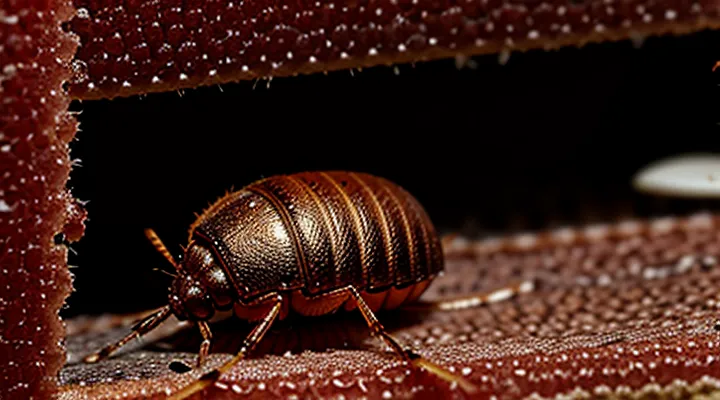
The Transformation of a Bedbug
Size and Shape Alterations
Initial Size
Adult bedbugs (Cimex lectularius) that have not fed measure 4.5–5.5 mm in length, 1.5–2.0 mm in width, and weigh approximately 1 mg. Their dorsally flattened bodies give a reddish‑brown appearance, but the small size makes them difficult to detect without magnification.
Nymphal stages differ markedly in size:
- 1st instar: 1.5–2.0 mm long, <0.2 mg
- 2nd instar: 2.0–2.5 mm long, ~0.3 mg
- 3rd instar: 2.5–3.0 mm long, ~0.5 mg
- 4th instar: 3.0–3.5 mm long, ~0.7 mg
- 5th instar (pre‑adult): 3.5–4.0 mm long, ~0.9 mg
After ingesting a blood meal, an unfed adult expands to 6–7 mm in length and can increase its mass up to fivefold, reaching 4–5 mg. The abdomen swells, the cuticle stretches, and the overall silhouette becomes noticeably broader, yet the initial dimensions remain a reliable baseline for identification.
Post-Meal Expansion
After ingesting a blood meal, a bedbug’s body swells dramatically. The abdomen expands up to twice its unfed size, giving the insect a rounded, balloon‑like silhouette. Cuticle coloration darkens to a glossy, mahogany hue, while the previously visible dorsal pattern becomes obscured by the stretched exoskeleton. Legs appear shorter relative to the enlarged torso, and the head and antennae are pushed forward, creating a more compact overall profile.
Key visual changes include:
- Abdomen lengthening and widening, often exceeding 5 mm in total length.
- Surface texture becoming smoother as the cuticle stretches over the engorged gut.
- Color shift from light brown to deep reddish‑brown or blackish tones.
- Reduced visibility of the characteristic “X” or “H” markings on the dorsal surface.
These alterations persist for several days while the bug digests the blood, after which the abdomen gradually contracts and the insect returns to its pre‑feeding appearance.
Color Changes
Before Feeding
Before a blood meal, a bed bug is a small, flattened insect measuring 4–5 mm in length. The body is elongated and oval, with a dorsal surface that appears matte and light brown to reddish‑brown. The abdomen is slightly wider than the thorax, giving the insect a tapered silhouette toward the head. Antennae are long, segmented, and brown, while the six legs are slender, ending in tiny claws that aid in clinging to fabric.
Key visual characteristics of an unfed bed bug include:
- Color: Uniform light brown, lacking the reddish engorgement seen after feeding.
- Transparency: Slightly translucent cuticle, allowing faint visibility of internal organs.
- Body shape: Flat and streamlined, facilitating movement through cracks and seams.
- Eyes: Small, dark, bean‑shaped ocelli positioned on the head.
- Mouthparts: Visible beak‑like proboscis concealed beneath the head when not in use.
These traits contrast sharply with the swollen, dark‑red appearance that develops once the insect has ingested blood. Understanding the pre‑feeding morphology helps identify infestations early, before the bugs become visibly engorged.
After Blood Engorgement
After a blood meal, a bed bug expands dramatically. The abdomen swells to nearly twice its unfed length, taking on a rounded, balloon‑like shape. The cuticle stretches, revealing a glossy, reddish‑brown hue that contrasts with the darker, matte coloration of an unfed insect.
Key visual changes include:
- Color shift: from pale tan or light brown to deep mahogany or brick red.
- Size increase: body length grows from about 4 mm to 6–7 mm; width expands proportionally.
- Abdominal outline: becomes convex and smooth, losing the distinct segmental lines visible when empty.
- Leg posture: legs may appear slightly tucked against the swollen abdomen, giving a compact appearance.
- Surface sheen: engorged cuticle reflects more light, producing a shiny, wet look.
These characteristics persist for several days while the insect digests the blood, after which the abdomen gradually contracts and the color fades toward the original shade.
Physical Characteristics
Abdominal Distention
After ingesting blood, a bed bug’s abdomen expands dramatically. The cuticle stretches to accommodate the meal, producing a rounded, balloon‑like silhouette that can increase the insect’s length by up to 50 percent. The swelling is most evident along the dorsal midline, where the abdomen becomes markedly convex.
The engorged abdomen also changes color. Initially translucent, it darkens to a mahogany or reddish hue as the blood fills the gut. The ventral surface may appear glossy due to the fluid’s surface tension. The abdomen’s edges lose their crisp definition, merging with the thorax to create a seamless, swollen profile.
Key visual indicators of abdominal distention:
- Uniform expansion from the anterior to posterior abdomen
- Rounded, dome‑shaped dorsal surface
- Darkening from light tan to deep brown or red
- Glossy, moist appearance on the ventral side
- Loss of distinct segmentation lines
These characteristics enable rapid identification of a fed bed bug without magnification.
Segment Visibility
After ingesting blood, a bedbug’s abdomen expands dramatically, making the thoracic and abdominal segments clearly distinguishable. The dorsal surface of the abdomen swells to a rounded, balloon‑like shape, often turning a translucent reddish‑brown as the blood fills the hemocoel. The segmentation of the abdomen becomes more pronounced; each of the nine visible tergites can be seen as faint lines separating the expanded areas. The cuticle over the abdomen thins, allowing the underlying blood to give the abdomen a glossy appearance.
The head and thorax remain relatively unchanged in size and color, but their borders become more evident against the enlarged abdomen. The pronotum and mesonotum retain their dark brown hue, creating a sharp contrast with the lighter, swollen abdominal segments.
Key visual changes include:
- Abdomen enlargement to several times its unfed size.
- Color shift to reddish‑brown or pinkish translucency.
- Enhanced visibility of abdominal tergite boundaries.
- Contrast between unchanged dark thorax and expanded lighter abdomen.
These segment‑visibility characteristics are reliable indicators that a bedbug has recently fed.
Leg and Antennae Appearance
After ingesting blood, a bedbug’s legs become noticeably engorged. The femora, tibiae, and tarsi swell proportionally, giving each leg a thicker, more rounded silhouette. The cuticle stretches but retains its glossy, reddish‑brown hue, while the joints remain clearly articulated. The dilation is especially evident at the distal segments, which may appear slightly flattened against the body as the abdomen expands.
The antennae also exhibit distinct changes. Each antenna, composed of four segments, expands slightly, particularly the basal segment, which elongates to accommodate the increased internal pressure. The surface of the antennae darkens, shifting from a pale tan to a deeper amber tone. Sensory hairs on the flagellum remain upright, but their spacing becomes tighter due to the overall enlargement of the head capsule.
The Physiological Impact of a Blood Meal
Digestion Process
Blood Absorption
After ingesting blood, a bedbug’s abdomen expands dramatically, often increasing its length by 30–50 %. The cuticle stretches, revealing a glossy, reddish‑brown hue that contrasts with the insect’s normally matte, tan coloration. The engorged state persists for 5–10 days before the abdomen gradually contracts as digestion proceeds.
Key aspects of blood absorption:
- Volume: An adult can intake up to 5 µL of blood, roughly 10 % of its body weight.
- Rate: In the first 30 minutes, the midgut fills rapidly; subsequent minutes involve peristaltic movement that distributes the meal throughout the digestive tract.
- Physiological changes: Hemoglobin is broken down into amino acids and lipids; excess water is excreted via the Malpighian tubules, causing the abdomen to appear less swollen after several days.
- Visual cues: Swollen abdomen, darkened coloration, and a slightly flattened dorsal surface indicate a recent feeding event.
The combination of size increase, color shift, and abdominal tension provides a reliable visual indicator of a recent blood meal.
Waste Excretion
After ingesting a blood meal, a bed bug’s digestive system processes the protein‑rich fluid rapidly. The insect converts the majority of the nutrients into growth tissue, while excess water and nitrogenous waste are eliminated as dark, liquid excreta. This excretion appears as small, reddish‑brown spots on the host’s skin or bedding, often mistaken for blood stains. The coloration results from hemoglobin breakdown products, primarily hemoglobin‑derived pigments that darken as they oxidize.
Key aspects of post‑meal waste excretion:
- Volume increases sharply within the first 12 hours, reflecting the large fluid intake.
- Excreted droplets contain uric acid crystals, a low‑solubility waste product that conserves water.
- The visible stains fade over several days as the pigments oxidize and the droplets evaporate.
The pattern of excretion provides a reliable indicator of recent feeding. Concentrated spots near the bite site suggest a recent meal, whereas dispersed, lighter stains indicate older excreta that has begun to degrade. Understanding these waste signatures assists in identifying bed‑bug activity and differentiating it from other skin lesions.
Behavioral Changes
Increased Lethargy
After ingesting a blood meal, a bed bug exhibits a marked reduction in activity. The insect’s metabolic rate rises to process the protein-rich intake, but locomotor drive declines, resulting in prolonged periods of stillness. This lethargic state serves two functions: it conserves energy for digestion and minimizes exposure to predators while the abdomen expands.
Observable signs of increased lethargy include:
- Minimal movement for several hours post‑feeding, often limited to occasional adjustments of body position.
- A flattened, elongated posture as the abdomen swells, reducing the need for active locomotion.
- Decreased response to tactile or vibrational stimuli compared to unfed individuals.
The duration of this inactivity varies with temperature and the size of the blood meal, typically lasting 4–12 hours before the bug resumes foraging behavior. During this phase, the bed bug’s coloration may appear darker due to the engorged abdomen, but the primary indicator of post‑feeding status is the pronounced, temporary lethargy.
Hiding Tendencies
After a blood meal, a bedbug’s abdomen swells and its cuticle darkens, but the insect retains a flattened profile that permits entry into the smallest fissures. The enlarged body increases the need for secure concealment; the insect actively seeks locations that protect it from disturbance and reduce the risk of detection.
Typical refuges include:
- Crevices in mattress seams and box‑spring frames
- Gaps behind headboards, footboards, and wall panels
- Upholstery folds, button holes, and cushion seams
- Baseboard cracks and floorboard joints
- Behind picture frames, electrical outlets, and wallpaper edges
The expanded abdomen limits the bug’s ability to move quickly, prompting prolonged periods of immobility. During this interval the insect remains motionless, relying on its cryptic coloration to blend with the surrounding substrate. The darkened, engorged appearance often matches the shadows within tight spaces, further obscuring visual cues.
Chemical signals released after feeding suppress the activity of nearby conspecifics, preventing crowding and encouraging each individual to maintain an isolated hideout. This behavior minimizes competition for shelter and reduces the likelihood that predators or human inspection will locate the bug.
Reproduction and Development
Egg Laying Cycle
After a blood meal, a female bedbug’s abdomen swells dramatically, turning a bright reddish‑brown hue as it fills with digested blood. This physiological state triggers the onset of oviposition. Within 3–5 days, the insect begins to deposit eggs, each measuring about 1 mm in length and appearing as smooth, ivory‑white ovals.
The egg‑laying cycle proceeds as follows:
- Initiation: Females lay the first batch after the digestive process reaches peak volume; the abdomen remains distended.
- Frequency: One to two eggs are released every 2–3 hours, continuing for 4–6 weeks while the host remains available.
- Quantity: A single female can produce 200–500 eggs over her lifespan, distributed in multiple clutches.
- Incubation: Eggs hatch in 6–10 days at typical indoor temperatures (22–26 °C). Hatchlings emerge as nymphs, immediately seeking a blood source.
During the interval between meals, the female’s coloration gradually fades to a dull brown as the blood is metabolized, yet she remains capable of laying additional clutches until mortality. The correlation between post‑feeding appearance and reproductive output provides a reliable indicator for monitoring infestations.
Nymph Stages
After a blood meal, each nymphal instar of Cimex lectularius shows a distinct visual shift that aids identification. The insects enlarge rapidly, their cuticle becomes glossy, and the body color deepens from a pale, translucent hue to a reddish‑brown tone as the ingested blood fills the abdomen.
The first instar, measuring roughly 1 mm, appears almost translucent before feeding. Post‑ingestion, the abdomen swells, the cuticle gains a faint amber sheen, and the overall length increases to about 1.5 mm.
The second instar, initially pale brown, expands to 2 mm after a meal. The abdomen turns a bright, rusty red, and the dorsal surface shows a slight gloss. The head capsule remains proportionally larger than in later stages.
The third instar reaches 2.5 mm. After feeding, the body exhibits a uniform mahogany coloration, with the abdomen fully engorged and the cuticle noticeably slick. Legs become more defined, and the antennae appear darker.
The fourth instar grows to 3 mm. Following a blood intake, the insect displays a deep, almost black abdomen, while the thorax retains a lighter brown. The overall silhouette becomes more robust, and the eyes appear more prominent.
The fifth instar, the final nymphal stage before adulthood, measures up to 4 mm. After feeding, the abdomen is fully distended, showing a vivid, blood‑red hue that contrasts with a darkened thorax. The cuticle is markedly glossy, and the insect’s posture is slightly flattened, preparing for the molt to the adult form.
Key visual cues per nymphal stage after feeding
- 1st instar: 1–1.5 mm, translucent to amber abdomen, slight swelling.
- 2nd instar: ~2 mm, bright rusty red abdomen, glossy dorsal surface.
- 3rd instar: 2.5 mm, uniform mahogany color, fully engorged abdomen.
- 4th instar: 3 mm, deep black abdomen, lighter thorax, robust silhouette.
- 5th instar: up to 4 mm, vivid blood‑red abdomen, dark thorax, glossy cuticle.
