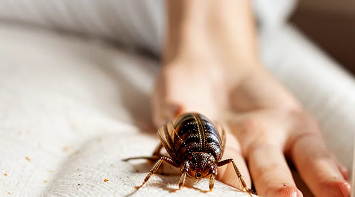
Initial Presentation and Characteristics
Common Visual Cues
Bedbug bites present a distinct set of visual characteristics that allow reliable identification. The lesions are typically erythematous, raised papules measuring 2–5 mm in diameter. Color ranges from pink to deep red, often intensifying after several hours. Swelling remains mild; the central area may appear slightly pale compared to the surrounding rim. Itching is common, with a delayed onset of 12–48 hours in many cases.
- Small, round or oval red welts
- Slight elevation above the skin surface
- Central pallor surrounded by a darker halo
- Arrangement in linear rows, clusters, or a “breakfast‑cereal” pattern
- Delayed pruritus, sometimes accompanied by a faint burning sensation
- Absence of necrosis or ulceration unless secondary infection occurs
Progression Over Time
Bedbug bites typically begin as small, reddish‑pink macules that appear within a few hours after the insect feeds. The lesions are often clustered in linear or zig‑zag patterns, reflecting the bug’s movement across the skin.
- 0–24 hours: Light erythema, slight swelling, intense itching. Occasionally a central punctum is visible where the mouthparts entered.
- 24–72 hours: Redness deepens, papules may become raised and firm. Edema can increase, and a halo of paler skin may surround the central spot.
- 3–7 days: Lesions may develop into vesicles or pustules if the reaction is strong. Crusting can occur as itching leads to excoriation.
- 1–2 weeks: Inflammation subsides; discoloration fades to a brownish or gray‑blue hyperpigmented stain that can persist for several weeks.
- Beyond 2 weeks: Most bites resolve without scarring, though some individuals retain residual hyperpigmentation for months, especially on darker skin tones.
The progression follows a predictable timeline, allowing clinicians to differentiate bedbug bites from other arthropod reactions based on the sequence of visual changes and symptom intensity.
Varied Reactions Based on Individual Sensitivity
Bed‑bug bites do not produce a uniform skin lesion; the visible response depends largely on each person’s immune sensitivity.
The most frequently reported appearance is a tiny, erythematous, raised spot, often 2–5 mm in diameter. Lesions commonly occur in a line, a cluster, or a “breakfast‑at‑the‑café” pattern, reflecting the insect’s feeding behavior.
Individual reactions diverge sharply. Some hosts show no discernible change despite confirmed feeding. Others develop pronounced pruritus, edema, or a wheal that expands to several centimeters. In rare cases, a vesicle or bullous eruption forms, indicating a heightened hypersensitivity.
Typical categories of cutaneous response:
- No visible reaction – microscopic puncture without erythema.
- Mild erythema – small red papule, limited itching, resolves in 2–3 days.
- Moderate inflammation – larger papule with surrounding redness, itching persists for 5–7 days.
- Severe hypersensitivity – extensive swelling, urticarial plaques, possible blistering, may last up to two weeks.
Onset ranges from immediate (within minutes) to delayed (24–48 hours). Duration correlates with reaction severity; mild lesions fade within days, while severe inflammation can persist for weeks.
Accurate assessment of a bite’s appearance therefore requires consideration of the patient’s known allergenic profile and the temporal pattern of the lesion.
Differentiating Bed Bug Bites from Other Skin Conditions
Comparison with Mosquito Bites
Bedbug bites appear as small, raised welts, typically 2–5 mm in diameter. The central area is often pale or slightly red, surrounded by a darker, inflamed halo. Bites frequently occur in linear or clustered patterns, reflecting the insect’s feeding habit of moving along the skin. The reaction develops within a few hours and may intensify over 24‑48 hours, producing intense pruritus and occasional swelling.
Mosquito bites manifest as round, raised papules, usually 3–7 mm across. The lesion is uniformly red with a prominent central punctum where the proboscis penetrated. Itching begins almost immediately after the bite and peaks within an hour. Mosquito bites are typically isolated, though multiple bites can appear when several insects feed in the same area.
Key comparative points:
- Size: Bedbug welts are slightly smaller than mosquito papules.
- Color pattern: Bedbug lesions show a pale center with a darker rim; mosquito lesions are uniformly red.
- Distribution: Bedbug bites often form rows or clusters; mosquito bites are generally single or randomly scattered.
- Onset of symptoms: Bedbug reactions may be delayed; mosquito itching starts promptly.
- Duration of swelling: Bedbug swelling can persist for several days; mosquito swelling usually subsides within 24 hours.
Distinction from Flea Bites
Bedbug bites typically appear as small, red welts that are grouped in a linear or clustered pattern. The lesions are often raised, slightly swollen, and may develop a central punctum where the insect inserted its mouthparts. Itching intensifies after several hours, and the reaction can persist for days.
Flea bites differ markedly. They are usually isolated, punctate papules surrounded by a narrow halo of erythema. The lesions are commonly found on the lower legs and ankles, reflecting the insect’s jumping behavior, and they tend to appear one at a time rather than in rows or clusters.
Key distinguishing points:
- Arrangement: bedbug bites – linear or clustered; flea bites – solitary.
- Location: bedbug bites – any exposed skin, often on the torso or arms; flea bites – lower extremities.
- Size and shape: bedbug bites – slightly larger, rounded welts; flea bites – small, pinpoint papules.
- Duration of itching: bedbug bites – delayed, lasting several days; flea bites – immediate, may subside sooner.
Identifying Other Insect Bites
Bedbug bites can be confused with those of fleas, mosquitoes, and spiders, making accurate identification essential for appropriate treatment.
-
Size and shape: Bedbug lesions are typically 2–5 mm, raised, and round. Flea bites are smaller, often with a sharp central puncture. Mosquito bites are larger, irregular, and may develop a central blister. Spider bites vary widely but often present with a necrotic center.
-
Distribution: Bedbug feeds in linear or clustered patterns on exposed skin, such as the forearms, neck, and face. Flea bites appear as single or scattered points, frequently on the ankles and legs. Mosquito bites are random across the body, without a discernible pattern.
-
Timing of appearance: Bedbug reactions emerge within several hours after feeding and may intensify over 24–48 hours. Flea bites produce immediate itching, while mosquito bites may take up to a day to swell. Spider bites can cause delayed necrosis, appearing days after the incident.
-
Associated signs: Presence of live insects, shed skins, or dark spotting on bedding indicates a bedbug infestation. Flea infestations are linked to pet hair and carpet debris. Mosquito activity correlates with standing water or outdoor exposure.
Identification relies on careful visual assessment of lesion morphology, arrangement, and surrounding environment. Documentation of bite chronology and any co‑resident insects strengthens diagnostic confidence.
If lesions progress rapidly, develop ulceration, or are accompanied by systemic symptoms such as fever, seek medical evaluation to rule out secondary infection or alternative envenomation.
Recognizing Allergic Reactions and Rashes
Bedbug bites typically appear as small, red papules arranged in clusters or linear patterns. The lesions are often raised, slightly swollen, and may develop a central punctum where the insect fed.
When the immune system reacts strongly, the bite can evolve into an allergic response. Symptoms extend beyond the usual redness, including intense itching, spreading edema, and the formation of larger, confluent welts. Some individuals develop hives or a urticarial rash that migrates away from the original bite sites.
Key indicators of an allergic reaction or secondary rash:
- Pruritus that intensifies within hours and persists despite antihistamine use.
- Swelling that exceeds the immediate bite area, sometimes affecting adjacent skin.
- Development of vesicles or bullae on the surface of the lesion.
- Presence of erythematous streaks or a “crawling” sensation indicating possible secondary infection.
- Systemic signs such as low‑grade fever, malaise, or lymphadenopathy.
Management requires prompt identification and treatment. Initial steps include thorough cleansing of the affected area with mild antiseptic, application of topical corticosteroids to reduce inflammation, and oral antihistamines for itch control. If vesicles form or the rash expands rapidly, a short course of systemic steroids may be warranted. Persistent or worsening symptoms should prompt medical evaluation to rule out secondary infection or severe hypersensitivity.
Symptoms and Associated Discomfort
Common Sensations
Bedbug bites typically produce a range of immediate and delayed sensations that help distinguish them from other insect bites. The initial feeling often appears within minutes to a few hours after the bite, though some individuals notice no reaction until the next day. Sensations vary with personal skin sensitivity, bite location, and the number of insects involved.
- Prickling or stinging: A brief, sharp sensation at the moment of penetration, comparable to a tiny needle.
- Burning: A mild heat that may persist for several minutes, sometimes intensifying as the bite site inflames.
- Itching: A persistent, sometimes intense urge to scratch that can develop hours after the bite and last several days.
- Tenderness: A dull, uncomfortable pressure when the area is touched, indicating localized inflammation.
- Swelling: A subtle puffiness that accompanies the itching, creating a raised, firm spot on the skin.
The intensity of these sensations can differ markedly between people. Some experience only mild irritation, while others report pronounced itching and swelling that interfere with sleep. The combination of prickling, burning, and prolonged itching is characteristic of bedbug bites and aids in clinical identification.
Secondary Symptoms and Complications
Bedbug bites often trigger a cascade of additional reactions beyond the initial red welts. After the primary lesion forms, patients may experience itching that intensifies over 24–48 hours, swelling that extends beyond the bite margin, and the development of secondary lesions such as papules, vesicles, or bullae. In some cases, the skin may become lichenified after repeated exposure.
- Persistent pruritus lasting several days
- Erythema that spreads or coalesces into larger patches
- Development of pustules or crusted lesions from scratching
- Hyperpigmentation or hypopigmentation persisting for weeks
- Localized edema extending several centimeters from the bite site
Complications arise when the skin barrier is breached. Bacterial superinfection, most commonly with Staphylococcus aureus or Streptococcus pyogenes, can produce purulent discharge, increased pain, and systemic signs such as fever. Allergic sensitization may lead to urticaria, angioedema, or, rarely, anaphylaxis. Chronic exposure can provoke psychosomatic effects, including heightened anxiety, insomnia, and reduced quality of life, which may exacerbate the physical symptoms. Prompt identification of secondary signs and early intervention—topical corticosteroids for inflammation, antihistamines for itching, and antibiotics for confirmed infection—mitigate the risk of long‑term sequelae.
Factors Influencing Bite Appearance
Location on the Body
Bedbug bites are most frequently found on body parts that are exposed while sleeping or are covered by thin clothing. The insects tend to target areas where they can easily pierce the skin and feed without being disturbed.
Common locations include:
- Face, especially around the eyes, nose, and mouth
- Neck and shoulders
- Arms, particularly the forearms and wrists
- Hands, including the backs of the hands
- Upper back and chest
- Abdomen and waistline
- Thighs and legs, especially the lower legs and ankles
Bites often appear in clusters or linear patterns, reflecting the insect’s movement across the skin. The distribution may vary with sleeping posture, clothing style, and the presence of personal items that provide access points for the pests.
Number of Bites
Bedbug bites typically appear as small, red welts that may itch. The quantity of lesions on a single host provides a key diagnostic clue.
- A solitary bite is uncommon; most infestations produce two or more marks.
- Bites often occur in clusters of 3‑5 spots, reflecting the insect’s feeding habit of moving short distances before resuming blood intake.
- Linear or “break‑line” patterns, consisting of 2‑6 puncta spaced a few centimeters apart, indicate multiple feedings along a host’s exposed skin.
- In severe cases, dozens of welts can cover a limited area, especially on the neck, shoulders, arms, or legs, where the insect has easy access.
The number of lesions correlates with the duration of exposure and the density of the infestation. A light infestation may generate only a few isolated bites, while a heavy infestation can produce extensive, overlapping clusters within a single night. Monitoring bite count, distribution, and pattern assists clinicians and pest‑control professionals in confirming bedbug activity.
Stage of Bite Development
Bedbug bites progress through distinct visual stages that clinicians and pest‑control professionals use to identify infestations.
-
Initial reaction (minutes to 12 hours): The bite appears as a faint, flat red spot (erythema) without swelling. Occasionally a tiny puncture mark is visible where the insect inserted its mouthparts.
-
Early development (12 hours to 48 hours): The lesion enlarges into a raised, pink papule. It becomes more noticeable as a small, raised bump surrounded by a slightly wider halo of redness. Pruritus typically intensifies during this phase.
-
Intermediate stage (2 days to 1 week): The papule may develop into a wheal or hive‑like swelling, often 3–5 mm in diameter. Central clearing can create a target‑shaped pattern, especially when several bites cluster together. The surrounding erythema may darken, and itching reaches its peak.
-
Late stage (1 week to several weeks): The lesion flattens, leaving a lingering reddish‑brown or hyperpigmented spot. In some individuals the discoloration persists for months, while others experience complete resolution. Secondary bacterial infection can appear as a pus‑filled pustule if the skin is broken by scratching.
Understanding these sequential changes enables accurate differentiation of bedbug bites from other arthropod reactions and guides appropriate medical or eradication measures.
When to Seek Medical Advice
Signs of Infection
Bed bug bites typically appear as small, red, raised welts that may be clustered in a line or a zig‑zag pattern. The lesions often itch intensely and can become swollen, but the bite itself is not normally dangerous. When a secondary infection develops, the skin’s response changes noticeably.
Signs that an infected bite is present include:
- Increasing pain rather than just itching
- Redness spreading outward from the original spot, forming a larger, warm area
- Swelling that does not subside within a few days or continues to enlarge
- Pus or clear fluid oozing from the lesion
- Fever, chills, or general malaise accompanying the skin changes
- Presence of yellow or green discoloration around the bite, indicating tissue breakdown
Prompt medical evaluation is advised if any of these indicators appear, as untreated infection can lead to cellulitis, abscess formation, or systemic involvement. Early antibiotic therapy and proper wound care reduce complications and accelerate recovery.
Severe Allergic Reactions
Bedbug bites typically present as small, red papules that may develop into raised, itchy welts. In most individuals the reaction is mild, but a subset of people experience severe allergic responses that differ markedly from the usual presentation.
A severe allergic reaction can include:
- Extensive erythema that spreads beyond the immediate bite area, forming large, confluent patches of inflamed skin.
- Intense pruritus accompanied by swelling (edema) that may involve the surrounding tissue and persist for several days.
- Formation of bullae or vesicles, indicating a hypersensitivity response that compromises the epidermal barrier.
- Systemic symptoms such as fever, headache, malaise, or, in rare cases, anaphylaxis characterized by difficulty breathing, throat swelling, and rapid pulse.
Laboratory evaluation may reveal elevated serum IgE levels and eosinophilia, confirming a heightened immunologic reaction. Management requires prompt intervention: topical corticosteroids to reduce local inflammation, oral antihistamines for itch control, and, when systemic involvement is evident, systemic corticosteroids or epinephrine administration. Early recognition of these severe manifestations is essential to prevent complications and to differentiate them from ordinary bite reactions.
Persistent or Worsening Symptoms
Bedbug bites typically begin as small, red papules that may appear in clusters or linear patterns. When the reaction does not subside within a few days, the skin can exhibit persistent redness, swelling, and itching that intensify rather than fade. In such cases, the following signs warrant closer attention:
- Expanding erythema that spreads beyond the original bite site.
- Development of raised, firm nodules that persist for weeks.
- Formation of vesicles or pustules indicating secondary infection.
- Persistent pruritus leading to excoriation, which may cause ulceration or scarring.
- Systemic manifestations such as fever, malaise, or enlarged lymph nodes, suggesting an allergic or infectious complication.
Continued irritation can trigger a hypersensitivity response, resulting in larger, more inflamed lesions that may last months. Individuals with atopic conditions or weakened immune systems are especially prone to prolonged or worsening reactions. Prompt medical evaluation is advised when any of the listed symptoms emerge, to confirm the cause, rule out secondary bacterial infection, and initiate appropriate treatment such as antihistamines, topical corticosteroids, or antibiotics. Early intervention reduces the risk of chronic skin changes and prevents unnecessary discomfort.
