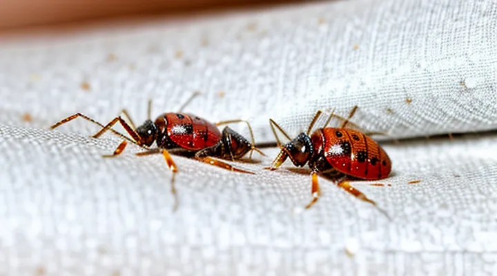
Understanding Bed Bug Bite Characteristics
Typical Appearance of Bed Bug Bites
Size and Shape
Bedbug bite marks are typically small, ranging from 1 mm to 5 mm across. The most frequent diameter falls between 2 mm and 3 mm, comparable to the tip of a needle. Size variations reflect feeding time and the individual’s skin sensitivity.
The marks are generally round, occasionally appearing slightly oval. A faint central puncture often marks the point of penetration. When multiple bites occur, they may form a linear or zig‑zag cluster, reflecting the insect’s movement across the skin.
Key dimensions
- Diameter: 1 mm – 5 mm (common: 2 mm – 3 mm)
- Shape: round to mildly oval, with a central point
- Arrangement: isolated or grouped in linear patterns
Color and Texture
Bedbug bite marks typically present as small, circular lesions with a pink‑to‑red hue shortly after feeding. The coloration often deepens to a darker red or purplish shade within 24–48 hours, especially on lighter skin tones. In some cases, the surrounding area may develop a faint, pale halo as the inflammatory response expands.
The texture of the lesions is generally raised and firm to the touch. A central puncture point may be discernible, indicating the feeding site. When scratched, the surface can become flat or develop a crusted layer, but the underlying swelling usually remains palpable. A surrounding area of mild swelling may accompany the central bump, creating a slightly uneven topography.
Distribution Patterns («Breakfast, Lunch, and Dinner»)
Bedbug bite marks typically present as small, red, raised welts that may appear in rows or clusters. The spatial arrangement of these lesions often reflects the feeding behavior of the insect, which can be linked to the host’s daily routine.
Patterns observed during different meals:
- «Breakfast»: bites concentrate on exposed areas such as the neck, face, and forearms, where clothing is often loose or partially removed for morning activities.
- «Lunch»: lesions shift toward the torso and upper arms, corresponding to mid‑day attire that leaves these regions uncovered.
- «Dinner»: clusters appear on the lower legs, ankles, and feet, aligning with evening footwear and the tendency to keep lower garments open while preparing for rest.
The progression from upper to lower body parts throughout the day suggests that bedbugs exploit periods when specific skin regions are most accessible. Recognizing these temporal distribution patterns aids in accurate identification and timely intervention.
Distinguishing Bed Bug Bites from Other Insect Bites
Mosquito Bites
Mosquito bites appear as small, raised papules surrounded by a faint red halo. The central puncture is often visible as a pinpoint depression. Itching intensifies within minutes and may persist for several days. Bites frequently occur on exposed areas such as arms, legs, and the face, and they may appear singly or in small clusters.
Key differences between mosquito and bed‑bug reactions include:
- Mosquito lesions: isolated or loosely grouped, round, central punctum, pronounced pruritus, duration up to a week.
- Bed‑bug lesions: often arranged in a linear or zig‑zag pattern, larger erythematous welts, may contain multiple puncta, itching variable, may persist longer.
Understanding these visual and distribution characteristics enables accurate identification of insect‑related skin reactions.
Flea Bites
Flea bites appear as small, pinpoint punctures, typically 1‑2 mm in diameter. The surrounding erythema is often a faint red halo that may develop into a raised wheal. Lesions frequently occur on the lower legs, ankles, and feet, reflecting the insect’s tendency to jump onto exposed skin. Itching is immediate and can be intense, sometimes accompanied by a brief burning sensation.
Key differences between flea and bed‑bug marks include:
- Size: flea bites are generally smaller than the 2‑5 mm welts produced by bed‑bugs.
- Arrangement: flea bites are isolated; bed‑bug bites often form linear or clustered patterns.
- Timing: flea bites manifest within minutes of contact, whereas bed‑bug reactions may emerge several hours later.
- Location: flea bites concentrate on lower extremities; bed‑bug bites favor exposed areas such as the face, neck, and forearms.
Diagnostic guidance: examine the bite’s dimensions, distribution, and onset interval. Assess the environment for signs of flea activity—pet bedding, carpets, and indoor plants. Absence of the characteristic “break‑fast‑bunch” pattern typical of bed‑bug infestations further supports a flea etiology.
Spider Bites
Spider bites often appear as single puncture wounds surrounded by a reddish halo. The central point may be slightly raised, while the surrounding area can become swollen and tender. In some cases, a faint white ring develops around the red zone, indicating mild inflammation.
Bedbug bites typically present as clusters of small, pruritic papules arranged in a line or zig‑zag pattern. The lesions are usually flat, with a bright red center and a lighter surrounding ring. Unlike spider bites, bedbug marks rarely exhibit significant swelling or necrotic tissue.
Key differences between the two types of lesions include:
- Number of lesions: spider bites are usually solitary; bedbug bites often occur in groups.
- Distribution: spider bites appear randomly; bedbug bites follow a linear or clustered arrangement.
- Swelling: spider bites may cause pronounced edema; bedbug bites produce minimal swelling.
- Pain level: spider bites can be painful or produce a burning sensation; bedbug bites are primarily itchy.
- Healing time: spider bite marks may persist for several days with possible scarring; bedbug marks usually resolve within a week without lasting marks.
Rash vs. Bites
Bedbug bites typically appear as small, red, raised welts arranged in linear or clustered patterns. The lesions often measure 2‑5 mm in diameter and may develop a central punctum where the insect inserted its mouthparts. Swelling can increase within a few hours, and itching intensifies as the reaction progresses.
A rash, by contrast, presents without the characteristic alignment of lesions. Common forms such as contact dermatitis, allergic reactions, or viral exanthems generate diffuse erythema that may cover large skin areas. Rash lesions frequently lack a distinct punctum and seldom follow a straight‑line distribution.
Key differences:
- Arrangement: bites align in rows or groups; rash spreads irregularly.
- Central point: bites show a pinpoint entry site; rash does not.
- Size variation: bite welts remain uniform; rash lesions vary in shape and size.
- Onset timing: bite swelling peaks within hours; rash may develop more gradually or appear suddenly depending on etiology.
- Associated symptoms: bites provoke localized itching and occasional burning; rash may accompany systemic signs such as fever or malaise.
Recognition of these features enables accurate identification of bedbug bite marks and distinguishes them from unrelated dermatological eruptions.
Symptoms and Reactions to Bed Bug Bites
Common Symptoms
Itching and Irritation
Bedbug bites commonly provoke intense itching that may begin within hours of the bite and persist for several days. The sensation is typically described as a sharp, pricking discomfort that evolves into a persistent, irritating itch. Scratching often intensifies the irritation and can lead to secondary skin lesions.
Key characteristics of the irritation include:
- Red, raised welts forming a linear or clustered pattern.
- Swelling that may fluctuate in size, reaching a maximum within 24 hours.
- A burning or stinging sensation accompanying the itch.
- Possible development of a darkened spot as the bite heals.
Prolonged scratching can break the skin, increasing the risk of bacterial infection. Signs of infection comprise increased redness, warmth, pus formation, and escalating pain. Prompt cleansing with mild soap and antiseptic, followed by topical corticosteroids or antihistamines, alleviates inflammation and reduces itching. In severe cases, oral antihistamines or prescription-strength steroids may be required.
Managing the irritation effectively reduces discomfort, prevents complications, and supports faster resolution of the bite marks.
Swelling and Redness
Bedbug bites commonly produce a localized reaction characterized by swelling and redness. The inflammatory response appears within minutes to hours after the bite and may persist for several days. Swelling manifests as a raised, firm area surrounding the puncture site, while redness presents as a circular, erythematous halo that can expand outward.
Typical features of the swelling and redness include:
- Firm, raised bump that may feel tender to the touch.
- Red halo that varies in diameter, often matching the size of the surrounding bite cluster.
- Gradual increase in size during the first 24 hours, followed by a slow decline.
- Possible coalescence of multiple bites, forming larger, irregularly shaped patches.
The severity of these signs depends on individual sensitivity and the number of bites received. In highly sensitive individuals, the reaction can intensify, leading to pronounced edema and vivid erythema that may mimic other arthropod bites.
Blistering (Rare)
Blistering represents an uncommon reaction to Cimex lectularius feeding. The skin response appears as a raised, fluid‑filled vesicle that may develop several hours after the initial bite. The vesicle typically measures 2–5 mm in diameter, exhibits a clear or slightly yellowish serum, and is surrounded by a reddened halo. Unlike the more frequent maculopapular lesions, blistering may persist for up to a week before rupturing and resolving.
Key diagnostic features of this rare presentation include:
- Sudden onset of a localized bubble at the bite site, often accompanied by a mild burning sensation.
- Absence of widespread inflammation; the reaction remains confined to one or two adjacent bites.
- Progression from vesicle to shallow ulceration if the blister ruptures, followed by gradual re‑epithelialisation without scarring.
Recognition of blister formation aids in distinguishing bedbug bites from other arthropod assaults, such as flea or mosquito bites, which seldom produce true vesicles. Prompt identification allows clinicians to advise patients on appropriate wound care and to consider antihistamine or topical corticosteroid therapy if discomfort is significant.
Individual Variability in Reactions
Allergic Reactions
Bedbug bites can trigger immune‑mediated responses that differ from the typical red, swollen welts. Allergic reactions manifest as intensified erythema, larger edema, and pronounced itching that persists beyond the usual 24‑hour period. In some cases, a secondary rash resembling a hive or urticaria develops, often spreading from the original bite sites.
Key characteristics of an allergic response include:
- Erythema extending several centimeters beyond the bite margin
- Swelling that may coalesce into a confluent area of inflammation
- Pruritus severe enough to cause excoriation or secondary infection
- Possible systemic signs such as low‑grade fever, malaise, or lymphadenopathy
The onset of these symptoms typically follows a delayed hypersensitivity pattern, appearing 48–72 hours after exposure. Laboratory evaluation may reveal elevated eosinophil counts, supporting an IgE‑mediated mechanism.
Management focuses on symptom control and prevention of complications. First‑line therapy consists of topical corticosteroids to reduce inflammation and oral antihistamines to alleviate itching. In severe cases, short courses of systemic corticosteroids may be prescribed. Maintaining a clean environment and eliminating infestations are essential to prevent recurrence.
Recognizing allergic manifestations enables timely intervention, limiting discomfort and reducing the risk of secondary skin infections.
Delayed Reactions
Bedbug bites may not produce immediate visible signs; reactions can appear several hours to days after exposure. The delayed response often manifests as enlarged, reddish‑purple welts that develop slowly and persist longer than the initial puncture marks. Swelling may increase over 24–48 hours, sometimes accompanied by a central clearing or a faint, raised ridge.
- Red or pink macules expanding outward from the bite site
- Central pallor or slight depression surrounded by a darker halo
- Persistent itching that intensifies after the first day
- Swelling that peaks between 48 and 72 hours, then gradually subsides
These characteristics differ from immediate reactions, which typically present as small, flat, itchy spots within minutes. Delayed welts tend to be larger, more raised, and may mimic allergic dermatitis. If lesions enlarge, become painful, or show signs of infection such as pus or increased warmth, professional medical evaluation is recommended.
No Visible Reaction
Bedbug bites often produce small, red welts, but in many cases the skin shows no discernible change. Absence of a visible mark does not eliminate the possibility of an infestation; the bite may remain imperceptible for hours or days, especially on individuals with low sensitivity or when the bite occurs on thickened skin.
Factors contributing to a non‑observable reaction include:
- High personal tolerance to the insect’s saliva, which reduces inflammation.
- Bite location on areas with naturally darker pigmentation, masking subtle redness.
- Early stage of the bite, before the immune response generates visible swelling.
Detection strategies when no mark appears involve monitoring secondary symptoms and environmental clues. Persistent itching, localized swelling after a delay, or a pattern of bites in line or cluster formations suggests bedbug activity. Regular inspection of mattress seams, headboards, and furniture crevices for live insects, shed skins, or fecal spots provides additional confirmation. Early identification despite a lack of skin evidence prevents widespread infestation and reduces discomfort.
Secondary Complications
Skin Infections
Bedbug bites appear as small, raised welts that are often grouped in a linear or clustered pattern. The lesions are typically red to pink, may develop a central punctum, and can vary in size from a few millimeters to a centimeter. Pruritus intensifies within hours, prompting repeated scratching.
Repeated trauma to the bite sites creates an entry point for opportunistic bacteria, most commonly Staphylococcus aureus and Streptococcus pyogenes. When secondary infection occurs, the skin exhibits additional signs that differ from the primary reaction.
Key indicators of bacterial involvement include:
- Erythema that expands beyond the original bite margin
- Purulent drainage or crust formation
- Increased warmth and tenderness at the affected area
- Swelling that progresses despite antihistamine use
- Systemic symptoms such as fever or malaise
Management focuses on controlling the primary inflammatory response and preventing infection. Topical corticosteroids reduce itching, while oral antibiotics are indicated when purulent discharge or systemic signs develop. Proper wound hygiene—gentle cleansing with mild antiseptic solution and avoidance of aggressive scratching—reduces bacterial colonization. Early identification of infectious changes prevents complications such as cellulitis or abscess formation.
Psychological Impact
Bedbug bite marks typically appear as small, red, raised welts arranged in clusters or linear patterns. Their sudden emergence on exposed skin often provokes immediate concern about infestation and personal hygiene.
The visual evidence of these bites triggers heightened anxiety. Persistent worry about ongoing exposure leads to sleep disturbances, including difficulty falling asleep and frequent nocturnal awakenings. Hypervigilance develops as individuals repeatedly inspect bedding and clothing for new lesions.
Extended exposure to bite‑related stress correlates with depressive symptoms. Persistent feelings of helplessness, reduced motivation, and loss of interest in daily activities may arise. In severe cases, trauma‑like responses emerge, characterized by intrusive thoughts about infestation and avoidance of environments perceived as contaminated.
Social ramifications include embarrassment over visible welts, prompting withdrawal from social gatherings and workplace interactions. Stigmatization may result from misconceptions about personal cleanliness, further isolating affected individuals.
Effective mitigation combines psychological and environmental interventions. Professional counseling, particularly cognitive‑behavioral therapy, addresses maladaptive fear responses and restructures catastrophic thinking. Education about bite morphology and realistic infestation risk reduces uncertainty. Prompt eradication of bedbugs through integrated pest management eliminates the source of distress, facilitating recovery of mental well‑being.
