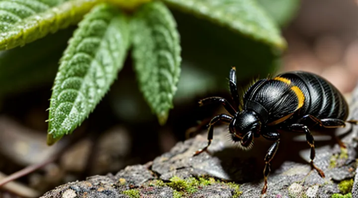
The Immediate Aftermath: Initial Biological Responses
Salivary Gland Activity and Secretion Changes
Anesthetic and Anticoagulant Release
Ticks inject a complex cocktail of salivary proteins the moment their mouthparts penetrate host skin. Two critical components of this cocktail are anesthetic agents and anticoagulants, which together enable prolonged blood ingestion without detection or clot formation.
Anesthetic molecules act on peripheral nerve endings and inflammatory mediators. Salivary proteins such as histamine‑binding proteins, Ixolaris‑like peptides, and salivary lipocalins bind histamine, serotonin, and other pain‑inducing substances, preventing the host from feeling the bite. By neutralizing these mediators, the tick remains undetected while feeding.
Anticoagulant factors interfere with the host’s hemostatic system at multiple points:
- Apyrase hydrolyzes ADP, a platelet activator, reducing platelet aggregation.
- Tick anticoagulant peptide (TAP) inhibits factor Xa, blocking the coagulation cascade.
- Salp14 and related proteins bind thrombin, preventing fibrin formation.
- Kunitz‑type protease inhibitors target serine proteases involved in clotting and inflammation.
These agents operate synergistically: anesthetics suppress sensory signals, while anticoagulants keep blood fluid. The combined effect creates a stable feeding site, allowing the tick to ingest large volumes of blood over days without triggering host defenses.
Immunomodulatory Proteins
When a tick attaches to a host, its salivary glands undergo rapid transcriptional activation, producing a suite of proteins that alter the host’s immune environment. This response is driven by the need to secure a blood meal while preventing detection and rejection by the host’s defenses. The shift includes up‑regulation of genes encoding immunomodulatory factors, secretion of anti‑hemostatic agents, and modification of glandular secretory pathways.
Immunomodulatory proteins in tick saliva act on multiple components of the host’s immune system. They suppress inflammatory cytokine release, inhibit complement activation, and impair antigen presentation. By dampening cellular and humoral responses, these molecules extend feeding duration and reduce the likelihood of host grooming or immune‑mediated wound closure.
Key immunomodulatory proteins identified in several tick species include:
- Salp15: binds to CD4⁺ T‑cell receptors, reducing interleukin‑2 production.
- Ixolaris: blocks the tissue factor–factor VIIa complex, preventing initiation of the coagulation cascade and downstream complement activation.
- Anticoagulant peptide (Amblyomin-X): interferes with factor Xa, limiting clot formation and associated inflammatory signaling.
- Sialostatin L: inhibits cysteine proteases in dendritic cells, blocking maturation and cytokine secretion.
Collectively, these proteins constitute the primary molecular strategy by which ticks remodel the host’s immune landscape after a bite, ensuring prolonged attachment and successful blood acquisition.
Cement Secretion for Attachment
Cement secretion is the primary mechanism that secures a tick to the host after the initial bite. Salivary glands release a protein‑rich, adhesive matrix that rapidly polymerizes on the epidermal surface. The hardened cement anchors the hypostome and chelicerae, preventing dislodgement throughout the blood meal.
The adhesive composition changes as feeding progresses. Early secretion contains high‑molecular‑weight glycoproteins that bind tightly to collagen fibers. Mid‑feeding layers incorporate enzymes that degrade host tissue proteins, allowing deeper penetration of the feeding canal. Late secretion adds structural proteins that reinforce the attachment site, compensating for the expanding mouthpart volume.
Key functional outcomes of cement deposition include:
- Mechanical fixation of the mouthparts, reducing shear forces from host movement.
- Concealment of tick mouthparts, limiting exposure of antigenic salivary proteins to the host immune system.
- Creation of a microenvironment that stabilizes the feeding lesion, facilitating prolonged blood intake.
Molecular regulation involves up‑regulation of cement‑specific genes within the salivary gland transcriptome. Proteomic analyses reveal a shift from predominantly adhesive peptides to a mixture of proteases, lipases, and structural proteins as the blood meal advances. This dynamic secretion pattern ensures both immediate attachment and sustained feeding efficiency.
Gut Development and Blood Meal Processing
Initiating Digestion and Nutrient Uptake
When a tick attaches to a host and inserts its hypostome, the feeding process triggers immediate physiological adjustments in the salivary glands. Salivary secretions shift from anticoagulant‑rich fluid to a cocktail enriched with proteolytic enzymes such as cathepsin L, serine proteases, and metalloproteases. These enzymes begin breaking down host proteins within the feeding cavity, creating a pool of soluble peptides and amino acids ready for absorption. Simultaneously, the glands up‑regulate transporter proteins (e.g., amino‑acid permeases and peptide transporters) that move digested nutrients into the tick’s hemolymph.
The ingested material passes into the midgut, where further enzymatic activity continues. Key changes include:
- Activation of midgut cathepsins B and D, accelerating protein hydrolysis.
- Expansion of microvilli surface area to increase absorptive capacity.
- Induction of glucose and lipid transporters (GLUT‑like proteins, fatty‑acid binding proteins) facilitating rapid uptake of carbohydrates and lipids.
- Elevation of intracellular pH to optimal levels for enzyme function.
These coordinated digestive and absorptive responses convert the blood meal into usable nutrients, supporting engorgement, molting, and subsequent reproductive processes.
Gut Microbiome Changes
After a tick attaches to a host and ingests blood, its gut microbial community undergoes rapid restructuring. The influx of nutrients triggers proliferation of opportunistic bacteria that thrive on blood-derived proteins and lipids, while obligate symbionts that specialize in nutrient synthesis may decrease in relative abundance. Simultaneously, the gut environment becomes more aerobic, favoring aerobes and facultative anaerobes over strict anaerobes.
Key alterations include:
- Expansion of Rickettsia and Anaplasma species that exploit the blood meal to establish infection.
- Increased representation of Enterobacteriaceae members, which metabolize hemoglobin breakdown products.
- Suppression of Coxiella-like endosymbionts that normally provide B‑vitamins during the unfed state.
- Elevated expression of genes involved in iron acquisition, oxidative stress response, and carbohydrate transport within the microbial population.
These shifts affect the tick’s physiological processes, such as digestion efficiency and immune modulation, and create a transient window during which pathogen transmission is most likely.
Hemoglobin Breakdown
After a tick attaches to a host and ingests blood, hemoglobin enters the midgut lumen where it is rapidly degraded. Proteolytic enzymes, primarily cathepsin L and aspartic proteases, cleave the globin chains into short peptides and free amino acids that feed the tick’s metabolism. Simultaneously, heme groups are liberated and subjected to heme‑oxygenase activity, producing biliverdin, carbon monoxide, and free iron.
The free iron is promptly bound by ferritin and other iron‑storage proteins to prevent oxidative damage. Biliverdin, a potent antioxidant, accumulates in the midgut lining and contributes to the characteristic reddish‑brown coloration of engorged ticks. The peptide fragments generated from globin degradation serve as substrates for the synthesis of vitellogenin, supporting egg development in engorged females.
Key steps in hemoglobin breakdown:
- Cathepsin L and aspartic proteases hydrolyze globin chains.
- Heme‑oxygenase converts heme to biliverdin, CO, and Fe²⁺.
- Ferritin sequesters Fe²⁺, mitigating toxicity.
- Biliverdin accumulates, providing antioxidant protection.
- Amino‑acid pools derived from globin support vitellogenesis and energy production.
These processes transform a potentially toxic protein into usable nutrients while protecting the tick from heme‑induced oxidative stress, enabling successful blood‑feeding and subsequent reproductive development.
Physiological Adaptations and Pathogen Transmission
Reproductive Organ Development
Oogenesis Stimulation
After a tick attaches and ingests a blood meal, the reproductive axis is rapidly activated. The influx of nutrients triggers a hormonal cascade that stimulates oogenesis, converting the acquired protein resources into developing oocytes.
The blood meal elevates hemolymph amino‑acid concentration, prompting the fat body to synthesize vitellogenin, the major yolk precursor. Vitellogenin production is regulated by insulin‑like peptides (ILPs) that bind to the TOR signaling pathway, enhancing transcription of vitellogenin genes.
Concomitantly, the ovaries increase ecdysteroid synthesis. Elevated ecdysteroid levels activate the vitellogenin receptor on oocyte membranes, facilitating yolk protein uptake and oocyte growth. The combined action of ILPs, TOR, and ecdysteroids drives the transition from previtellogenic to vitellogenic stages within 24–48 hours after feeding.
Key molecular events include:
- Up‑regulation of vitellogenin gene expression in the fat body.
- Activation of the TOR pathway by ILPs.
- Increased ecdysteroid production in the ovarian tissue.
- Enhanced expression of vitellogenin receptors on oocyte surfaces.
- Rapid accumulation of yolk granules within developing oocytes.
Morphologically, oocytes enlarge visibly by day 3–4 post‑feeding, and mature eggs are ready for oviposition by day 5–7, depending on species and environmental conditions. This coordinated response ensures that the tick converts the blood meal into a full complement of fertilizable eggs.
Spermatogenesis and Mating Readiness
A blood meal initiates a cascade of endocrine signals that shift a tick from a quiescent state to reproductive activity. The influx of protein and lipids raises hemolymph concentrations of insulin‑like peptides, which stimulate the production of ecdysteroids in the corpora allata. Elevated ecdysteroid levels trigger the activation of spermatogenic stem cells in the testes, accelerating the progression through mitosis, meiosis, and spermiogenesis. Mature sperm accumulate in the seminal vesicles within 48–72 hours after attachment, rendering males capable of copulation.
Simultaneously, the same hormonal surge induces expression of vitellogenin genes in the fat body, directing yolk protein synthesis for oocyte development in females. Accessory gland secretions increase in volume, providing the lubricating medium required for successful mating. The coordinated changes produce a defined window of mating readiness that aligns with the engorged tick’s questing behavior, ensuring that both sexes are physiologically prepared to exchange gametes before detachment.
Key physiological events after a bite:
- Insulin‑like peptide release → ecdysteroid synthesis
- Ecdysteroid rise → spermatogonial proliferation and spermiogenesis
- Vitellogenin up‑regulation → oocyte maturation
- Accessory gland expansion → mating fluid production
- Behavioral shift to seek mates during the post‑feeding period
These processes link nutrient acquisition directly to the onset of reproductive competence, allowing ticks to maximize fecundity during the limited time between feeding and egg laying.
Molting and Growth Hormones
Ecdysone Production
After a tick attaches to a host and ingests blood, the insect’s endocrine system initiates a rapid increase in ecdysteroid synthesis. The primary source of this hormone is the synganglion, which releases ecdysone into the hemolymph. Elevated ecdysone levels trigger a cascade of transcriptional events that prepare the tick for post‑feeding development.
Key physiological effects of the ecdysone surge include:
- Activation of ecdysone‑responsive genes that encode cuticular proteins and enzymes for chitin polymerisation.
- Stimulation of epidermal cell proliferation, leading to expansion of the integument to accommodate the engorged body.
- Initiation of metabolic reprogramming that redirects stored nutrients toward tissue remodeling and future molting.
- Coordination with juvenile hormone pathways to fine‑tune timing of the subsequent developmental stage.
The hormone’s concentration peaks within 24–48 hours after detachment, then declines as the tick completes its molt. This temporal pattern ensures that cuticle formation and hardening occur only after the blood meal is fully processed, preventing premature exoskeleton formation that would impede expansion.
Cuticle Synthesis
After a tick attaches to a host and begins ingesting blood, the organism initiates rapid remodeling of its exoskeleton. The cuticle, composed mainly of chitin and protein matrix, must expand to accommodate dramatic weight gain and to preserve structural integrity during prolonged feeding.
The expansion is driven by hormonal cues, chiefly ecdysteroids that rise within hours of attachment. Elevated ecdysteroid levels activate transcription factors that increase expression of genes encoding chitin synthase, cuticular proteins, and enzymes involved in cross‑linking.
Key molecular events include:
- Up‑regulation of chitin synthase isoforms, catalyzing polymerization of N‑acetylglucosamine into chitin fibers.
- Synthesis of cuticular proteins rich in glycine, proline, and aromatic residues, which embed within the chitin scaffold.
- Activation of phenoloxidase and laccase enzymes that catalyze sclerotization, strengthening the newly formed cuticle.
- Increased secretion of chitin deacetylases that modify chitin to enhance flexibility.
The resulting cuticle exhibits:
- Greater thickness to resist mechanical stress from host movement.
- Enhanced permeability control, limiting water loss while allowing gas exchange.
- Improved barrier against microbial invasion during the vulnerable feeding period.
Collectively, these changes enable the tick to sustain a rapid increase in body mass, maintain protection, and complete the blood meal successfully.
Pathogen Acquisition and Transmission
Replication in Tick Tissues
After a tick attaches and ingests blood, the pathogen introduced during the bite begins to multiply within the arthropod’s internal environment. Replication is driven by the sudden increase in nutrient availability, especially proteins and lipids derived from the host’s blood, which provide the energy and building blocks required for pathogen proliferation.
The primary sites of replication include:
- Midgut epithelial cells, where the pathogen first encounters a nutrient‑rich lumen and initiates division.
- Salivary gland acini, where replication continues to ensure transmission to subsequent hosts.
- Hemocoel, serving as a conduit for dissemination between tissues.
Cellular changes accompany replication. Midgut cells undergo hypertrophy to accommodate expanding pathogen loads, while the tick’s immune response is modulated; antimicrobial peptide expression is down‑regulated, reducing clearance pressure. In the salivary glands, secretory vesicles increase in number, facilitating the packaging and release of pathogen particles during later feeding stages.
These tissue‑specific replication events transform the tick from a passive carrier into an active vector, directly linking the blood meal to enhanced pathogen transmission potential.
Salivary Gland Colonization
After a tick attaches to a host, the blood meal triggers rapid expansion of the salivary glands. Cellular hypertrophy and increased secretory activity provide the volume of saliva required for prolonged feeding. Simultaneously, the glandular tissue becomes a niche for microorganisms transmitted by the host.
Pathogen colonization follows a defined sequence:
- Acquisition: microbes enter the tick’s midgut during ingestion of infected blood.
- Migration: specific surface proteins bind to midgut receptors, allowing translocation across the gut epithelium.
- Entry into salivary glands: bacteria exploit hemolymph circulation and interact with salivary gland receptors, establishing a foothold.
- Replication and adaptation: within the glands, pathogens up‑regulate genes that facilitate survival in the immunologically active environment of tick saliva.
Colonization induces structural and molecular alterations in the glands. Epithelial cells exhibit increased expression of anti‑coagulative factors, immunomodulatory peptides, and enzymes that degrade host antibodies. The glandular lumen shows accumulation of pathogen‑derived outer‑surface proteins that modulate host immune responses during subsequent transmission.
These changes optimize the tick’s ability to feed uninterrupted while preparing the salivary apparatus for efficient delivery of infectious agents to the next host.
Transmission to Subsequent Hosts
After a tick attaches and begins feeding, its physiology shifts to support rapid blood ingestion and pathogen dissemination. Salivary gland cells proliferate, and gene expression pivots toward the production of anti‑hemostatic, anti‑inflammatory, and immunomodulatory proteins. Simultaneously, the midgut epithelium becomes more permeable, allowing microorganisms acquired from the first host to cross into the hemocoel.
These internal adjustments directly influence the tick’s capacity to infect subsequent vertebrate hosts. Key points include:
- Pathogen migration: Once in the hemocoel, spirochetes, rickettsiae, or viruses travel to the salivary glands, where they concentrate for delivery during the next blood meal.
- Enhanced salivary secretion: Up‑regulated salivary proteins increase the volume and rate of saliva injected, providing a larger inoculum of infectious agents.
- Reduced immune detection: Elevated expression of immunosuppressive molecules dampens the host’s early immune response, facilitating pathogen establishment.
- Cuticular remodeling: Expansion of the feeding apparatus and softening of the cuticle improve attachment stability, ensuring prolonged feeding periods that maximize transmission opportunities.
Collectively, these changes transform the tick from a passive carrier into an active vector, ensuring that pathogens acquired from one host are efficiently passed to the next during subsequent feedings.
