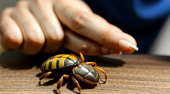
Preparation for Tick Removal
Essential Tools and Materials
To extract a tick without increasing infection risk, the following items are required:
- Fine‑point tweezers or small forceps with smooth, non‑slipping jaws
- Disposable gloves (latex, nitrile, or vinyl)
- Antiseptic solution (70 % isopropyl alcohol, iodine, or chlorhexidine)
- Sterile gauze pads or cotton swabs
- Small, sealable plastic bag or container for tick disposal
- Adhesive bandage for post‑removal wound care
The tweezers must grasp the tick as close to the skin as possible, avoiding compression of the body. Gloves protect the handler from potential pathogens. Antiseptic wipes the bite site before and after removal, while gauze provides a clean surface for handling the tick and applying pressure if bleeding occurs. The sealed container prevents accidental release of the parasite into the environment. An adhesive bandage secures the site after cleaning. All tools should be sterile or disinfected before use to minimize secondary infection.
Understanding the Risks of Improper Removal
Improper extraction of a tick can lead to immediate and delayed health problems. When the mouthparts remain embedded, the wound may become infected, and the tick can continue to transmit pathogens. Incomplete removal also increases the likelihood of local inflammation, swelling, and secondary bacterial infection.
Risks associated with incorrect removal:
- Retention of mouthparts – fragments left in the skin serve as a conduit for disease agents and may cause chronic irritation.
- Enhanced pathogen transmission – squeezing the tick’s body raises internal pressure, forcing saliva and infected material into the host’s bloodstream.
- Secondary infection – rough handling or unsterile tools introduce bacteria, leading to cellulitis or abscess formation.
- Allergic reaction – improper removal can trigger a severe local or systemic allergic response to tick saliva.
- Delayed diagnosis – failure to recognize a retained fragment postpones treatment for tick‑borne illnesses such as Lyme disease, Rocky Mountain spotted fever, or babesiosis.
Avoiding these hazards requires precise technique: use fine‑point tweezers to grasp the tick as close to the skin as possible, apply steady upward pressure, and disinfect the site before and after removal. If any part of the tick remains, seek medical assistance promptly to prevent complications.
Step-by-Step Tick Removal Process
Positioning for Optimal Access
Proper positioning is the first factor that determines whether a tick can be grasped and extracted without breaking its mouthparts. The target area must be fully visible, well‑lit, and reachable without straining the remover’s arms or wrists.
Expose the skin by removing clothing or lifting folds. Sit or stand on a stable surface; avoid leaning over the body part. Keep the affected limb supported on a firm object (e.g., a chair arm, a table edge) to prevent movement. Use a magnifying lens if the tick is small or located in a hair‑dense region. Ensure the hand holding the tweezers rests on a steady surface to maintain control.
- Position the patient so the tick lies at eye level; this reduces the need to look down and improves precision.
- Align the forearm or leg so the tick is on the outer side, allowing the dominant hand to approach from the same direction.
- Place a clean towel or disposable pad beneath the area to catch any blood or dropped debris.
- Keep the non‑dominant hand free to gently stretch the skin, creating a taut surface for the tweezers.
Maintain a calm posture; sudden movements can cause the tick to embed deeper. Have fine‑point tweezers, antiseptic wipes, and a sealed container ready before beginning. After removal, cleanse the bite site and monitor for signs of infection.
Grasping the Tick Correctly
Using Fine-Tipped Tweezers
Fine‑tipped tweezers provide the precision needed to grasp a tick’s head without compressing its body, reducing the risk of pathogen transmission.
Before beginning, wash hands thoroughly, clean the tweezers with alcohol, and place a clean surface nearby. Keep a small container with antiseptic solution ready for the tick after removal.
- Grip the tick as close to the skin as possible, using the tips of the tweezers to hold the mouthparts securely.
- Pull upward with steady, even pressure; avoid twisting, jerking, or squeezing the body.
- Continue pulling until the tick releases completely.
After extraction, disinfect the bite area with iodine or alcohol. Observe the site for several days; if redness, swelling, or a rash develops, seek medical advice. Store the removed tick in a sealed container with alcohol for identification if needed.
Dispose of the tick by immersing it in a disinfectant solution, then placing it in a sealed bag before discarding in household waste. Maintain a record of the removal date and any symptoms that appear thereafter.
Avoiding Crushing the Tick’s Body
When extracting a tick, the primary goal is to keep the parasite’s body intact. A ruptured tick can release saliva and gut contents that contain pathogens, increasing the risk of infection for the host.
Use fine‑point tweezers or a specialized tick‑removal tool. Grip the tick as close to the skin as possible, securing the head and mouthparts without squeezing the abdomen. Apply steady, upward pressure until the organism separates from the skin. Do not twist, jerk, or pinch the body, as these actions compress internal structures.
After removal, place the tick in a sealed container for identification or disposal. Clean the bite area with antiseptic and wash hands thoroughly. Monitor the site for signs of redness, swelling, or fever over the next several weeks; seek medical advice if symptoms develop.
Key practices to prevent crushing:
- Position tweezers parallel to the skin surface.
- Maintain a firm but gentle grip on the tick’s head.
- Pull straight upward without bending the legs.
- Avoid using fingers or blunt objects that can compress the body.
Gentle and Steady Pulling Technique
Straight Upward Motion
Straight upward motion is the preferred technique for extracting a tick from human skin. The method minimizes the risk of breaking the parasite’s mouthparts, which can remain embedded and cause infection.
- Clean the bite area with an antiseptic.
- Use fine‑point tweezers or a specialized tick‑removal tool.
- Grip the tick as close to the skin as possible, avoiding compression of the body.
- Apply steady, gentle pressure upward, parallel to the skin surface, until the tick releases.
- Transfer the tick to a sealed container for identification if needed.
- Disinfect the bite site again and wash hands thoroughly.
- Observe the area for several days; seek medical advice if redness, swelling, or fever develop.
The upward force aligns with the tick’s hypostome, allowing the barbed anchoring structures to disengage without tearing. Maintaining a constant motion prevents the tick from rotating, which could detach the head while leaving the mouthparts behind. After removal, cleaning and monitoring reduce the likelihood of pathogen transmission.
Avoiding Twisting or Jerking
When a tick is attached, the mouthparts embed deeply into the skin. Pulling the parasite with a twisting or jerking motion can break the head, leaving fragments that may continue to release pathogens. Maintaining a straight, steady grip on the tick’s body prevents this risk.
Use fine‑point tweezers to grasp the tick as close to the skin as possible. Apply even pressure and lift upward in a single motion. Do not rock the instrument, do not wiggle the tick, and do not squeeze its abdomen.
Key points for a clean extraction:
- Position tweezers at the tick’s head, not the body.
- Grip firmly, avoiding slippage.
- Pull upward with constant force, no sudden movements.
- After removal, clean the bite area with antiseptic and wash hands thoroughly.
If any part of the mouth remains embedded, consult a healthcare professional. Proper technique eliminates the chance of retained fragments and reduces the likelihood of disease transmission.
Post-Removal Care and Monitoring
Cleaning the Bite Area
After the tick is detached, the bite site must be decontaminated immediately to reduce the risk of infection. Use a sterile gauze or a clean cloth soaked in an antiseptic solution such as 70 % isopropyl alcohol, povidone‑iodine, or chlorhexidine. Apply gentle pressure for 30 seconds, then allow the area to air‑dry. Do not scrub aggressively; excessive friction can damage skin and increase irritation.
If an antiseptic pad is unavailable, wash the site with mild soap and lukewarm water for at least 20 seconds, then rinse thoroughly. Pat the skin dry with a disposable towel before applying an antiseptic.
After cleaning, cover the wound with a sterile adhesive bandage to protect it from external contaminants. Change the dressing daily and monitor for signs of inflammation, such as redness, swelling, warmth, or pus. Should any of these symptoms appear, seek medical evaluation promptly.
Disposing of the Tick Safely
Options for Tick Disposal
After extracting a tick, the first priority is to prevent any remaining pathogen from escaping the specimen. Secure containment eliminates the risk of accidental contact and allows for proper disposal.
- Place the tick in a small, sealable plastic bag, add a few drops of isopropyl alcohol, and close the bag tightly. The alcohol kills the arthropod within minutes.
- Submerge the tick in a container of 70 % ethanol for at least 10 minutes, then discard the liquid according to local hazardous‑waste guidelines.
- Drop the tick into a disposable cup filled with water and a pinch of dish soap, then flush the mixture down the toilet. The soap reduces surface tension, preventing the tick from adhering to plumbing.
- Wrap the tick in a piece of tissue, seal it inside a zip‑lock bag, and place the bag in an outdoor trash bin that is collected weekly.
If local regulations permit, incineration of the sealed bag provides an irreversible method. In regions where medical waste facilities are available, hand the sealed container to the appropriate disposal service. Avoid crushing the tick with fingers; use tweezers or a disposable tool to transfer it into the chosen medium.
When to Save the Tick for Testing
When a tick is removed, preserve it if any of the following conditions apply:
- The bite occurred in an area where tick‑borne diseases are common.
- The tick appears engorged or has been attached for more than 24 hours.
- The person develops fever, rash, joint pain, or other symptoms within weeks after the bite.
- A healthcare provider requests the specimen for laboratory analysis.
- The species cannot be identified visually and may belong to a medically significant group.
Store the specimen in a sealable container (e.g., a small plastic tube or zip‑lock bag). Place a damp paper towel inside to prevent desiccation, then refrigerate at 4 °C. Deliver the tick to the laboratory within 48 hours; if longer storage is necessary, keep it frozen at –20 °C.
Testing the tick can confirm the presence of pathogens such as Borrelia burgdorferi, Anaplasma, or Rickettsia. Positive results guide early treatment decisions and improve clinical outcomes.
Recognizing Symptoms Requiring Medical Attention
Localized Reactions
When a tick is detached at home, the skin around the bite often shows a confined response. The reaction typically appears within minutes to a few hours and remains limited to the immediate area of attachment.
- Redness that forms a small, well‑defined halo around the puncture site.
- Swelling that may raise the skin a few millimeters, resembling a raised bump.
- Itching or mild burning sensation confined to the bite margin.
- Small, localized bruising if the tick’s mouthparts caused minor trauma.
If the area becomes increasingly painful, expands beyond a few centimeters, or develops pus, these signs indicate secondary infection rather than a simple localized reaction. In such cases, professional medical evaluation is required.
For uncomplicated localized responses, clean the skin with soap and water, apply an antiseptic, and monitor for change. A short course of over‑the‑counter antihistamine can reduce itching; topical corticosteroid creams may lessen swelling. Document the size and appearance of the reaction, and seek medical advice if it persists beyond 48 hours or worsens.
Systemic Symptoms of Tick-Borne Illnesses
After a tick has been detached at home, attention must shift to the body’s overall response. Systemic manifestations often indicate infection and require prompt evaluation.
Common systemic signs include:
- Fever or chills
- Severe headache, sometimes with neck stiffness
- Muscle or joint aches, especially in large joints
- Fatigue or malaise that persists beyond a day
- Nausea, vomiting, or abdominal pain
- Generalized rash (e.g., erythema migrans, maculopapular eruptions)
- Swollen lymph nodes
- Dizziness or faintness
The specific pattern depends on the pathogen transmitted. Lyme disease typically presents with a gradually expanding erythema migrans and migratory arthralgia, while Rocky Mountain spotted fever often produces high fever, a petechial rash, and rapid neurologic decline. Ehrlichiosis and Anaplasmosis may begin with flu‑like symptoms and progress to thrombocytopenia or hepatic dysfunction. Symptoms usually emerge within 3 – 14 days after the bite, but some illnesses, such as babesiosis, can develop later.
Seek medical care if any systemic symptom appears, especially fever above 38 °C, a spreading rash, severe headache, or joint swelling. Early diagnosis and targeted antimicrobial therapy reduce the risk of complications and improve outcomes.
Prevention and Awareness
Best Practices for Tick Bite Prevention
Preventing tick attachment eliminates the need for emergency removal and reduces the risk of disease transmission.
Wear long sleeves and trousers when entering wooded or grassy areas; tuck pants into socks to create a barrier. Apply EPA‑registered repellents containing DEET, picaridin, or IR3535 to exposed skin and treat clothing with permethrin according to label directions. Choose light‑colored garments to spot ticks more easily.
Perform a thorough body inspection after outdoor activities. Use a hand mirror or partner assistance to examine hard‑to‑see sites such as the scalp, behind ears, underarms, groin, and behind knees. Remove any attached tick promptly with fine‑tipped tweezers, grasping as close to the skin as possible and pulling upward with steady pressure.
Maintain yard hygiene to deter ticks. Keep grass trimmed to a maximum of 4 inches, remove leaf litter, and create a mulch barrier between wooded zones and recreational spaces. Reduce wildlife hosts by discouraging deer and rodents; install fencing or use deer‑resistant plants where feasible.
Schedule regular veterinary care for pets, applying tick preventatives and checking them after outdoor exposure. Pets can transport ticks into the home, increasing human exposure.
Adopt these measures consistently to minimize tick encounters and protect health.
Understanding Common Tick-Borne Diseases
Ticks transmit a limited set of pathogens that cause serious illness in humans. Recognizing these agents clarifies why prompt, correct extraction matters.
- Lyme disease – bacterium Borrelia burgdorferi; early sign often a expanding erythema migmatum; later joint, cardiac or neurologic involvement; prevalent in northeastern, mid‑Atlantic and upper‑midwestern United States.
- Rocky Mountain spotted fever – Rickettsia rickettsii; fever, headache, rash that begins on wrists and ankles; highest risk in southeastern and south‑central states.
- Anaplasmosis – Anaplasma phagocytophilum; fever, muscle pain, leukopenia; common in the upper Midwest and New England.
- Ehrlichiosis – Ehrlichia chaffeensis; fever, nausea, thrombocytopenia; concentrated in the south‑central and south‑eastern regions.
- Babesiosis – Babesia microti; hemolytic anemia, fever, chills; endemic in the Northeast and upper Midwest.
- Powassan virus – flavivirus; encephalitis, meningitis; rare but severe, reported in the Great Lakes, Northeast and Canada.
Each pathogen requires a different therapeutic approach, yet all share a common preventive measure: removal of the attached tick before transmission can occur. The longer a tick remains attached, the greater the chance of pathogen transfer; many agents require ≥24 hours of feeding to reach infectious levels.
After extraction, observe the bite site and overall health for at least four weeks. Symptoms such as fever, rash, headache, muscle aches, or joint pain warrant medical evaluation. Early laboratory testing, guided by the known regional disease profile, improves treatment outcomes.
Accurate knowledge of tick-borne diseases equips individuals to act swiftly, apply proper removal technique, and seek timely care if illness develops.
