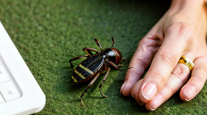
Understanding Tick-Borne Risks
Why Proper Removal is Crucial
Removing a tick without following proper technique can introduce serious health hazards. An incomplete extraction often leaves mouthparts embedded in the skin, creating a portal for bacteria and viruses that the parasite carries. Pathogens such as Borrelia burgdorferi (Lyme disease), Anaplasma phagocytophilum (anaplasmosis), and Rickettsia species can enter the bloodstream within minutes of attachment; any remaining fragments increase the likelihood of infection.
Improper handling also raises the risk of tissue injury. Rough pulling or squeezing the tick’s body may crush its abdomen, forcing infected fluids into the host. Damage to surrounding skin can lead to secondary bacterial infection, prolonged healing, and scarring.
Legal and clinical consequences follow when removal is performed incorrectly. Health professionals may be held liable for preventable disease transmission, and patients may require extensive diagnostic testing and prolonged antibiotic courses, increasing medical costs and burden on healthcare systems.
Key reasons for meticulous removal:
- Guarantees complete extraction of the parasite, eliminating residual mouthparts.
- Minimizes mechanical disruption of the tick, reducing pathogen release.
- Prevents secondary skin infection and promotes rapid wound closure.
- Avoids unnecessary medical interventions and associated expenses.
Adhering to established removal protocols—using fine‑point tweezers, grasping the tick as close to the skin as possible, and applying steady, upward pressure—directly addresses these risks and safeguards the individual's health.
Potential Health Complications
Ticks attach firmly to skin, delivering saliva that can contain bacteria, viruses, and parasites. An incomplete or rough removal can introduce additional health risks beyond the bite itself.
Potential complications include:
- Transmission of Lyme disease, caused by Borrelia burgdorferi, leading to joint pain, neurological symptoms, and cardiac involvement.
- Anaplasmosis and ehrlichiosis, presenting with fever, headache, and muscle aches.
- Rocky Mountain spotted fever, characterized by rash, high fever, and potential organ failure.
- Babesiosis, a malaria‑like illness causing hemolytic anemia.
- Tick‑borne encephalitis, resulting in meningitis or encephalitis.
- Secondary bacterial infection at the bite site, producing redness, swelling, and pus formation.
- Allergic reactions ranging from localized urticaria to systemic anaphylaxis.
Improper extraction can cause:
- Retained mouthparts embedded in tissue, serving as a nidus for infection and prolonging pathogen exposure.
- Mechanical injury to skin and underlying structures, increasing inflammation and risk of necrosis.
- Enhanced pathogen transmission due to squeezing of the tick’s body, forcing infected saliva deeper into the wound.
Accurate removal with fine‑point tweezers, steady upward traction, and immediate cleansing of the area reduces the likelihood of these outcomes. Prompt medical evaluation is advised if symptoms of infection or allergic response develop after removal.
Preparation Before Extraction
Essential Tools and Materials
Removing a tick safely depends on using the right instruments and supplies. The goal is to grasp the parasite close to the skin, apply steady pressure, and avoid crushing its body, which can release pathogens.
- Fine‑point tweezers (flat or curved tip) – provide a secure grip without slippage.
- Small, sterile needle or hook – useful for lifting the tick’s mouthparts if they remain embedded.
- Antiseptic wipes or alcohol swabs – disinfect the bite area before and after extraction.
- Disposable gloves – protect the handler from potential infection and prevent cross‑contamination.
- Sealable plastic bag or container – store the removed tick for identification or testing, if required.
- Bandage or sterile gauze – cover the wound after removal to protect against secondary infection.
Additional materials enhance the procedure:
- Magnifying lens – improves visibility of the tick’s attachment point, especially on hair‑covered skin.
- Clean towel or disposable pad – provides a stable surface and contains any debris.
- Documentation sheet – records date, location of bite, and tick description for medical follow‑up.
All items should be sterile, single‑use when possible, and readily accessible in a first‑aid kit. Proper preparation ensures the tick is removed in one motion, minimizing tissue trauma and reducing the risk of disease transmission.
Locating the Tick
When a tick attaches to skin, the first step is to identify its exact position before removal. Visual inspection should begin with the area where the bite was felt, often the scalp, neck, armpits, groin, or behind the knees. Use a well‑lit environment and, if necessary, a magnifying lens to distinguish the small, dark body from surrounding hair or skin folds.
- Gently part hair or clothing with fingertips; avoid pulling or crushing the tick.
- Look for a rounded, engorged shape, usually 2–5 mm in diameter; early‑stage ticks may appear as a tiny speck.
- Note the direction of the mouthparts; they embed at an angle toward the body surface.
- If the tick is partially hidden, press a clean, gloved finger against the skin to raise a small bulge that reveals the parasite.
Once the parasite’s outline is visible, confirm that no additional ticks are present nearby. A thorough search reduces the risk of leaving an attached tick unnoticed, which can increase the chance of pathogen transmission.
The Extraction Process
Grasping the Tick
Grasping the tick requires a firm, precise hold that prevents the mouthparts from breaking off in the skin. Use fine‑pointed, non‑slipping tweezers; avoid thumb‑finger pinch that can crush the body. Position the tweezers as close to the skin surface as possible to capture the entire organism without squeezing.
- Place the tweezer tips on opposite sides of the tick’s head, just above the skin.
- Apply steady, gentle pressure to lift the tick straight upward.
- Maintain the grip until the tick releases completely; do not twist or jerk.
- After removal, disinfect the bite area with an antiseptic and store the tick in a sealed container if testing is needed.
Pulling Technique
Straight Pull
The straight‑pull technique removes a tick by grasping the mouthparts as close to the skin as possible and applying steady, upward traction without twisting. This method minimizes the risk of leaving mouthparts embedded, which can cause local inflammation or infection.
Use fine‑point tweezers or a specialized tick‑removal tool. Position the instrument so the tip encircles the tick’s head, not the body, to avoid crushing the abdomen and forcing fluids into the host.
- Pinch the tick’s head firmly.
- Pull straight upward with constant force.
- Continue until the tick releases from the skin.
- Inspect the bite site; if any part remains, repeat the grip and pull.
Disinfect the area with an antiseptic solution after removal. Store the tick in a sealed container if identification or testing is required. Monitor the site for signs of redness, swelling, or rash over the next several days; seek medical advice if symptoms develop.
Avoiding Twisting or Jerking
When a tick is attached to skin, the mouthparts embed deeply into tissue. Applying a twisting or jerking motion can fracture the mouthparts, leaving fragments that may continue to feed and increase the risk of infection. Maintaining a steady, straight pull ensures the entire organism separates intact.
A proper extraction technique includes:
- Grasp the tick as close to the skin surface as possible with fine‑point tweezers.
- Apply gentle, constant pressure directly outward, avoiding any rotational force.
- Continue the pull until the tick releases without resistance.
- Disinfect the bite area and wash hands thoroughly after removal.
If resistance occurs, pause and reassess the grip rather than increasing force or twisting. Re‑gripping higher on the body may provide a better angle for a smooth extraction. Once removed, inspect the tick; if the head remains embedded, seek medical advice rather than attempting additional manipulation.
Post-Removal Actions
Cleaning the Bite Area
After a tick has been taken out, the skin surrounding the attachment point must be treated promptly to reduce infection risk.
- Wash the area with mild soap and running water, scrubbing gently to remove any residual saliva or debris.
- Apply an antiseptic such as povidone‑iodine, chlorhexidine, or alcohol swab; allow it to dry before covering.
- If a bandage is necessary, choose a sterile, non‑adhesive dressing that does not compress the wound.
Observe the site for the next 24–48 hours. Redness expanding beyond the immediate perimeter, swelling, pus, or increasing pain may indicate bacterial involvement and require medical evaluation. Document the date of removal and any changes in the lesion for reference during follow‑up.
Disposing of the Tick
After a tick is removed, it must be dealt with promptly to eliminate any chance of pathogen transmission. The following actions ensure safe disposal and appropriate follow‑up.
- Place the tick in a sealed container (e.g., a zip‑lock bag) with a damp cotton ball.
- Add a few drops of 70 % isopropyl alcohol or submerge the insect in alcohol to kill it instantly.
- Label the container with the date, location of the bite, and species if known; retain for up to 30 days if testing for disease is required.
- If testing is unnecessary, discard the sealed bag in household waste; do not flush alive ticks down the toilet.
Disinfect the bite site with an antiseptic (e.g., povidone‑iodine or chlorhexidine) and cover with a clean bandage. Observe the area for several weeks, noting any rash, fever, or flu‑like symptoms, and seek medical evaluation if they appear.
Finally, reduce future exposure by inspecting clothing and skin after outdoor activities, using tick‑repellent treatments on attire, and maintaining a tidy yard free of leaf litter and tall grass.
Aftercare and Monitoring
Observing for Symptoms
When a tick has been attached, the first step after removal is to monitor the bite site and the person’s overall condition. Early detection of adverse reactions reduces the risk of complications.
Key symptoms to watch for include:
- Redness or swelling that expands beyond the immediate bite area.
- A circular rash, often called a “bull’s‑eye,” that appears within days.
- Fever, chills, or sweats without an obvious cause.
- Headache, fatigue, or muscle aches.
- Joint pain or swelling, especially if it develops a week after the bite.
- Nausea, vomiting, or abdominal discomfort.
Observe the individual for at least two weeks. Record any new signs daily, noting the date of onset and severity. If any listed symptom emerges, contact a healthcare professional promptly; early treatment can prevent severe disease progression.
Consistent observation provides the necessary data for clinicians to diagnose tick‑borne illnesses accurately and to initiate appropriate therapy without delay.
When to Seek Medical Attention
If a tick is removed but any of the following conditions appear, professional medical evaluation is required.
- The bite site becomes red, swollen, or develops a bull’s‑eye rash within a few days.
- Fever, chills, headache, muscle aches, or fatigue emerge after the bite.
- The tick was attached for longer than 24 hours, was engorged, or could not be fully extracted.
- The individual has a weakened immune system, is pregnant, or has a history of severe allergic reactions.
- The tick is identified as a species known to transmit serious pathogens (e.g., Ixodes scapularis, Dermacentor variabilis).
Prompt consultation reduces the risk of complications such as Lyme disease, Rocky Mountain spotted fever, or anaphylaxis. Seek care immediately if symptoms progress rapidly or if uncertainty exists about the completeness of removal.
