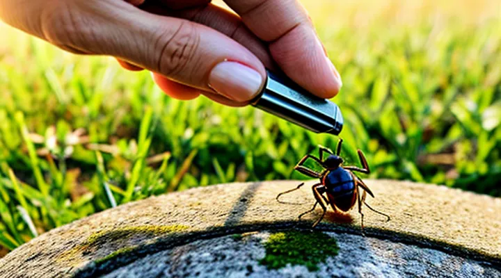
Understanding Tick Removal «Best Practices»
Why Proper Removal is Crucial?
Preventing Disease Transmission
Effective removal of attached ticks is essential to interrupt pathogen transmission. Immediate action reduces the time pathogens have to migrate from the tick’s salivary glands into the host’s bloodstream. The following measures guarantee complete extraction and minimize infection risk:
- Grasp the tick as close to the skin surface as possible with fine‑pointed tweezers.
- Apply steady, upward pressure without twisting or crushing the body.
- Maintain traction until the mouthparts detach fully; avoid leaving any fragment embedded.
- Disinfect the bite area with an antiseptic solution after removal.
- Preserve the tick in a sealed container for identification if disease surveillance is required.
Post‑removal monitoring should include observation for local erythema, fever, or flu‑like symptoms within several weeks. Prompt medical evaluation is advised if any signs develop, enabling early treatment for tick‑borne illnesses such as Lyme disease, anaplasmosis, or babesiosis.
Avoiding Further Skin Irritation
After a tick is taken out, the surrounding skin is vulnerable to irritation. Immediate care reduces inflammation and prevents secondary infection.
- Clean the bite site with mild soap and lukewarm water.
- Pat dry with a sterile gauze; avoid rubbing.
- Apply an alcohol‑based antiseptic or a povidone‑iodine solution.
- Use a sterile, flat‑edge instrument (e.g., a tweezers with smooth jaws) to press gently around the wound, not into it.
- Cover the area with a breathable, non‑adhesive dressing if friction is likely.
Post‑removal treatment focuses on soothing the skin and monitoring healing. Apply a thin layer of a hypoallergenic moisturizer or a zinc‑oxide ointment to maintain barrier function. Refrain from scratching; use a cold compress to alleviate itching. Observe the site for redness extending beyond a few millimeters, swelling, or pus formation—indicators of infection.
Seek medical evaluation if symptoms progress rapidly, if a rash develops, or if fever appears. Prompt professional assessment prevents complications and ensures complete resolution of the bite.
Step-by-Step Guide to Tick Removal
Preparation «Before You Start»
Gathering Necessary Tools
To remove a tick completely, the right instruments reduce the risk of leaving mouthparts embedded and minimize tissue trauma. Selecting appropriate implements before beginning the procedure ensures control and sterility.
- Fine‑pointed, non‑slipping tweezers – grip the tick close to the skin without crushing the body.
- Magnifying lens or portable loupe – reveal the attachment site and verify complete extraction.
- Antiseptic solution (e.g., povidone‑iodine or chlorhexidine) – cleanse the area before and after removal.
- Disposable gloves – protect both the handler and the patient from possible pathogens.
- Sterile gauze pads or cotton swabs – apply pressure to stop bleeding and cover the wound.
- Small container with sealable lid – store the tick for identification or testing, if required.
- Alcohol wipes – sanitize tools between uses when multiple removals are performed.
Each item contributes to a systematic approach that eliminates the tick while preserving skin integrity and preventing secondary infection.
Sanitizing Equipment and Hands
Proper sanitization of tools and hands is a critical component of guaranteeing complete tick extraction. Contaminated instruments can retain mouthparts, increasing the risk of infection.
- Clean tweezers with hot, soapy water immediately after use. Rinse thoroughly and dry with a disposable paper towel.
- Submerge tweezers in a 70 % isopropyl alcohol solution for at least one minute. Replace the solution daily in high‑use settings.
- Disinfect reusable containers or trays with a chlorine‑based sanitizer (0.5 % sodium hypochlorite) before and after each tick removal session.
- When single‑use instruments are unavailable, sterilize metal tools in an autoclave at 121 °C for 15 minutes.
Hand hygiene prevents pathogen transfer from the tick to the practitioner’s skin.
- Perform hand washing with antimicrobial soap for a minimum of 20 seconds, covering all surfaces, before and after each removal.
- Apply an alcohol‑based hand rub (minimum 60 % ethanol) if soap and water are not immediately accessible.
- Wear disposable nitrile gloves; discard them after each tick is removed. If gloves become torn or contaminated, replace them without delay.
Regular monitoring of sanitizing procedures ensures that no residual tick fragments remain on equipment, thereby supporting effective removal and reducing the likelihood of disease transmission. «CDC guidelines state that strict disinfection protocols are essential for safe tick extraction».
The Removal Process «Technique Matters»
Grasping the Tick Correctly
Grasping a tick firmly is the first critical step in guaranteeing its complete extraction. The mouthparts must be secured as close to the skin as possible to prevent the barbed hypostome from tearing during removal.
- Use fine‑point tweezers or a dedicated tick‑removal tool; avoid blunt instruments that slip.
- Position the tips at the tick’s head, directly above the mouthparts, and apply steady, even pressure.
- Pull upward in a smooth motion without twisting or jerking; twisting can detach the mouthparts and leave them embedded.
- Maintain the grip until the tick releases entirely; a detached body indicates incomplete removal.
After extraction, inspect the site for any remaining parts. If fragments are visible, repeat the grip and pull method until the area is clear. Disinfect the bite area with an antiseptic and store the tick in a sealed container for potential pathogen testing. Proper handling eliminates the risk of residual mouthparts and reduces the chance of infection.
Pulling Motion «Slow and Steady»
A steady, controlled pull is the most reliable method for extracting a tick without leaving mouthparts embedded. Grasp the tick as close to the skin as possible with fine‑pointed tweezers. Apply a continuous force using the «Slow and Steady» pulling motion; avoid jerking, twisting, or squeezing the body, which can cause the mouthparts to break off.
- Position tweezers parallel to the skin.
- Secure a firm grip on the tick’s head.
- Initiate a slow, uniform traction until the tick releases.
- Inspect the bite site for any remaining fragments.
- Disinfect the area with antiseptic after removal.
If any part of the mouth remains, repeat the pulling motion with the same controlled force. Monitoring the site for redness or swelling over the next 24‑48 hours helps identify potential infection early.
Post-Removal Care «After the Tick is Out»
Cleaning the Bite Area
After a tick is extracted, the bite site requires immediate attention to prevent infection and reduce irritation. First, wash hands thoroughly with soap and water before touching the area. Then, cleanse the skin using a mild antiseptic solution such as povidone‑iodine or chlorhexidine. Apply the antiseptic with a sterile cotton swab, moving outward from the center of the wound to avoid re‑contaminating the site.
After disinfection, dry the area with a clean gauze pad. If minor redness persists, apply a thin layer of topical antibiotic ointment to create a protective barrier. Cover the bite with a sterile adhesive bandage only if the wound is open or if friction is expected; otherwise, leave it uncovered to allow airflow and natural healing.
Monitor the bite for signs of infection, including increasing redness, swelling, warmth, or discharge. Seek medical evaluation if any of these symptoms develop, or if a rash characteristic of Lyme disease appears. Maintaining proper hygiene during the first 24 hours after removal significantly reduces the risk of secondary complications.
Disposing of the Tick Safely
After removal, the tick must be destroyed to prevent disease transmission and accidental re‑attachment. Place the specimen in a sealable plastic bag, expel excess air, and close tightly. Submerge the bag in a container of 70 % isopropyl alcohol for at least five minutes, then discard the bag in a household waste bin. Alternatively, place the tick in a small, labeled glass jar and freeze at –20 °C for a minimum of 24 hours before disposal.
Key precautions:
- Do not crush the tick with fingers; use tweezers or a tick‑removal tool.
- Avoid flushing the tick down the toilet; it may survive in sewage systems.
- Wash hands thoroughly with soap and water after handling the container.
- Record the removal date and tick species, if known, for medical reference.
If a professional medical facility is accessible, hand the sealed container to staff for autoclave sterilization or incineration. This ensures complete destruction and eliminates the risk of residual pathogens.
Monitoring for Symptoms
After extracting a tick, systematic observation of the bite site and overall health is essential to verify complete removal. Early detection of adverse reactions prevents complications and guides timely treatment.
Key indicators to monitor include:
- Redness or swelling that expands beyond the immediate bite area
- Development of a target‑shaped rash (erythema migrans)
- Fever, chills, or unexplained fatigue
- Joint pain, muscle aches, or neurological disturbances such as facial weakness or tingling sensations
- Symptoms appearing within 3 – 30 days post‑removal, with most cases emerging within the first two weeks
If any of these signs arise, immediate medical consultation is warranted. Diagnostic evaluation may involve serologic testing for tick‑borne pathogens and, when indicated, antibiotic therapy. Continuous documentation of symptom onset, duration, and progression assists healthcare providers in determining the appropriate management plan.
Common Mistakes to Avoid
Incorrect Removal Methods
Twisting or Jerking
Removing a tick completely requires a technique that disengages the parasite without breaking its mouthparts. Twisting or jerking the attached organism is a common approach, but it must be executed with precision.
The method involves grasping the tick as close to the skin as possible, then applying a steady rotational force while pulling outward. Sudden jerks increase the risk of leaving fragments embedded in the tissue and should be avoided.
- Use fine‑point tweezers or a specialized tick‑removal tool.
- Position the instrument at the tick’s head, near the skin surface.
- Rotate the tick clockwise or counter‑clockwise with a smooth motion.
- Maintain constant tension; do not release grip until the tick detaches fully.
- Inspect the extracted specimen; ensure the mouthparts are intact.
Improper twisting can cause the hypostome to fracture, leading to infection or inflammation. If resistance is felt, cease rotation and reassess grip before continuing. After removal, cleanse the bite area with antiseptic and monitor for signs of local reaction.
Using Home Remedies «Heat, Petroleum Jelly, etc.»
A tick that remains partially attached can transmit pathogens; complete extraction prevents further exposure.
Effective home‑based techniques focus on immobilizing the parasite, facilitating grasp, and minimizing tissue damage.
- Apply a warm compress («heat») to the bite site for several minutes. The temperature increase causes the tick’s mouthparts to relax, allowing easy removal with fine‑tipped tweezers.
- Coat the tick with a thin layer of petroleum jelly («petroleum jelly»). The barrier suffocates the arthropod, prompting it to detach without aggressive pulling.
- Use a small piece of adhesive tape, pressed gently over the tick. The tape adheres to the body, enabling removal in one motion while keeping the head intact.
- Submerge the area in warm water (approximately 40 °C) for five minutes. Hydration expands the tick’s cuticle, reducing grip strength and easing extraction.
After any method, grasp the tick as close to the skin as possible, pull upward with steady pressure, and avoid twisting. Disinfect the wound, store the specimen for identification if needed, and observe the site for signs of infection over the next week.
Not Inspecting Thoroughly
When a tick is removed without a careful visual check, remnants may remain embedded in the skin. Incomplete extraction often results from the failure to inspect the bite site and surrounding area thoroughly. The presence of mouthparts or a small portion of the tick can trigger local inflammation, infection, or transmission of pathogens such as Borrelia burgdorferi.
Neglecting a detailed examination increases the likelihood of secondary complications. Residual fragments serve as a nidus for bacterial growth, prolong healing time, and may necessitate medical intervention. Early detection of retained parts prevents escalation to systemic illness.
Practical measures to eliminate the risk associated with «Not Inspecting Thoroughly»:
- Use a magnifying lens or a bright light source immediately after removal.
- Scan the entire bite region, including surrounding hair and skin folds, for any visible tick fragments.
- Compare the extracted specimen with reference images to confirm that the body, legs, and head are intact.
- If doubt remains, cleanse the area with antiseptic and seek professional evaluation.
Consistent application of these steps ensures that the removal process leaves no trace of the arthropod, thereby safeguarding health.
When to Seek Medical Attention
Incomplete Removal «Remaining Parts»
When a tick is pulled from the skin, any portion that remains attached constitutes an incomplete removal. The retained mouthparts or abdomen can embed in tissue, creating a gateway for pathogens and provoking localized inflammation.
Risks increase if fragments stay embedded. Bacterial or viral agents may travel from the tick’s salivary glands into the host, raising the probability of disease transmission. Inflammation may persist, causing redness, swelling, or pain at the bite site.
Verification of complete extraction requires systematic observation and, when possible, magnification:
- Inspect the bite area with a magnifying lens or handheld microscope. Look for any visible fragments, especially the capitulum (mouthparts).
- Compare the extracted specimen with reference images that show the full body length and shape. Absence of the anterior segment indicates incomplete removal.
- Gently cleanse the area with antiseptic solution and re‑examine after a few minutes to detect any residual tissue that may have become more visible.
- If doubt remains, apply sterile tweezers to grasp any suspected fragment and remove it with steady, upward traction, avoiding squeezing the tick’s body.
After confirming removal, disinfect the site with an appropriate antiseptic, apply a sterile dressing if necessary, and monitor for signs of infection over the following 24–48 hours. Persistent redness, swelling, or fever warrants medical evaluation.
Signs of Infection or Illness
Rash Development
After a tick is extracted, the skin at the attachment site may exhibit a localized reaction. The reaction frequently begins within hours and can evolve over several days, indicating the body’s response to residual mouthparts or pathogen exposure.
Typical progression follows a predictable pattern. An initial erythema appears as a faint pink area, often accompanied by mild itching. Within 24–48 hours, the erythema may expand, forming a raised border. In some cases, the lesion adopts a target‑like configuration, with a central clearing surrounded by concentric rings. Persistence beyond one week, enlargement, or the emergence of systemic symptoms such as fever or fatigue warrants further evaluation.
Key clinical features include:
- Uniform redness that enlarges gradually
- Central clearing or a bull’s‑eye appearance
- Absence of purulent discharge
- Lack of immediate severe pain
Management focuses on observation and early intervention. Recommended actions are:
- Clean the area with mild antiseptic solution.
- Document size, shape, and any changes daily.
- Apply a topical corticosteroid to reduce inflammation if itching is pronounced.
- Initiate oral antibiotics when signs of infection or a bull’s‑eye rash suggest Lyme disease or other tick‑borne illnesses.
- Seek medical assessment if the rash expands rapidly, becomes painful, or is accompanied by fever, joint pain, or neurological signs.
Prompt recognition of rash development and adherence to these steps minimize complications and support full recovery after tick removal.
Flu-like Symptoms
Flu‑like symptoms may appear after a tick bite, signalling that the parasite was not completely extracted or that a pathogen has been transmitted. Recognising these signs enables timely medical intervention and reduces the risk of complications.
Typical manifestations include:
- fever of 38 °C or higher
- chills and sweating
- headache
- muscle or joint aches
- fatigue
- mild nausea
When any of these signs develop within two weeks of removal, the following actions are recommended. First, inspect the bite site for residual mouthparts; a small, dark fragment may remain embedded in the skin. Second, document the date of the bite, the geographic area, and any known exposure to tick‑borne diseases. Third, seek professional evaluation promptly; laboratory testing can identify early infection, and appropriate antibiotic therapy can be initiated.
Continuous monitoring for at least four weeks after extraction is advisable. Persistent or worsening symptoms warrant re‑examination, as delayed treatment may increase the likelihood of severe outcomes. Maintaining a record of symptom onset and progression assists healthcare providers in determining the most effective therapeutic approach.
