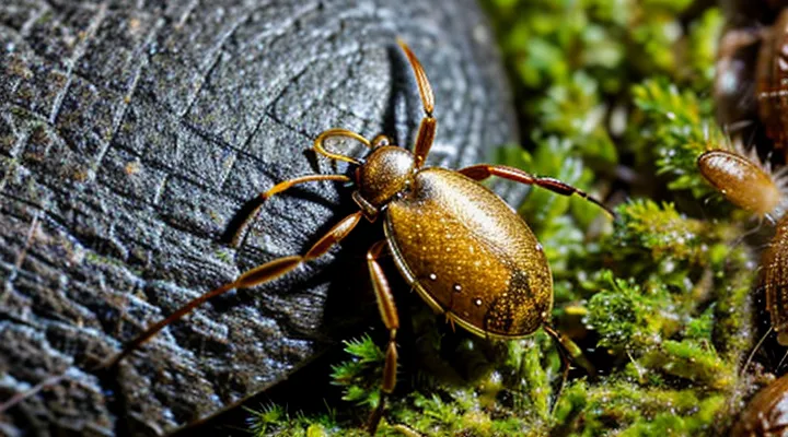
«The Misconception of Rotation in Tick Removal»
«Why Rotation is Not Recommended»
«Understanding Tick Anatomy and Attachment»
Ticks are obligate blood‑feeding arachnids that attach to hosts for extended periods. Successful detachment depends on knowledge of their mouth‑part structure and the mechanics of attachment.
Key anatomical components involved in attachment:
- Capitulum (mouth‑part complex) housing the feeding apparatus.
- Hypostome, a barbed tube that penetrates the skin.
- Chelicerae, small cutting structures that assist entry.
- Palps, sensory organs that help locate a suitable feeding site.
- Cement glands, which secrete a proteinaceous glue that stabilizes the hypostome.
The hypostome’s barbs interlock with host tissue, while cement reinforces the bond. Both elements create a one‑way grip: the barbs resist upward motion, and the cement solidifies around the embedded portion. Consequently, any force applied opposite to the direction of barbed entry risks breaking the mouthparts or leaving fragments in the skin.
Removal technique derived from anatomy:
- Grasp the tick as close to the skin as possible using fine‑point tweezers.
- Apply steady, gentle pressure to disengage the cement.
- Rotate the tick in the direction opposite to the orientation of the hypostome barbs—typically a counter‑clockwise motion—until the mouth‑part releases.
- Pull straight upward without squeezing the body to avoid contaminating the bite site with pathogen‑laden fluids.
Understanding the tick’s attachment architecture clarifies why a counter‑clockwise twist aligns with the barbs and reduces the chance of mouth‑part rupture. The described method minimizes tissue trauma and prevents residual tick parts from remaining embedded.
«The Risk of Leaving Parts Behind»
Removing a tick with improper technique can leave mouthparts embedded in the skin, creating a portal for infection. The feeding apparatus of a tick consists of barbed hypostome structures that anchor firmly to tissue; if the tick is twisted or squeezed, these structures may fracture.
- Retained parts serve as a nidus for bacterial colonization, increasing the likelihood of local cellulitis or systemic illness such as Lyme disease.
- Incomplete extraction complicates serological testing because antigenic material remains in the host, potentially altering immune response interpretation.
- Surgical removal of residual fragments may be required, leading to additional tissue trauma, scarring, and healthcare costs.
Evidence from clinical studies shows that steady, unidirectional traction, applied without rotation, minimizes the chance of mouthpart detachment. The force should be constant and sufficient to overcome the tick’s attachment, but not so excessive as to crush the body. When removal is performed correctly, the risk of leaving any portion behind drops dramatically, reducing subsequent complications.
«Potential for Infection and Complications»
Removing a tick improperly raises the likelihood of bacterial transmission and local tissue damage. The primary concerns are:
- Pathogen transmission – pathogens such as Borrelia burgdorferi (Lyme disease), Anaplasma phagocytophilum, Babesia microti, and tick‑borne encephalitis virus reside in the tick’s salivary glands. A prolonged attachment increases the probability of transfer.
- Retention of mouthparts – crushing or twisting the tick can detach the hypostome, leaving fragments embedded in the skin. Residual parts serve as a nidus for bacterial colonization and may provoke chronic inflammation.
- Secondary bacterial infection – broken skin and retained fragments provide entry points for skin flora. Symptoms include erythema, swelling, purulent discharge, and fever.
- Allergic or hypersensitivity reactions – mechanical irritation or saliva proteins can trigger local edema, urticaria, or systemic anaphylaxis in sensitized individuals.
- Delayed wound healing – excessive pressure or rotation of the tick may cause tissue necrosis, increasing scar formation and risk of infection.
The direction of extraction (counter‑clockwise versus clockwise) does not influence pathogen transfer if the tick is grasped close to the skin with fine‑pointed tweezers and lifted steadily without rotation. The critical factor is avoiding compression of the body, which forces saliva and potentially infected gut contents back into the host. A smooth, vertical pull minimizes tissue trauma, reduces the chance of mouthpart loss, and therefore limits the infection and complication risk.
«The Proper Method for Tick Removal»
«Essential Tools for Safe Removal»
«Fine-Tipped Tweezers»
Fine‑tipped tweezers are the preferred instrument for extracting ticks because their narrow, pointed jaws grasp the parasite close to the skin without crushing the body. The design allows precise control of force and angle, which is essential when applying the rotational motion required for safe removal.
When using fine‑tipped tweezers, follow these steps:
- Position the tips as close to the skin as possible, gripping the tick’s head or mouthparts.
- Apply steady, gentle pressure to lift the tick upward.
- Rotate the tick counter‑clockwise (or clockwise, depending on the tick’s orientation) while maintaining upward traction; the rotation dislodges the mouthparts from the skin.
- Continue the motion until the tick separates completely; avoid squeezing the abdomen to prevent pathogen release.
- Disinfect the tweezers after each use and store them in a clean container.
The fine tip minimizes tissue damage, and the ability to rotate the instrument mirrors the natural twisting motion needed to break the attachment. Compared with blunt forceps or finger removal, fine‑tipped tweezers reduce the risk of leaving mouthparts embedded and lower the chance of pathogen transmission. Proper sterilization between procedures maintains hygiene and prevents cross‑contamination.
«Antiseptic Wipes or Alcohol Swabs»
When a tick is detached, the bite site must be disinfected promptly to reduce bacterial contamination. The choice between pre‑moistened antiseptic wipes and individual alcohol swabs influences both effectiveness and practicality.
- Antiseptic wipes contain a broad‑spectrum agent (often chlorhexidine or povidone‑iodine) that remains active for several minutes, offering sustained antimicrobial protection. They cover a larger area, useful when the surrounding skin is also exposed to potential pathogens.
- Alcohol swabs deliver 70 % isopropyl alcohol, providing rapid bactericidal action limited to the immediate contact point. Their low viscosity allows precise application to the puncture wound without excess moisture.
Use the selected product after removing the tick: apply a single wipe or several swab strokes directly onto the bite, allow the surface to remain wet for at least 15 seconds, then let it air‑dry. Both methods achieve comparable reduction of skin flora, but antiseptic wipes may be preferable when extended coverage is required, while alcohol swabs suit situations demanding quick, targeted disinfection.
«Step-by-Step Guide to Removing a Tick»
«Grasping the Tick Correctly»
Grasping the tick correctly is the first prerequisite for safe removal. Use fine‑point tweezers or a specialized tick‑removal tool; position the instrument as close to the skin as possible to capture the head without compressing the body. Apply firm, steady pressure and lift the parasite in a straight line outward.
- Place tweezers at the tick’s mouthparts, not the abdomen.
- Maintain a horizontal grip parallel to the skin surface.
- Pull upward with constant force; avoid jerking or twisting motions.
- Release the tick into a container with alcohol or soap and water for disposal.
The extraction direction does not involve rotating the tick. Rotational movements risk breaking the mouthparts, leaving fragments embedded in the skin and increasing the chance of infection. A linear, upward traction ensures the entire organism separates cleanly, minimizing tissue trauma and pathogen transmission.
«Applying Steady, Upward Pressure»
When a tick is attached, the goal is to detach it without compressing its mouthparts. The most reliable method combines a gentle twist with a constant upward force.
- Grip the tick as close to the skin as possible using fine‑point tweezers.
- Rotate the tick slowly in the direction opposite to the way its legs are oriented; this typically means a counter‑directional twist.
- Maintain steady upward pressure throughout the rotation, avoiding squeezing the body.
- Once the tick releases, pull it straight out, keeping the motion smooth and continuous.
- Disinfect the bite area and store the removed tick for identification if needed.
Applying a uniform upward pull prevents the tick’s mouthparts from breaking off and embedding in the skin, reducing the risk of infection. The brief rotational motion disengages the anchoring barbs, while the sustained lift ensures complete removal.
«Avoiding Twisting, Jerking, or Squeezing»
When a tick is attached, the priority is to extract it without damaging its body. Any deformation—twisting, jerking, or squeezing—can cause the mouthparts to break off and remain embedded, increasing the risk of infection.
Key actions to prevent damage
- Grip the tick with fine‑point tweezers as close to the skin as possible.
- Apply steady, linear traction directly away from the host.
- Avoid any rotational movement; a smooth pull eliminates the chance of the mandibles separating.
- Do not pinch the abdomen; compression can force internal fluids into the bite site.
Consequences of improper handling
- Fragmented mouthparts may stay lodged, creating a portal for bacteria and viruses.
- Excessive force can rupture the tick’s gut, releasing pathogens into the wound.
- Repeated attempts increase tissue trauma and prolong healing.
By maintaining a controlled, straight pull and refraining from twisting, jerking, or squeezing, the entire tick can be removed intact, minimizing health hazards. After removal, clean the area with antiseptic and monitor for signs of infection.
«Aftercare and Monitoring»
«Cleaning the Bite Area»
After a tick is removed, the skin surrounding the attachment point requires immediate decontamination. Residual saliva and tick excretions can introduce pathogens; thorough cleaning reduces infection risk.
Use a single‑use sterile swab or gauze moistened with 70 % isopropyl alcohol, povidone‑iodine solution, or chlorhexidine. Apply firm, circular motions for at least 10 seconds to eradicate surface microbes. Discard the swab without reusing it.
Following chemical disinfection, rinse the area with sterile saline or clean water to remove residual antiseptic that may irritate tissue. Pat the skin dry with a fresh sterile gauze pad.
Document the cleaning procedure in the patient’s record, noting the antiseptic used and the time of application. Observe the site for signs of erythema, swelling, or discharge over the next 24–48 hours; report any abnormal changes promptly.
«Disposing of the Tick Safely»
After extracting the tick, immediate containment prevents pathogen transmission. Place the specimen in a sealable plastic bag, a puncture‑proof container, or a tightly closed jar. If a syringe or tweezers were used, wash them with soap and hot water or disinfect with 70 % isopropyl alcohol before storing them.
Dispose of the sealed container by one of the following methods:
- Submerge the bag or jar in a household disinfectant solution (e.g., 10 % bleach) for at least ten minutes, then discard in regular trash.
- Freeze the sealed container at –20 °C (0 °F) for 24 hours, then dispose of it in trash.
- Burn the container in a controlled outdoor fire, ensuring complete combustion.
Finally, cleanse the bite area with antiseptic and wash hands thoroughly. Record the removal date and monitor the site for signs of infection for the next two weeks.
«When to Seek Medical Attention»
When a tick is removed, medical evaluation is warranted under specific circumstances. Immediate consultation is recommended if any of the following conditions are present:
- The bite site shows a rash that expands beyond the initial area, especially a bull’s‑eye pattern.
- Fever, chills, headache, muscle aches, or joint pain develop within weeks of the bite.
- The tick remains attached after an attempt at removal, or only part of its mouthparts are visible in the skin.
- The individual belongs to a high‑risk group (children, elderly, immunocompromised, or pregnant persons).
- The removal technique involved excessive twisting or force, increasing the likelihood of tissue damage.
Even in the absence of these signs, a follow‑up with a healthcare professional is advisable if the tick was identified as a known disease carrier in the region or if the bite occurred in an area where tick‑borne illnesses are prevalent. Prompt assessment enables early diagnosis and treatment, reducing the risk of complications.
