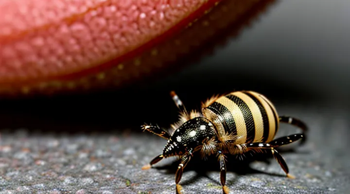
Understanding Subcutaneous Tick Infestation
What Are Subcutaneous Ticks?
Types of Ticks that Burrow
Ticks capable of penetrating beyond the epidermis and establishing a subcutaneous position are limited to a few species that exhibit aggressive attachment behavior and a propensity for prolonged feeding. Their ability to burrow under the skin increases the risk of delayed diagnosis and systemic complications.
-
Ixodes scapularis (deer tick) – prevalent in eastern North America; nymphs and adults frequently attach to the scalp or torso, sometimes embedding deeply enough to become subcutaneous. Associated with Lyme disease, anaplasmosis, and babesiosis.
-
Ixodes ricinus (castor bean tick) – common throughout Europe and parts of Asia; adult females can remain attached for up to ten days, occasionally migrating into the hypodermis. Vectors of Lyme borreliosis, tick‑borne encephalitis, and rickettsial infections.
-
Amblyomma americanum (lone‑star tick) – distributed across the southeastern United States; adults often feed on the lower extremities and may burrow into the dermis, producing a subcutaneous nodule. Transmits ehrlichiosis, Southern tick‑associated rash illness, and can induce alpha‑gal allergy.
-
Haemaphysalis longicornis (Asian long‑horned tick) – invasive in the United States and native to East Asia; larvae and nymphs attach to a wide range of hosts and have been documented forming subcutaneous granulomas. Carries severe fever with thrombocytopenia syndrome virus and various bacterial pathogens.
-
Dermacentor variabilis (American dog tick) – found throughout the United States; adult females occasionally embed deeply, especially when feeding on the groin or axilla. Transmits Rocky Mountain spotted fever and tularemia.
These species share traits that facilitate subcutaneous migration: a robust hypostome equipped with barbs, prolonged feeding periods, and a preference for concealed body sites where host grooming is limited. Early removal is often hindered by the tick’s deep placement, necessitating surgical excision or imaging‑guided extraction. Recognition of the specific tick type guides clinical management, including appropriate antimicrobial therapy and surveillance for associated vector‑borne diseases.
Common Locations for Infestation
Medical literature identifies specific body regions where subcutaneous tick attachment most frequently occurs. Ticks preferentially select sites that provide easy access to thin skin, warmth, and moisture, facilitating prolonged feeding and deeper penetration.
- Scalp and hairline, especially behind the ears
- Neck folds and posterior cervical region
- Axillary (underarm) area
- Groin and inguinal creases
- Abdomen, particularly around the waistline
- Inner thighs and popliteal fossa (behind the knee)
- Lower back and lumbar region
- Feet, especially between the toes and on the heel
These locations correspond to areas where clothing or hair may conceal the tick, reducing detection during the early feeding phase. Prompt inspection of these regions after outdoor exposure decreases the risk of unnoticed subcutaneous migration.
Initial Contact and Attachment
Preferred Host Areas
Subcutaneous ticks embed beneath the epidermis, often escaping detection during routine skin examinations. Their attachment sites reflect the parasite’s preference for regions where the skin is thin, vascularized, and protected from frequent disturbance.
- Scalp and hairline – dense hair masks the tick, while the scalp provides a warm, moist environment.
- Retro‑auricular area (behind the ears) – limited movement and thin skin facilitate insertion.
- Neck and supraclavicular region – proximity to the head and reduced friction support prolonged feeding.
- Axillary folds – humidity and skin folds create a sheltered niche.
- Inguinal and genital folds – warmth, moisture, and limited exposure reduce host grooming.
- Abdominal skin near the waistline – thin dermis and frequent contact with clothing allow concealment.
These locations collectively maximize the tick’s access to blood while minimizing the chance of removal by host grooming or clothing friction.
Mechanism of Attachment
Ticks attach to human skin through a sequence of mechanical and biochemical actions that enable the parasite to penetrate the epidermis and reach the dermal layer. The mouthparts, composed of chelicerae and a barbed hypostome, are positioned against the host surface while the tick secretes a cocktail of saliva containing enzymes and anticoagulants. These secretions soften the stratum corneum, dissolve keratin, and prevent blood clotting, facilitating deeper insertion.
The attachment process proceeds as follows:
- Questing – the tick climbs onto the host and seeks a suitable site.
- Gripping – forelegs grasp the skin, and the tick draws its body forward.
- Insertion – the hypostome pierces the epidermis, anchoring with backward‑facing barbs.
- Salivation – enzymatic saliva digests tissue and suppresses local immune responses.
- Engorgement – the tick expands its body while remaining securely attached.
The combination of physical anchoring and pharmacologically active saliva ensures a stable, subcutaneous position that can persist for days, allowing the tick to feed and potentially transmit pathogens.
The Transmission Process
Factors Influencing Transmission
Environmental Conditions
Environmental conditions determine the likelihood that a tick will embed beneath the skin of a person. Warm temperatures accelerate tick metabolism, increase questing activity, and shorten the developmental cycle, thereby raising the number of actively seeking nymphs and adults. High relative humidity prevents desiccation, allowing ticks to remain on vegetation for extended periods and to climb to the host‑seeking height.
- Temperature ≥ 10 °C (50 °F) for several consecutive days
- Relative humidity ≥ 80 % near ground level
- Dense understory or leaf litter providing microclimate stability
- Presence of suitable wildlife hosts (rodents, deer) that sustain tick populations
Seasonal patterns reflect these parameters. In temperate zones, peak subcutaneous tick infections occur during late spring and early summer when temperatures rise and vegetation is dense. In subtropical regions, activity may persist year‑round, with a secondary peak during the rainy season when humidity spikes.
Human exposure correlates with outdoor activities that place individuals in tick‑infested habitats. Hiking, gardening, or working in fields during peak questing periods increases contact risk. Wearing protective clothing, using repellents, and limiting time in high‑risk microhabitats reduce the probability of subcutaneous tick entry.
Human Behavior
Human actions directly affect the likelihood of a tick embedding beneath the skin. Outdoor recreation in wooded or grassy areas increases exposure because ticks quest on vegetation awaiting a host. Wearing short sleeves, shorts, or sandals reduces the barrier between skin and questing ticks, facilitating penetration. Contact with domestic animals that roam in tick‑infested habitats can transfer ticks to human skin during petting or grooming. Neglecting regular body inspections after exposure allows attached ticks to remain undetected, giving them time to embed subcutaneously. Lack of prompt removal, especially when the tick’s mouthparts remain attached, can promote deeper tissue invasion.
Preventive behaviors reduce subcutaneous entry:
- Dress in long sleeves, pants, and closed shoes; tuck clothing into socks.
- Apply approved repellents containing DEET, picaridin, or permethrin to skin and clothing.
- Perform thorough tick checks within 30 minutes of leaving high‑risk environments; use a mirror for hard‑to‑see areas.
- Shower promptly after outdoor activity to dislodge unattached ticks.
- Inspect and groom pets regularly; treat them with veterinarian‑recommended acaricides.
- Remove attached ticks with fine‑pointed tweezers, grasping close to the skin and pulling straight upward without crushing the body.
These behaviors shape the pathway by which ticks can breach the epidermis and establish a subcutaneous position in humans.
Stages of Subcutaneous Migration
Entry Point and Initial Burrowing
Ticks attach to the host’s skin by locating a suitable site—often a thin‑skinned area such as the scalp, armpit, or groin. The tick’s forelegs grasp the epidermis, and its chelicerae pierce the outer layer, creating a pinpoint opening. Salivary secretions containing anesthetic and anticoagulant compounds facilitate painless penetration and prevent clot formation at the entry site.
Once the opening is established, the tick inserts its hypostome, a barbed feeding organ, into the dermis. The barbs anchor the tick, while a cement‑like substance secreted from the salivary glands hardens around the mouthparts, securing a stable attachment. The hypostome then advances beneath the epidermis, forming a tunnel that extends into the subcutaneous tissue. This initial burrowing creates a concealed feeding channel, allowing the tick to remain attached for days while it ingests blood and potentially transmits pathogens.
Progression Through Dermal Layers
Ticks attach to the host’s skin and insert their hypostome into the superficial epidermis. The mouthparts, equipped with backward‑directed barbs, anchor the tick while cutting through the stratum corneum and viable epidermal cells. Salivary secretions containing anticoagulants and immunomodulators facilitate tissue dissolution and suppress local inflammation.
Once the hypostome breaches the epidermis, it progresses into the papillary dermis. In this layer, the tick’s chelicerae separate collagen fibers, creating a channel that extends toward the deeper reticular dermis. Mechanical pressure and enzymatic activity allow the feeding apparatus to reach the subcutaneous fat layer, where blood vessels are more accessible. The tick remains anchored by the barbs, preventing dislodgement during prolonged feeding.
The final stage places the tick’s feeding tube within the subcutaneous tissue, directly adjacent to capillaries. This position maximizes blood intake and creates an efficient route for pathogen transmission. Key points of progression:
- Epidermis: penetration of stratum corneum and viable layers.
- Papillary dermis: separation of collagen, formation of feeding channel.
- Reticular dermis: deeper advancement toward vascular structures.
- Subcutaneous fat: proximity to capillaries, optimal blood acquisition.
The depth of insertion correlates with the likelihood of transmitting bacteria, viruses, or protozoa, as the tick’s saliva is deposited close to the bloodstream. Early removal before the hypostome reaches the subcutaneous layer reduces the risk of infection.
Symptoms and Diagnosis
Early Signs of Infestation
Early signs of a subcutaneous tick infestation appear shortly after the arthropod penetrates the dermis. The bite site typically presents as a small, raised papule or nodule that may be tender to pressure. Redness often surrounds the lesion, and a faint halo can develop as the tick’s mouthparts embed deeper.
Additional indicators include:
- Persistent itching or localized burning sensation
- Slight swelling that does not resolve within 24‑48 hours
- Visible movement or a palpable lump beneath the skin surface
- Low‑grade fever, headache, or malaise in the absence of other infection sources
Recognition of these symptoms enables prompt removal of the tick and reduces the risk of pathogen transmission associated with subcutaneous attachment.
Diagnostic Methods
Subcutaneous tick infestations in humans often present as a painless, firm nodule beneath the skin. Early detection relies on systematic assessment and laboratory confirmation.
Physical inspection identifies a localized swelling; palpation may reveal a movable, slightly raised mass. Dermatoscopy enhances visualization, allowing clinicians to observe the tick’s mouthparts or engorged body through the epidermal layer.
Imaging techniques provide deeper insight:
- High‑frequency ultrasound detects hypoechoic structures consistent with a tick, delineates depth, and guides removal.
- Magnetic resonance imaging distinguishes soft‑tissue lesions from cysts or tumors when ultrasound results are inconclusive.
Laboratory analysis confirms tick species and potential pathogen carriage:
- Polymerase chain reaction (PCR) amplifies tick‑specific DNA from tissue or blood samples, enabling species identification.
- Serologic tests detect antibodies against common tick‑borne agents (e.g., Borrelia, Anaplasma) to assess co‑infection risk.
- Histopathologic examination of excised tissue reveals inflammatory response and may capture residual tick fragments.
Combining visual assessment, imaging, and molecular testing yields accurate diagnosis and informs appropriate therapeutic measures.
Prevention and Control
Personal Protective Measures
Repellents and Protective Clothing
Repellents applied to skin or clothing create a chemical barrier that deters ticks from attaching. Effective agents include DEET (20‑30 % concentration for moderate duration, 30‑50 % for extended protection), picaridin (20 % for up to 8 hours), and IR3535 (10‑20 %). Permethrin, applied to fabric at 0.5 % concentration, remains active after several washes and kills ticks that contact treated material. Reapplication is required after swimming, heavy sweating, or after the labeled exposure time. Apply repellents evenly to exposed areas, avoiding eyes and mucous membranes, and allow the product to dry before dressing.
Protective clothing reduces the surface area available for tick attachment. Recommended items:
- Long‑sleeved shirts and full‑length trousers, preferably made of tightly woven fabric.
- Pants tucked into socks or boots to eliminate gaps.
- Light‑colored garments to facilitate visual detection of attached ticks.
- Clothing pre‑treated with permethrin or treated on‑site with a spray kit following manufacturer instructions.
Combining a high‑efficacy repellent with permethrin‑treated attire provides layered defense, markedly lowering the risk of subcutaneous tick penetration.
Post-Exposure Checks
After a tick attaches beneath the skin, the bite site must be examined promptly. Visual inspection should include the entire body, focusing on concealed areas such as scalp, behind ears, armpits, groin and between toes. If a tick is found, grasp it with fine‑point tweezers as close to the skin as possible, pull upward with steady pressure, and avoid crushing the body. Preserve the specimen in a sealed container for identification if disease testing is required.
Following removal, perform a series of post‑exposure checks:
- Record the date and estimated duration of attachment; exposure risk rises sharply after 24 hours of feeding.
- Observe the bite area for erythema, expanding rash, or a target‑shaped lesion (erythema migrans).
- Monitor systemic signs such as fever, headache, myalgia, fatigue or joint pain for up to 30 days.
- Document any known tick‑borne pathogens prevalent in the region (e.g., Borrelia burgdorferi, Anaplasma phagocytophilum, Rickettsia spp.) and consider prophylactic antibiotics when indicated by local guidelines.
- Seek medical evaluation if the tick was engorged, removal was incomplete, or if symptoms develop.
Regular self‑examination for at least two weeks after exposure enhances early detection of transmitted infections. Timely reporting to a healthcare professional facilitates appropriate laboratory testing and treatment, reducing the likelihood of severe complications.
Environmental Management
Habitat Modification
Habitat modification directly influences the likelihood of subcutaneous tick exposure in people by altering the environmental conditions that support tick development and host activity. Reducing dense underbrush, maintaining short grass, and removing leaf litter diminish the microclimate preferred by nymphs and larvae, limiting their capacity to attach to humans.
Effective measures include:
- Trimming vegetation to create open, sun‑exposed areas that lower humidity levels essential for tick survival.
- Installing physical barriers such as wood chips or gravel around residential zones to discourage wildlife movement into yards.
- Managing wildlife populations through targeted exclusion techniques, reducing the availability of primary blood‑meal hosts.
- Applying acaricidal treatments to perimeter zones where complete vegetation removal is impractical.
Implementing these strategies consistently lowers tick density on the ground surface, thereby decreasing the probability that a tick will penetrate the skin and migrate subcutaneously. Monitoring tick counts before and after habitat changes provides quantitative evidence of intervention success and guides further adjustments.
Pest Control Strategies
Ticks embed their mouthparts into the skin while feeding, allowing saliva and pathogens to enter the subcutaneous tissue. Transmission occurs when a tick remains attached for several hours, during which it secretes anticoagulants and infectious agents directly into the host’s bloodstream.
Effective pest control strategies reduce the likelihood of such contact:
- Habitat modification: Trim vegetation, remove leaf litter, and maintain a clear perimeter around residential areas to decrease tick habitats.
- Chemical barriers: Apply acaricides to lawns and high‑risk zones following label instructions; reapply at recommended intervals.
- Personal protection: Wear long sleeves and trousers, treat clothing with permethrin, and use EPA‑registered repellents containing DEET or picaridin on exposed skin.
- Host management: Treat domestic animals with tick‑preventive products; control wildlife access to yards by installing fencing or using repellents.
- Biological agents: Introduce entomopathogenic fungi (e.g., Metarhizium anisopliae) or nematodes that target tick larvae and nymphs.
- Integrated approach: Combine environmental, chemical, and biological measures, monitor tick populations regularly, and adjust tactics based on surveillance data.
