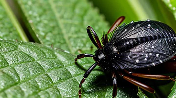
Understanding Tick Respiration: An Overview
The Importance of Respiration in Ticks
Respiration supplies the oxygen required for cellular processes that sustain tick activity, growth, and reproduction. Oxygen supports ATP production, which powers muscle contraction during host seeking, attachment, and blood‑feeding. Efficient energy metabolism enables rapid engorgement and subsequent egg development, directly influencing population dynamics.
Metabolic waste removal depends on the same respiratory pathway. Carbon dioxide generated during glycolysis diffuses out of the body, preventing acid‑base imbalance that could impair neural signaling and muscular coordination. Maintaining internal pH is essential for the proper functioning of sensory organs that detect host cues.
The respiratory system also influences pathogen transmission. Many tick‑borne microorganisms rely on the vector’s metabolic state for replication. Adequate oxygen levels promote bacterial proliferation within the tick, increasing the likelihood of pathogen transfer to vertebrate hosts during feeding.
Key aspects of tick respiration include:
- A single spiracular opening on each side of the body, leading to a tracheal network that penetrates the cuticle.
- Tracheal tubes lined with a cuticular sheath, providing a rigid conduit for gas diffusion.
- Absence of a dedicated respiratory pump; diffusion across the tracheal walls drives gas exchange, sufficient for the tick’s low metabolic rate.
- Adaptations for prolonged fasting periods, allowing survival without active gas exchange for months.
Overall, respiration underpins energy production, waste elimination, and vector competence, establishing it as a fundamental physiological process for tick survival and disease ecology.
General Principles of Arthropod Respiration
Arthropod respiration relies on an external air‑filled network that delivers oxygen directly to tissues. The system is open, with the external environment connected to internal tubes through cuticular openings called spiracles. Air enters the tracheal trunks, which branch into progressively finer tracheae, ending in terminal tracheoles that surround individual cells. Gas exchange occurs by simple diffusion across the thin tracheolar walls; no circulatory transport of oxygen is required. In many terrestrial arthropods, spiracles can be opened or closed to limit water loss, while some aquatic forms possess gill structures that extract dissolved oxygen.
Key structural elements include:
- «Spiracles»: cuticular pores regulated by muscular or valve mechanisms.
- «Tracheae»: thick‑walled tubes reinforced with cuticle, providing structural support.
- «Tracheoles»: fine, flexible extensions that increase surface area for diffusion.
- Hemolymph: serves primarily for nutrient and waste transport; does not carry respiratory pigments in most species.
- Ventilation: passive diffusion dominates; active ventilation via body movements or muscular pumping appears in larger or more active taxa.
Ticks exemplify a simplified version of this design. They possess a single pair of spiracles located laterally on the dorsal surface. The spiracles open into a reduced tracheal system that supplies the body cavity and salivary glands. Absence of book lungs or gills reflects adaptation to a terrestrial parasitic lifestyle. Limited tracheal branching constrains metabolic rate, aligning with the tick’s slow, intermittent activity. The reliance on diffusion through a minimal network explains the tick’s tolerance for prolonged periods of low oxygen availability while attached to a host.
The Tick Respiratory System
Unique Adaptations for Gas Exchange
Spiracles: The External Openings
Ticks respire through a tracheal system that opens to the external environment via spiracles. These structures serve as the sole portals for gas exchange between the arthropod’s internal tissues and atmospheric oxygen.
Spiracles are situated on the dorsal surface of the idiosoma, typically forming a bilateral pair near the coxae of the fourth pair of legs. Their placement allows direct access to the surrounding air while minimizing exposure of the ventral regions that contact the host.
The external openings consist of cuticular sclerites that form a protective rim. Each spiracle possesses a muscular valve capable of closing the aperture. Valve closure reduces water loss during prolonged periods of desiccation or when the tick is attached to a host with limited airflow.
Key characteristics of tick spiracles:
- Paired arrangement on each side of the body
- Position adjacent to the fourth leg coxae
- Cuticular rim providing structural support
- Muscular valve enabling reversible closure
- Small diameter that limits evaporative loss
Through these features, spiracles maintain a balance between efficient oxygen uptake and preservation of internal moisture, supporting the tick’s survival in diverse microhabitats.
Tracheal System: Internal Air Delivery
Ticks possess a closed tracheal network that transports atmospheric gases directly to internal tissues. Air enters through a pair of dorsal spiracles, each linked to a primary tracheal trunk. The trunks branch into progressively finer tracheae, terminating in tracheoles that permeate the hemocoel and contact individual cells.
Key characteristics of the internal air‑delivery system include:
- Spiracular valves that open only when the tick is active, reducing desiccation risk.
- Rigid cuticular lining of the tracheae, preventing collapse under external pressure.
- Absence of a diaphragm; gas flow relies on passive diffusion driven by concentration gradients.
- Extensive tracheolar arborization within the opisthosoma, ensuring oxygen supply to the digestive and reproductive organs.
The tracheal architecture enables ticks to maintain metabolic function during prolonged periods of host attachment. Gas exchange occurs without a circulatory lung, allowing efficient oxygen delivery even at low ambient humidity. The system’s simplicity supports the arthropod’s ability to survive in diverse microhabitats, from leaf litter to mammalian skin.
Main Trunks and Branches
Ticks respire through a system of tracheae that begins with two principal trunks extending from the posterior end of the body. Each trunk runs dorsally toward the anterior region, where it bifurcates into a network of finer tubes delivering oxygen directly to tissues.
- Primary trunks: paired, longitudinal, anchored to the exoskeleton, provide the main conduit for gas exchange.
- Major branches: lateral extensions from each trunk, penetrate the cuticle of the legs, mouthparts, and genital capsule.
- Secondary branches: subdivide from the major branches, form a dense mesh surrounding muscle fibers and internal organs.
- Terminal tracheoles: microscopic tubes ending in the hemocoel, facilitate diffusion of oxygen into the hemolymph and removal of carbon dioxide.
The arrangement ensures rapid distribution of gases despite the tick’s low metabolic rate. The trunks remain open to the external environment through spiracular openings located on the ventral surface of each segment, allowing continuous airflow without the need for a circulatory pump. This architecture reflects an adaptation to the arthropod’s parasitic lifestyle, where efficient gas exchange must be maintained while the organism remains attached to a host for extended periods.
Tracheoles: Sites of Gas Exchange
Tracheoles constitute the terminal elements of the tick respiratory network, directly interfacing with body tissues to facilitate oxygen uptake and carbon‑dioxide release. Each tracheole forms a thin, flexible tube whose walls are permeable to gases, allowing diffusion across a minimal diffusion distance. The extensive branching of tracheoles creates a dense mesh that reaches virtually every cell, ensuring that metabolic demands are met even in the arthropod’s relatively low‑oxygen environment.
Key characteristics of tick tracheoles include:
- Diameter on the order of 0.5–1 µm, optimizing surface‑to‑volume ratio for efficient diffusion.
- Cuticular lining reinforced with chitin, providing structural stability while remaining sufficiently thin for gas exchange.
- Absence of active ventilation mechanisms; respiratory flow relies on passive diffusion driven by concentration gradients between hemolymph and ambient air.
- Integration with the peritrophic membrane in the midgut, permitting direct gas exchange with digestive tissues.
- Connection to larger tracheae via spiral valves that regulate the direction of airflow and prevent back‑flow.
The arrangement of tracheoles in the integument and internal organ systems reflects a design that maximizes respiratory efficiency without the need for muscular pumping, a feature that distinguishes ticks from many other arthropods.
Mechanisms of Gas Exchange
Passive Diffusion
Ticks respire through a minimalist system that lacks a tracheal network. Gas exchange occurs across the body surface and a pair of openings called spiracles. The process depends entirely on concentration gradients, without active pumping mechanisms.
Key aspects of the diffusion‑based respiratory system:
- Spiracles open directly to the external environment, providing a pathway for oxygen entry and carbon‑dioxide exit.
- Thin cuticle layers adjacent to the hemolymph allow gases to pass by «passive diffusion».
- Hemolymph circulation distributes dissolved oxygen throughout internal tissues, while metabolic activity maintains the gradient that drives diffusion.
- Absence of muscular ventilation means that respiratory efficiency is limited by surface area and the size of the openings, which is sufficient for the low metabolic demands of ticks.
The reliance on «passive diffusion» defines the physiological constraints of tick respiration, shaping their behavior and habitat preferences.
Active Ventilation (if applicable)
Ticks respire through a system of spiracles and tracheae that functions primarily by diffusion. In most species the respiratory apparatus lacks musculature capable of generating a pressure gradient, so active ventilation is generally absent. When active ventilation occurs, it involves rhythmic contraction of body walls that forces air through the tracheal network, a mechanism documented in certain hard‑tick (Ixodidae) stages adapted for rapid metabolism during blood feeding.
Key characteristics of active ventilation in ticks:
- Limited to engorged females of large ixodid species; nymphs and males rely exclusively on passive diffusion.
- Initiated by coordinated expansion and compression of the opisthosomal cuticle, creating transient pressure changes that move air toward the ventral spiracles.
- Provides temporary augmentation of oxygen delivery to support heightened metabolic demands during blood meal digestion.
- Ceases once the tick returns to a quiescent state, reverting to the default diffusive mode.
Overall, the respiratory system of ticks is optimized for low‑energy gas exchange, with active ventilation representing a conditional, short‑term adaptation rather than a constitutive feature.
Environmental Influences on Respiration
Humidity and Oxygen Levels
Ticks respire through a pair of spiracles that open to a simple tracheal network. Gas exchange occurs by diffusion across the cuticle and through the tracheae, without a dedicated circulatory transport system. Ambient conditions directly modulate this process.
High humidity maintains cuticular moisture, preventing spiracle closure caused by dehydration. When relative humidity exceeds 80 %, spiracles remain open, allowing continuous oxygen uptake and carbon‑dioxide release. Below 60 %, cuticular water loss accelerates, spiracles constrict, and diffusion rates decline sharply.
Oxygen concentration in the surrounding air dictates metabolic activity. At atmospheric levels (~21 % O₂), tracheal diffusion sustains normal locomotion and blood‑feeding. Reduced O₂ (≤10 %) forces a metabolic slowdown; ticks remain viable by limiting movement and extending feeding intervals. Elevated O₂ does not increase respiratory capacity beyond the fixed spiracle aperture, but it supports higher activity levels when water balance permits.
Key adaptive responses:
- Spiracle regulation according to humidity gradients.
- Metabolic rate adjustment in hypoxic environments.
- Prolonged feeding periods under combined low‑humidity and low‑oxygen stress.
Impact of Temperature
Ticks respire through a pair of spiracles that open into a simple tracheal network. The tracheae deliver atmospheric oxygen directly to internal tissues, while carbon dioxide diffuses outward through the same openings. This system functions efficiently under the low‑oxygen conditions typical of the microhabitats ticks occupy.
Temperature exerts a direct influence on the respiratory apparatus. Elevated ambient heat raises metabolic demand, which in turn increases the rate of tracheal ventilation. Conversely, low temperatures depress metabolic activity, reducing the frequency of spiracular opening and the volume of gas exchange. The relationship can be summarized:
- Above 30 °C – metabolic rate peaks; spiracles remain open longer; risk of desiccation rises.
- 15–30 °C – optimal balance between oxygen supply and water loss; tracheal flow sufficient for normal activity.
- Below 15 °C – metabolic processes slow; spiracles close more rapidly; activity limited to brief periods.
Temperature‑dependent changes also affect developmental stages. Larvae and nymphs, possessing thinner cuticles, experience greater respiratory water loss at higher temperatures than adults. Adult females, especially when engorged, exhibit reduced tracheal efficiency due to increased body mass, making them more sensitive to thermal stress.
These physiological responses shape tick distribution. Regions with stable, moderate temperatures support continuous activity, while areas with extreme heat or cold impose seasonal constraints on population growth. Understanding the thermal thresholds of tick respiration informs predictive models of disease risk and guides timing of control measures.
Evolutionary Aspects and Survival Strategies
Comparison with Other Arachnids
Ticks respire through a pair of spiracles located on the ventral surface of the idiosoma, each opening into a simple tracheal tube that terminates in a network of fine tracheae delivering oxygen directly to tissues. This system lacks the extensive tracheal branching seen in many other arachnids.
- Spiders possess multiple book lungs—paired, lamellate respiratory organs situated within the abdomen. Air enters through external openings called slit pores, then diffuses across the lamellae before reaching the hemolymph. Some spider families supplement book lungs with a tracheal system, creating a dual respiratory arrangement.
- Scorpions rely exclusively on book lungs, typically two pairs, each consisting of a series of thin, stacked plates that increase surface area for gas exchange. The openings are situated on the ventral side of the opisthosoma, and ventilation occurs via rhythmic movements of the plates.
- Harvestmen (Opiliones) feature a primitive tracheal system comprising a few large, unbranched tubes that open through a single pair of spiracles near the anterior body region. Their tracheae are less extensive than those of spiders that possess both tracheae and book lungs.
- Mites, closely related to ticks, exhibit varied respiratory adaptations: many have reduced or absent tracheae, relying on cutaneous diffusion across the integument, while some retain simple spiracular openings similar to ticks.
Key distinctions include the number and type of respiratory openings, the presence or absence of book lungs, and the degree of tracheal branching. Ticks maintain a minimalistic tracheal layout, whereas other arachnids employ more complex structures to meet higher metabolic demands.
Survival in Diverse Environments
Submersion Tolerance
Ticks respire through a pair of posterior spiracles that open into a simple tracheal system. The tracheae branch minimally, delivering atmospheric oxygen directly to internal tissues. Cuticular respiration contributes a small proportion of gas exchange, especially when spiracles are closed.
Submersion tolerance depends on the capacity to limit water entry through spiracles and to extract dissolved oxygen across the cuticle. When immersed, spiracles close reflexively, preventing flooding of the tracheal lumen. Cuticular diffusion supplies enough oxygen to sustain metabolic processes for limited periods.
Key factors influencing underwater survival:
- Developmental stage: larvae and nymphs exhibit longer tolerance than adults due to lower metabolic demand.
- Ambient oxygen concentration: higher dissolved oxygen extends survival time.
- Temperature: lower temperatures reduce metabolic rate, increasing tolerance.
- Duration of spiracle closure: prolonged closure forces reliance on cuticular diffusion, shortening viable immersion time.
Typical immersion intervals range from a few minutes in warm, low‑oxygen water to several hours in cool, oxygen‑rich environments. Prolonged submersion leads to hypoxia, loss of motor function, and eventual mortality.
Desiccation Resistance
Ticks possess a respiratory system that must function while minimizing water loss, a prerequisite for effective «desiccation resistance». Gas exchange occurs through a pair of posterior spiracles that lead to a short tracheal network reaching the body’s interior. The spiracles are equipped with flexible valves that close rapidly when ambient humidity drops, limiting evaporative loss without interrupting oxygen uptake.
Cuticular modifications reinforce the barrier against dehydration. The exoskeleton surrounding the spiracular openings is coated with a waxy layer that reduces transepidermal diffusion of water molecules. Internal tracheal walls contain a thin film of lipid‑rich secretion that further impedes moisture escape.
Key physiological features that support survival in dry environments include:
- Rapid spiracle closure triggered by humidity sensors.
- Waxy cuticle on spiracle margins and adjacent integument.
- Tracheal lining enriched with hygroscopic proteins that retain water.
- Behavioral positioning in microhabitats with higher relative humidity during periods of low ambient moisture.
Collectively, these adaptations allow ticks to maintain respiration while preserving internal water balance, ensuring activity across a broad range of terrestrial habitats.
Future Research and Implications
Future investigations should prioritize molecular characterization of the tracheal system and associated ion channels. Advanced imaging techniques, such as micro‑CT and confocal microscopy, can resolve three‑dimensional architecture of the spiracular openings and internal air‑filled spaces, enabling precise correlation between structural variation and environmental tolerance.
Key research avenues include:
- Genomic screening for genes regulating cuticular permeability and spiracle regulation;
- Physiological assays measuring gas exchange rates under controlled humidity and temperature gradients;
- Comparative studies across acariform and ixodid lineages to identify evolutionary adaptations;
- Development of computational models integrating diffusion dynamics with metabolic demand during blood‑feeding cycles.
Implications extend to public health and pest management. Clarifying respiratory constraints may inform climate‑based risk assessments for tick‑borne disease transmission. Targeted disruption of respiratory pathways offers a potential avenue for novel acaricidal strategies, reducing reliance on broad‑spectrum chemicals.
