Recognizing a Tick Bite: Immediate Signs and Symptoms
Visual Confirmation of a Tick
Locating the Tick
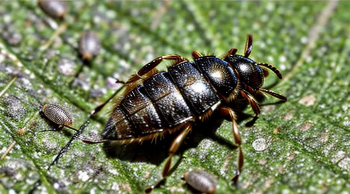
After a possible attachment, the first step is to examine the skin for the arthropod itself. Look for a small, rounded object ranging from 2 mm to 1 cm, depending on its stage of development. Ticks often appear as a dark or brown lump, sometimes resembling a tiny button.
- Inspect the entire body, paying special attention to hidden areas: scalp, behind ears, underarms, groin, and behind knees.
- Use a magnifying glass or a smartphone camera with zoom to enhance visibility.
- Run fingers gently over the skin; a live tick may feel like a firm bump, while a detached one may feel softer.
- If the animal is embedded, you may see a tiny protruding mouthpart (the hypostome) at the center of the body.
When the tick is not immediately visible, shave or trim hair in the suspect region, then repeat the visual inspection. If a bite site shows a red, expanding rash or a small puncture surrounded by a clear halo, it often indicates the tick’s feeding location even if the creature is no longer attached. In such cases, document the area and seek medical advice promptly.
Identifying the Tick Species
Identifying the tick species that has attached to you provides essential information for assessing disease risk and guiding appropriate medical response. Different species transmit distinct pathogens; recognizing the culprit enables targeted advice from health professionals.
Ticks can be distinguished by size, coloration, body shape, and the presence or absence of specific markings. Observe the engorgement level, the scutum (hard shield) on the dorsal surface, and the pattern of festoons (small rectangular areas) along the margin of the abdomen. Note the number of eyes on the anterior surface and the shape of the mouthparts.
- Ixodes scapularis (black‑legged or deer tick) – small, dark brown, with a distinctive black shield on the dorsal side; legs appear black; often found in wooded, humid regions.
- Dermacentor variabilis (American dog tick) – larger, reddish‑brown with white speckling on the scutum; legs are pale; commonly encountered in grassy fields.
- Amblyomma americanum (lone star tick) – medium size, reddish‑brown body, single white spot on the dorsal scutum of adult females; legs are striped; prevalent in southeastern United States.
- Rhipicephalus sanguineus (brown dog tick) – uniformly brown, oval scutum without distinct markings; legs match body color; thrives in indoor environments and warm climates.
After species determination, compare the known pathogen profile of that tick with your geographic exposure. If the identified species is a known vector for Lyme disease, Rocky Mountain spotted fever, or ehrlichiosis, seek medical evaluation promptly. Preserve the tick in a sealed container for laboratory confirmation if recommended by a clinician.
Size and Appearance of Ticks
Ticks vary markedly in size and visual characteristics, which are essential for recognizing an attachment. Unfed larvae measure 0.5–1 mm, appear translucent, and lack visible legs. Nymphs are 1.5–2 mm, dark brown to reddish, with six legs and a flattened body. Adult females range from 3–5 mm when unfed, expanding to 10–12 mm after a blood meal; they are brown to gray, with a rounded, engorged abdomen. Adult males remain 2–3 mm, stay relatively flat, and do not swell significantly after feeding.
Key visual indicators of a recent bite include:
- A small, dark spot at the bite site, often surrounded by a red halo.
- Presence of a tick body attached to the skin, typically near the scalp, armpits, groin, or waistline.
- An engorged tick that appears larger than the surrounding skin structures, with a distended abdomen.
- A visible mouthpart (hypostome) protruding from the skin, creating a pinprick sensation.
Detecting these size and appearance cues enables prompt removal and reduces the risk of disease transmission.
Physical Sensations and Skin Reactions
Itching and Irritation
Itching and irritation often appear soon after a tick attaches to the skin. The bite site may feel a mild to intense pruritus that intensifies when the tick is disturbed or when the host’s immune response activates.
Typical characteristics of the reaction include:
- A small, red papule surrounding the embedded tick.
- Localized swelling that can become raised or form a halo.
- A burning sensation that may persist for several hours.
- Secondary scratching that can lead to excoriation or secondary infection.
If the irritation spreads beyond the immediate area, forming a rash or a concentric ring (often called a target lesion), it may indicate pathogen transmission and warrants medical evaluation. Continuous or worsening symptoms, such as fever, fatigue, or joint pain, should also prompt professional assessment.
Rash Characteristics
A tick bite often manifests as a skin reaction that differs from ordinary insect bites. The most common sign is a small, red bump at the attachment site, typically 2–5 mm in diameter. This lesion may develop a dark center—known as a “target” or “bullseye” pattern—where the outer ring is reddish and the inner area is darker or purplish. The center can be a raised, raised, or flat spot, sometimes resembling a tiny vesicle.
Additional rash features include:
- A gradual increase in size over 24–48 hours.
- Persistent itching or mild burning sensation.
- Presence of a tiny, engorged tick still attached or a visible puncture mark after removal.
- Development of a secondary rash away from the bite, such as a diffuse erythematous patch, which may indicate early infection.
When the rash spreads, forms multiple lesions, or is accompanied by fever, headache, or joint pain, medical evaluation is warranted promptly.
Swelling and Redness
Swelling and redness are the most immediate visual cues that a tick has attached to the skin. The reaction typically appears within hours of the bite and may evolve over several days.
- Localized swelling forms a raised area around the attachment site; size can range from a few millimeters to several centimeters depending on individual sensitivity.
- Redness surrounds the swollen region, often presenting as a uniform halo. In some cases, the border may be sharply defined, resembling a target pattern.
- The skin may feel warm to the touch, indicating an inflammatory response.
- Accompanying itching or mild pain is common, but absence of discomfort does not rule out a bite.
Persistent or expanding redness, especially if it develops a central clearing (bullseye), warrants medical evaluation for potential infection such as Lyme disease. Early detection relies on prompt visual inspection of the affected area and recognition of these characteristic changes.
What to Do After a Tick Bite
Safe Tick Removal Techniques
Tools for Removal
When a tick attaches, the skin may show a small, often unnoticed puncture. Prompt removal reduces the risk of disease transmission, making the choice of proper instruments critical.
Effective removal tools include:
- Fine‑point tweezers with a flat, serrated tip: grip the tick close to the skin without crushing its body.
- Tick removal hooks (e.g., the “tick key”): slide under the mouthparts and lift straight upward.
- Small, curved forceps designed for veterinary use: allow precise control in tight areas.
- Commercial tick removal devices that combine a hook and a grip surface: provide a single‑step extraction method.
Supplementary items that assist the process:
- Disposable gloves: prevent direct contact with the tick’s saliva.
- Antiseptic wipes or alcohol pads: clean the bite site before and after removal.
- A sealed container or zip‑lock bag: store the extracted tick for identification if symptoms develop.
Technique matters as much as the instrument. Position the tool to grasp the tick’s head, apply steady upward pressure, and avoid twisting. After extraction, wash the area with soap and water, then monitor for redness, swelling, or a rash over the next several weeks. If any signs appear, seek medical evaluation promptly.
Step-by-Step Removal Process
Detecting a recent tick attachment involves observing a small, often unnoticed bump, a red halo, or a raised area where the parasite’s mouthparts remain embedded. Prompt removal reduces the risk of pathogen transmission.
- Gather fine‑point tweezers, alcohol swabs, and a clean container.
- Grasp the tick as close to the skin as possible, holding the head and mouthparts, not the body.
- Apply steady, upward pressure; pull straight without twisting or squeezing the abdomen.
- Release the tick into the container; submerge in alcohol or seal for disposal.
- Clean the bite site with an alcohol swab or antiseptic.
- Inspect the removed tick; ensure no parts remain attached. If fragments are visible, repeat the extraction process.
After removal, monitor the area for redness, swelling, or a rash over the next weeks. Record the date of the bite and any emerging symptoms; report persistent changes to a healthcare professional. This systematic approach minimizes infection risk and facilitates early detection of tick‑borne illness.
Post-Removal Care
After extracting a tick, the primary concern is preventing infection and recognizing any delayed reaction. Immediate cleaning, vigilant observation, and timely medical consultation form the core of effective post‑removal management.
- Wash the bite site with soap and water; apply an antiseptic such as povidone‑iodine or alcohol.
- Pat the area dry and cover with a sterile bandage only if the skin is broken or irritated.
- Record the removal date, tick size, and appearance; retain the specimen for identification if symptoms develop.
- Inspect the site twice daily for redness, swelling, a rash, or a expanding lesion resembling a bullseye.
- Note systemic signs—fever, headache, muscle aches, fatigue—within the next 30 days.
- Contact a healthcare professional promptly if any of the above symptoms appear, or if the tick was attached for more than 24 hours.
Maintain a log of observations for at least four weeks. Early detection of an emerging infection allows prompt treatment and reduces the risk of complications.
When to Seek Medical Attention
Persistent Symptoms
Ticks often go unnoticed during attachment, and the most reliable indication of a bite can be symptoms that persist after the insect is removed. Persistent manifestations usually develop days to weeks after exposure and may signal infection with a tick‑borne pathogen.
- Erythema migrans: expanding red rash, often oval, with central clearing, appearing 3‑30 days post‑bite.
- Fever, chills, headache, and muscle aches that last longer than a typical viral illness.
- Joint pain or swelling, especially in knees or elbows, that does not resolve within a few weeks.
- Neurological signs such as facial palsy, numbness, or tingling that endure beyond the acute phase.
- Fatigue and cognitive difficulties that persist for weeks or months.
These symptoms differ from the brief irritation caused by a bite site reaction. If any of them appear, medical evaluation is warranted promptly; laboratory testing can confirm infection and guide antibiotic therapy. Early treatment reduces the risk of long‑term complications, while delayed care may lead to chronic arthritis, persistent neurological deficits, or ongoing systemic fatigue.
Signs of Infection
A tick bite may go unnoticed, yet the body can reveal an infection through specific symptoms. Early local reactions often appear within hours to days and include a red, expanding area around the bite site, sometimes forming a target‑shaped rash. Persistent swelling or warmth at the attachment point suggests bacterial involvement.
Systemic indicators develop later and can signal more serious disease. Common signs are:
- Fever or chills without an obvious cause
- Headache, neck stiffness, or facial palsy
- Joint pain, particularly in the knees or elbows, accompanied by swelling
- Fatigue and malaise lasting several weeks
A characteristic rash known as erythema migrans typically emerges 3–30 days after exposure. It begins as a small red spot and enlarges to a diameter of 5 cm or more, often with a central clearing. Presence of this lesion strongly points to a Borrelia infection.
Laboratory confirmation may be required when symptoms are ambiguous. Elevated inflammatory markers, positive serology for tick‑borne pathogens, or a PCR test from blood or tissue can substantiate the diagnosis.
Prompt medical evaluation is essential once any of these signs appear. Early antibiotic therapy reduces the risk of chronic complications, including persistent arthritis, neurological deficits, and cardiac involvement. Monitoring the bite area and overall health for at least four weeks after removal helps ensure timely detection of infection.
Symptoms of Tick-Borne Diseases
A tick bite often goes unnoticed because the insect may detach before the host feels it. Early signs of a bite include a small, painless puncture site that may develop a red ring or a slight swelling. When a pathogen is transmitted, specific clinical manifestations emerge, varying with the disease agent.
Common symptoms indicating infection by tick‑borne pathogens:
- Fever: sudden onset, sometimes accompanied by chills.
- Headache: persistent, may be severe.
- Muscle or joint aches: especially in the neck, shoulders, or knees.
- Fatigue: pronounced tiredness not relieved by rest.
- Rash:
- Expanding erythema with a clear center (“bull’s‑eye”) suggests Lyme disease.
- Small red spots (petechiae) or maculopapular eruptions may appear with Rocky Mountain spotted fever.
- Neurological signs: facial palsy, meningitis‑like symptoms, or numbness.
- Cardiac involvement: palpitations, heart block, or chest discomfort in advanced Lyme disease.
- Gastrointestinal upset: nausea, vomiting, or abdominal pain, occasionally linked to ehrlichiosis or anaplasmosis.
If any of these manifestations develop within days to weeks after potential exposure, medical evaluation is warranted. Prompt laboratory testing and, when indicated, antimicrobial therapy reduce the risk of severe complications.
