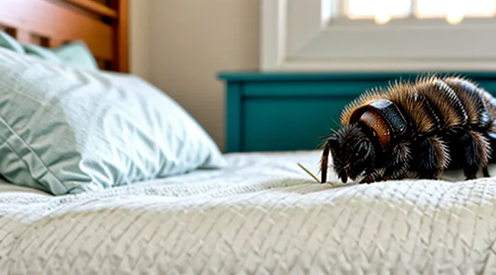
What Are Bed Mites?
Microscopic Appearance
Bed mites are microscopic arachnids measuring 0.2–0.4 mm in length. Under high‑magnification they appear oval, translucent to reddish, with a smooth dorsal surface. The body consists of a gnathosomal capsule housing the feeding apparatus, and an idiosoma bearing eight short legs arranged in four pairs. Each leg ends in a claw and a set of sensory hairs. The eyes are absent; sensory structures are limited to tactile setae on the dorsal and ventral plates. The abdomen is composed of a series of sclerotized plates that may be faintly visible, but the overall outline remains uniformly rounded.
Bedbugs are considerably larger, 4–5 mm in length, and visible to the naked eye. Microscopic examination reveals a flattened, dorsoventrally compressed body with a dark brown coloration that lightens after feeding. The thorax bears six legs in three pairs, each leg longer than those of mites and ending in a well‑defined claw. Prominent compound eyes are located on the dorsal surface near the head. The rostrum, a needle‑like mouthpart, extends forward from the head region. The abdomen is segmented into distinct, glossy tergites that give the insect a characteristic “shield” appearance.
Key microscopic differences:
- Size: 0.2–0.4 mm (mite) vs. 4–5 mm (bug).
- Leg count: eight legs (four pairs) vs. six legs (three pairs).
- Eye presence: absent in mites, visible compound eyes in bugs.
- Body shape: uniformly oval and smooth vs. flattened with segmented tergites.
- Mouthparts: gnathosomal capsule in mites, elongated rostrum in bugs.
Habitat and Behavior
Bed mites (Dermatophagoides spp.) inhabit the seams, folds, and edges of mattresses, box springs, and upholstered furniture. They thrive in environments with high humidity and temperatures between 20 °C and 30 °C. Adults and nymphs feed exclusively on skin flakes, hair, and fungal spores; they never bite humans. Their activity peaks at night, and they disperse by crawling across the surface of bedding rather than flying or jumping.
Bedbugs (Cimex lectularius) occupy similar locations—mattress seams, bed frames, headboards, and nearby cracks—but they are attracted to carbon‑dioxide and body heat. They prefer temperatures around 24 °C and can survive for months without feeding. Female bedbugs lay eggs in hidden crevices; nymphs and adults feed on blood, typically during nighttime hours, and leave visible bite marks. They move by crawling and can travel several meters to locate a host.
Key distinctions in habitat and behavior:
- Feeding: mites consume keratinous debris; bedbugs require blood.
- Mobility: mites crawl on surface layers; bedbugs can traverse larger distances and hide in deeper cracks.
- Survival without food: mites survive weeks without host material; bedbugs endure months without a blood meal.
- Environmental preferences: mites favor higher humidity; bedbugs tolerate drier conditions.
Health Implications
Bed-associated arthropods pose distinct health risks, and recognizing the specific organism is essential for effective management.
Dust‑type mites that inhabit mattresses and bedding thrive in humid environments and feed on human skin flakes. Their primary impact is allergic sensitization; exposure can trigger rhinitis, conjunctivitis, and asthma exacerbations. Symptoms arise from microscopic fecal particles and body fragments that become airborne during sleep. Chronic exposure may increase airway hyper‑responsiveness and reduce lung function in susceptible individuals.
Cimex species, commonly called bedbugs, feed on blood and produce visible bite lesions. Reactions range from mild erythema to intense pruritus, sometimes developing into wheals or bullae. Scratching can introduce bacterial pathogens, leading to secondary cellulitis or impetigo. Psychological effects include insomnia, anxiety, and reduced quality of life, which may persist even after eradication. Unlike mites, bedbugs are not known to transmit infectious diseases, but their feeding behavior can exacerbate existing dermatologic conditions.
Key health distinctions:
- Allergic response – mites: IgE‑mediated reactions; bedbugs: localized inflammatory response to saliva.
- Transmission potential – mites: no proven vector for pathogens; bedbugs: no confirmed disease transmission.
- Secondary infection risk – mites: minimal; bedbugs: high due to excoriation.
- Psychological impact – mites: limited; bedbugs: significant stress and sleep disturbance.
Accurate identification directs appropriate interventions, reduces unnecessary pesticide use, and minimizes long‑term health consequences.
What Are Bedbugs?
Macroscopic Appearance
Bed mites and bedbugs are easily confused when observed without magnification, yet their macroscopic features differ markedly.
Bed mites are microscopic to the naked eye, typically measuring 0.2–0.5 mm in length. Their bodies are elongated, translucent, and lack the distinct, hardened exoskeleton seen in larger insects. The abdomen appears smooth, and the legs are short, often invisible without close inspection. Coloration ranges from pale yellow to almost invisible against fabric.
Bedbugs are visible to the unaided eye, usually 4–5 mm long, with a flattened, oval shape. Their exoskeleton is dark brown to reddish after feeding and becomes lighter when unfed. The abdomen displays a pronounced, rounded silhouette, and the head is clearly defined with a short, pointed proboscis. Six legs are conspicuous, each ending in a small claw.
Key macroscopic distinctions:
- Size: mites < 0.5 mm; bugs ≈ 4–5 mm.
- Visibility: mites often require magnification; bugs are readily seen.
- Body texture: mites translucent, smooth; bugs hardened, glossy.
- Color change: mites remain pale; bugs shift from light to reddish after a blood meal.
- Leg visibility: mites’ legs hidden; bugs’ legs clearly displayed.
Observing these traits allows reliable visual separation without laboratory equipment.
Life Cycle and Reproduction
Bed mites (Dermatophagoides spp.) reproduce through a rapid, egg‑to‑adult cycle that can be completed in 7‑10 days under warm, humid conditions. Females lay 20‑30 eggs over several weeks, each egg deposited singly on bedding fibers. Development proceeds through six active stages: egg, larva, protonymph, deutonymph, tritonymph, and adult. All stages remain microscopic (≤ 0.5 mm) and do not require blood meals.
Bedbugs (Cimex lectularius) follow a slower, blood‑dependent cycle lasting 4‑6 weeks at typical indoor temperatures. A female deposits 200‑500 eggs in clusters within crevices near the host. The life cycle comprises five stages: egg, five nymphal instars, and adult. Each nymph must ingest a blood meal before molting to the next instar, resulting in visibly larger, reddish‑brown specimens after each feeding.
Key reproductive distinctions useful for identification:
- Egg placement: mites scatter eggs on fabric; bedbugs embed eggs in protected cracks.
- Development speed: mites reach maturity within a week; bedbugs require several weeks.
- Feeding requirement: mites never feed on blood; bedbugs obligatorily require blood at each nymphal stage.
- Size progression: mite stages remain microscopic; bedbug nymphs increase markedly in size after each meal.
Observing these life‑cycle traits—egg distribution, developmental timeline, and feeding behavior—provides reliable criteria for separating the two pests.
Feeding Habits and Bites
Bed mites (also called dermanyssids) feed primarily on skin flakes and sweat; they do not pierce the skin to draw blood. Their mouthparts are adapted for scraping, leaving no puncture marks. When they bite, the result is typically a fine, red irritation that appears after prolonged contact, often concentrated on the face, neck, and forearms. The reaction is usually mild and may be confused with an allergic response to dust or fabric.
Bedbugs (Cimex lectularius) are hematophagous insects. Their elongated proboscis penetrates the epidermis to ingest blood. Bites manifest as distinct, raised welts with a central puncture point. The lesions often appear in linear or clustered patterns, commonly on exposed areas such as the arms, legs, and torso. Reactions can range from negligible to intense swelling, depending on individual sensitivity.
Key distinctions in feeding behavior and bite presentation:
- Target: mites → skin debris; bedbugs → blood vessels.
- Method: scraping vs. piercing.
- Mark: diffuse redness vs. punctate welts.
- Pattern: random irritation vs. lines or groups.
- Timing: symptoms emerge after hours of exposure; bedbug bites become visible within minutes to a few hours.
Recognizing these differences enables accurate identification of the culprit and appropriate control measures.
Key Distinctions for Identification
Visual Differences
Bed mites and bedbugs differ markedly in appearance, allowing reliable identification without laboratory analysis.
- Size: Bed mites measure 0.2–0.4 mm, often invisible to the naked eye; adult bedbugs range from 4.5 to 7 mm, easily seen on skin or fabric.
- Body shape: Mites possess an oval, smooth, and often translucent body; bedbugs display a flattened, reddish‑brown, oval silhouette with a distinct swelling after a blood meal.
- Legs: Mites have eight short legs clustered near the front, giving a “spider‑like” impression; bedbugs have six relatively long legs extending from the thorax, each ending in a small claw.
- Antennae: Mites exhibit a pair of short, segmented antennae visible under magnification; bedbugs lack antennae altogether.
- Eyes: Mites may have simple eyespots or none; bedbugs possess two prominent compound eyes on the head region.
- Movement: Mites crawl slowly and tend to remain hidden in fabric fibers; bedbugs move quickly across surfaces, often leaving a trail of fecal spots.
These visual criteria enable accurate separation of the two pests during inspection.
«Bite» Patterns and Symptoms
Bed mites and bedbugs produce distinct bite manifestations that aid identification.
-
Bed mite bites are usually microscopic, leaving only a faint, red puncture that may go unnoticed. When visible, lesions appear as clustered, linear or irregularly spaced spots, often confined to the face, neck, or exposed skin. Itching is mild to moderate and develops within a few hours.
-
Bedbug bites are larger, 2–5 mm, with a raised, inflamed papule surrounded by a reddened halo. Bites commonly occur in rows or a “breakfast‑lunch‑dinner” pattern on arms, legs, and torso. Intense itching arises within minutes to a day, sometimes accompanied by swelling or secondary infection.
Additional symptoms support differentiation. Bed mite irritation may be accompanied by a lingering, dry sensation and occasional swelling of the surrounding tissue, but no visible excrement or odor. Bedbug infestations often present with dark‑brown fecal spots on bedding, a sweet, musty odor, and occasional allergic reactions that include hives or systemic symptoms such as fever.
Recognizing these bite patterns and associated signs enables accurate separation of the two pests.
Evidence in the Environment
Environmental evidence provides the most reliable basis for separating dust mites from common household insects that feed on blood.
Dust mite indicators appear primarily in the bedding and upholstered furniture.
- Microscopic skin scales and fecal pellets, each measuring 10–30 µm, accumulate on mattress surfaces and pillowcases.
- A fine, powdery residue becomes visible after shaking sheets or vacuuming.
- Absence of visible insects or egg clusters confirms a non‑insect infestation.
Bedbug evidence manifests differently.
- Dark, kidney‑shaped excrement spots, 1 mm in diameter, dot mattress seams and headboards.
- Molted exoskeletons, ranging from 2–5 mm, are found near cracks, baseboards, and furniture joints.
- Live or dead adult insects, reddish‑brown and 4–5 mm long, are discovered in concealed harborages.
- Bite patterns on skin appear in linear or clustered arrangements, often accompanied by a localized swelling.
Comparative assessment relies on size, visibility, and location of residues. Microscopic particles and powdery deposits point to a microscopic arthropod that does not bite, whereas macroscopic stains, shed skins, and live specimens indicate a hematophagous insect. Accurate identification follows from systematic inspection of these environmental clues.
Actionable Steps for Detection
Inspection Techniques
Effective identification begins with a systematic examination of the sleeping environment. Inspectors should follow a step‑by‑step protocol that isolates distinguishing characteristics of the two pests.
- Use a magnifying lens (10–20×) to view specimens on mattress seams, box‑spring folds, and headboard crevices. Bed mites appear as tiny, translucent, elongated bodies (0.2–0.5 mm) lacking distinct coloration, while bedbugs are larger (4–5 mm), reddish‑brown, and display a flattened, oval shape.
- Conduct a light‑box inspection. Place fabric samples on a backlit surface; mites remain nearly invisible, whereas bedbugs reflect light and reveal their characteristic “apple‑seed” silhouette.
- Deploy passive adhesive traps along the perimeter of the bed frame. Capture rates differ: mites are captured in greater numbers on fine‑mesh traps, while bedbugs are attracted to larger, rougher surfaces.
- Perform a tactile sweep with a gloved finger or soft brush. Bedbugs retreat quickly, leaving a visible exoskeleton fragment if disturbed; mites move sluggishly and may remain attached to the brush.
- Sample dust from bedding using a vacuum with a fine‑filter canister. Examine the collected debris under a microscope; mite fecal spots appear as tiny, dark specks, whereas bedbug feces are larger, dark‑red smears.
After collection, confirm identification through morphological keys or DNA barcoding when visual cues are ambiguous. Accurate differentiation relies on combining magnification, lighting, trapping, and microscopic analysis to avoid misdiagnosis and ensure appropriate control measures.
Tools for Confirmation
Accurate identification relies on objective tools rather than visual guesses.
A handheld digital microscope (magnification × 50–200) reveals distinguishing morphology: bed mites display elongated, translucent bodies with clear setae, while bedbugs show a flattened, reddish‑brown form with distinct segmented plates.
A laboratory slide microscope (magnification ≥ 400×) permits examination of mouthparts; mites possess chelicerae adapted for feeding on skin debris, whereas bedbugs have proboscises designed for piercing skin.
Molecular analysis provides definitive confirmation. Polymerase chain reaction (PCR) targeting mitochondrial COI gene differentiates species by comparing amplified sequences to reference databases.
Sticky monitoring traps equipped with pheromone lures capture active insects for subsequent microscopic or molecular assessment.
Professional pest‑diagnostic kits often combine adhesive cards, a magnifier, and a brief guide to key anatomical features, enabling rapid field verification before laboratory referral.
Management and Eradication Strategies
Professional Pest Control
Professional pest‑control technicians rely on precise identification to select effective interventions. Bed mites (Acari) and bedbugs (Cimex lectularius) differ markedly in size, anatomy, and activity patterns, allowing clear separation during inspections.
- Size: bed mites measure 0.2–0.5 mm, often invisible to the naked eye; bedbugs range from 4 to 7 mm, easily seen on linens.
- Body shape: mites possess oval, soft bodies with a smooth dorsal surface; bedbugs have a flattened, bean‑shaped exoskeleton with distinct ridges.
- Legs: mites display eight short legs clustered near the front; bedbugs have six robust legs extending from the thorax.
- Color: live mites appear translucent or pale yellow; bedbugs are reddish‑brown after feeding, turning darker when engorged.
Behavioral cues further aid discrimination. Mites remain in the mattress seam, wall cracks, or upholstery, feeding continuously on skin flakes and secretions; they do not bite. Bedbugs emerge at night, travel several meters to bite exposed skin, and leave visible blood spots on sheets. Detection tools such as a hand lens (30×) reveal mite morphology, while a flashlight and magnifying glass expose bedbug exoskeletons and fecal streaks.
Treatment protocols diverge accordingly. For mite infestations, professionals apply acaricides formulated for dust‑mite control, combined with thorough vacuuming and humidity reduction. Bedbug eradication requires insecticide sprays approved for Hemiptera, heat treatment (≥50 °C for 90 min), and extensive encasement of mattresses. Accurate identification prevents cross‑application of chemicals, reduces re‑infestation risk, and optimizes client outcomes.
DIY Prevention Methods
Effective home‑based strategies reduce the risk of confusing tiny bed mites with larger bedbugs and limit infestations.
First, maintain a regular cleaning schedule. Vacuum mattresses, box springs, bed frames, and surrounding floor areas weekly; discard the vacuum bag or clean the canister immediately to prevent relocation of organisms.
Second, wash all bedding, curtains, and removable upholstery in hot water (minimum 60 °C) and dry on high heat for at least 30 minutes. Heat destroys eggs and adults of both pests, eliminating a common source of misidentification.
Third, apply natural repellents. Sprinkle diatomaceous earth lightly across the sleep surface and along baseboards; the abrasive particles cause desiccation in arthropods without harming humans. For a botanical option, dilute tea tree oil (5 % solution) and spray lightly on fabric surfaces; the scent deters colonization.
Fourth, seal entry points. Use caulk to close gaps around headboards, wall joints, and furniture legs. Install fitted mattress encasements with zippered closures rated to block organisms as small as 0.2 mm; encasements also simplify visual inspection by containing any remaining insects.
Fifth, conduct routine visual checks. In low light, examine seams, folds, and crevices for the characteristic oval body of bedbugs (≈5 mm) versus the microscopic, elongated form of bed mites (≈0.3 mm). A magnifying glass assists in distinguishing size and shape differences, confirming whether preventive measures are effective.
Finally, limit clutter. Remove piles of clothing, books, and magazines from the bedroom; clutter creates hiding spots that obscure identification and facilitates breeding.
Combining these actions creates a proactive environment that both prevents infestations and clarifies the visual distinctions between the two arthropods.
