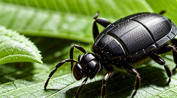
«Recognizing a Tick Bite»
«Visual Signs of a Tick»
«The Tick Itself»
The tick is a small, eight‑legged arachnid whose body consists of a capitulum (mouthparts) and an idiosoma (main body). Adult females enlarge dramatically after a blood meal, while males remain relatively unchanged. The species most commonly associated with human bites can be identified by a reddish‑brown scutum on the dorsal surface and a flattened, oval shape when unfed.
During attachment, the tick inserts its hypostome, a barbed feeding tube, into the skin. Saliva containing anticoagulants and anesthetic compounds is released, reducing immediate pain and preventing clot formation. This physiological mechanism often masks the initial bite, making early detection reliant on visual inspection of the attached organism.
Typical indicators of a recent encounter with «The Tick Itself» include:
- A small, darkened spot on the skin where the mouthparts were inserted.
- Presence of an engorged tick attached for several hours to a day.
- Localized redness or swelling surrounding the attachment site.
- A gradual increase in the size of the attached tick, visible as a bulge.
Prompt removal with fine‑tipped tweezers, grasping the tick close to the skin surface and pulling upward with steady pressure, reduces the risk of pathogen transmission. After extraction, the bite area should be cleaned, and the site monitored for signs of rash, fever, or expanding redness, which may warrant medical evaluation.
«Skin Reactions to a Bite»
Skin reactions provide the most reliable indication that a tick has attached and fed. The bite site often appears as a small, red papule with a central punctum where the mouthparts entered. In many cases the lesion expands over hours to a dome‑shaped erythema, sometimes referred to as a “target” or “bull’s‑eye” pattern. The surrounding skin may become warm, slightly swollen, and tender to touch.
Typical cutaneous responses include:
- Localized redness (erythema) surrounding the attachment point.
- A raised, firm bump (papule) with a visible puncture hole.
- Progressive enlargement of the lesion, forming a ring‑shaped rash.
- Minor swelling (edema) and mild itching or burning sensation.
Additional manifestations suggest an allergic or infectious complication. Rapidly spreading redness, increasing pain, or the appearance of pus indicate secondary bacterial infection. A diffuse rash, fever, joint pain, or a “flushed” appearance can signal a systemic reaction such as Lyme disease or anaphylaxis. In rare cases, a tick may release neurotoxic saliva, leading to progressive weakness and paralysis; skin changes are often accompanied by numbness or tingling in the affected limb.
Immediate medical evaluation is warranted when:
- The rash expands beyond the immediate bite area or develops a central clearing.
- Fever, chills, or severe headache accompany the skin changes.
- Signs of infection (pus, increasing warmth, or foul odor) emerge.
- Neurological symptoms (muscle weakness, difficulty swallowing) appear.
Monitoring the bite site for the described skin changes enables prompt identification of a tick attachment and timely intervention.
«Common Locations for Tick Bites»
«Hidden Areas»
Ticks frequently attach in regions that are difficult to see, allowing the bite to remain unnoticed until symptoms appear. Recognizing these concealed locations improves early detection and reduces the risk of disease transmission.
Typical concealed sites include:
- Scalp and hairline
- Behind the ears
- Under the arms
- In the groin and genital area
- Between the toes and on the feet
- Around the navel and abdominal crease
Signs observable in these zones are:
- Small, raised bump resembling a pinhead
- Red or pink halo surrounding the attachment point
- Slight swelling or tenderness without a clear wound
- Presence of a dark, engorged body partially embedded in the skin
Effective inspection protocol:
- Conduct a full‑body visual survey after outdoor exposure, focusing on the listed concealed sites.
- Use a fine‑toothed comb or magnifying glass to examine hair and skin folds.
- If a tick is identified, grasp it with fine tweezers as close to the skin as possible and pull upward with steady pressure.
- Disinfect the area and monitor for rash or fever over the next several days.
Awareness of «Hidden Areas» and systematic examination are essential for reliable identification of tick attachments.
«Accessible Areas»
Ticks attach in locations that are easy to examine without disassembly of clothing or removal of hair. Regular inspection of these sites after outdoor exposure reduces the risk of unnoticed attachment.
Typical «Accessible Areas» include:
- scalp and hairline, especially behind the ears
- neck and the base of the skull
- underarms and the inner side of the elbows
- groin and the inner thigh region
- waistline and the area around the belt buckle
Inspection should involve parting hair, lifting clothing, and using a magnifying lens if necessary. Presence of a small, engorged, dark spot or a raised, firm nodule indicates a possible tick attachment. Prompt removal of the organism and documentation of the bite site facilitate appropriate medical follow‑up.
«Symptoms of a Tick-Borne Illness»
«Early Symptoms»
«Flu-Like Symptoms»
Flu‑like manifestations often appear after a hidden tick attachment and may serve as an early clue that a bite has occurred. The body’s response typically mimics common viral illness, making recognition essential for timely intervention.
Typical elements of «Flu‑Like Symptoms» include:
- Fever ranging from low‑grade to high
- Chills and sweats
- Headache, often frontal or retro‑orbital
- Muscle aches and joint pain
- Generalized fatigue and malaise
These signs usually emerge within a few days to two weeks following the tick’s attachment, coinciding with the initial phase of pathogen transmission. Absence of a visible bite mark does not exclude exposure; ticks can remain undetected for several hours, especially in concealed body areas.
Immediate medical evaluation is warranted when any of the following occur:
- Persistent fever exceeding 38 °C (100.4 °F) for more than 48 hours
- Severe headache or neck stiffness
- Rapidly spreading rash, particularly an expanding erythema at the bite site
- Neurological symptoms such as numbness, tingling, or facial weakness
- Unexplained joint swelling or severe muscle pain
Prompt diagnosis and treatment reduce the risk of complications associated with tick‑borne infections. Monitoring for «Flu‑Like Symptoms» after potential exposure provides a practical strategy for early detection.
«Rash Characteristics»
Recognizing a tick bite frequently depends on observing the skin reaction at the attachment site.
- Localized redness that appears within hours of exposure.
- Erythema that expands outward, typically reaching a diameter of 5 cm or more.
- Central clearing that creates a concentric “bullseye” pattern.
- Small papule or wheal directly over the tick’s mouthparts.
- Itching, burning, or mild pain accompanying the lesion.
- Onset ranging from a few hours to several days after the bite.
The rash usually develops on exposed areas such as the scalp, neck, arms, or legs, but may appear anywhere the tick attached. Rapid expansion and a target‑shaped appearance differentiate it from simple irritation or allergic reactions, which tend to remain limited in size and lack concentric zones.
If the lesion enlarges beyond 10 cm, persists for more than a week, or is accompanied by fever, headache, fatigue, or joint pain, medical evaluation is advised. Early identification of these characteristics enables prompt treatment and reduces the risk of tick‑borne disease.
«Later, More Severe Symptoms»
«Neurological Symptoms»
Ticks can transmit pathogens that affect the nervous system. Early neurological manifestations may appear within days to weeks after exposure. Recognizing these signs helps differentiate a tick‑borne illness from unrelated conditions.
Typical neurologic indicators include:
- Severe headache unresponsive to usual analgesics
- Neck stiffness or pain suggesting meningitis
- Facial muscle weakness, often unilateral, causing drooping mouth or difficulty closing the eye
- Numbness, tingling, or burning sensations in the limbs
- Balance disturbances, unsteady gait, or coordination loss
- Visual disturbances such as blurred vision or double vision
- Cognitive changes, including confusion, memory lapses, or difficulty concentrating
Accompanying systemic symptoms—fever, rash, or fatigue—strengthen the suspicion of a tick‑borne infection. Prompt medical evaluation is essential when any of these neurologic findings emerge after potential tick exposure. Early treatment reduces the risk of long‑term complications.
«Joint Pain and Swelling»
Joint pain and swelling often appear after a tick attachment, especially when the vector carries Borrelia burgdorferi, the agent of Lyme disease. The inflammation typically manifests in large joints such as the knee, but can involve smaller joints as well. Pain may be sudden or gradually intensify over days, while swelling may be visible as joint distension or limited range of motion.
Key indicators that a tick bite may be responsible for the arthritic symptoms include:
- Erythema migrans rash near the bite site, often expanding over 24–48 hours.
- Flu‑like symptoms (fever, headache, fatigue) preceding joint involvement.
- Onset of joint discomfort within two to four weeks after exposure to tick‑infested areas.
- Persistent or recurrent swelling of one or more joints without an alternative explanation.
When «Joint Pain and Swelling» accompany the above signs, medical evaluation should focus on serologic testing for Lyme disease, imaging to assess joint effusion, and consideration of early antibiotic therapy. Prompt recognition reduces the risk of chronic arthritis and other systemic complications.
«What to Do After a Suspected Bite»
«Tick Removal Techniques»
«Tools for Removal»
When a possible tick attachment is identified, prompt removal reduces the risk of pathogen transmission. Effective extraction depends on appropriate instruments and proper technique.
«Tools for Removal» include:
- Fine‑tipped tweezers with smooth jaws
- Tick‑specific removal device (plastic hook or “tick key”)
- Small, curved forceps designed for arthropods
- Single‑use safety pin with a flat tip
- Disposable gloves to protect hands
The chosen instrument should grasp the tick as close to the skin as possible without compressing the abdomen. Apply steady, upward pressure to detach the mouthparts. Avoid twisting or jerking motions that could leave fragments embedded.
After extraction, cleanse the bite area with antiseptic solution and wash hands thoroughly. Preserve the removed specimen in a sealed container for potential laboratory analysis. Dispose of tools according to local biohazard guidelines.
«Proper Removal Steps»
Detecting a recent tick attachment often leads to the need for immediate removal to reduce disease transmission risk.
The following procedure ensures safe extraction while preserving the tick’s mouthparts:
1. Prepare a pair of fine‑tipped tweezers, a sterile needle or pin, and an antiseptic solution.
2. Grasp the tick as close to the skin’s surface as possible, securing the head and mouthparts without squeezing the abdomen.
3. Apply steady, downward pressure to pull the tick straight out, avoiding twisting or jerking motions.
4. If the mouthparts remain embedded, use the sterile needle to lift them gently; do not dig deeper.
5. Place the detached tick in a sealed container with alcohol or a resealable bag for identification, if required.
6. Disinfect the bite area and the tools with the antiseptic solution.
7. Monitor the site for several weeks; seek medical advice if redness, swelling, or flu‑like symptoms develop.
Proper removal minimizes the chance of pathogen entry and facilitates accurate identification when necessary.
«Post-Removal Care»
«Cleaning the Bite Area»
Proper sanitation of a suspected tick attachment site reduces infection risk and aids visual assessment of the bite. Immediate cleaning removes debris, blood, and potential pathogens, making it easier to identify the tick’s mouthparts and any surrounding erythema.
Steps for effective cleaning:
- Wash hands thoroughly with soap and water before touching the area.
- Rinse the bite site with lukewarm water to loosen surface particles.
- Apply a mild antiseptic solution (e.g., povidone‑iodine or chlorhexidine) using a sterile gauze pad.
- Gently pat the area dry with a clean towel; avoid rubbing, which could irritate the skin.
- Inspect the cleaned site for a small, red puncture mark, a halo of swelling, or a retained tick mouthpart.
If the wound shows signs of infection—persistent redness, swelling, or discharge—seek medical evaluation promptly. Regular monitoring for several days after cleaning is advisable, as delayed reactions can develop. The procedure described under «Cleaning the Bite Area» constitutes a critical component of post‑exposure care.
«Monitoring for Symptoms»
Monitoring for symptoms after a possible tick attachment requires systematic observation of the bite site and overall health. Examine the skin daily for redness, swelling, or a characteristic bullseye pattern. Record any changes in size, color, or texture.
Key indicators to watch for include:
- Localized itching, burning, or pain at the attachment point.
- Development of a rash, especially one resembling a target.
- Fever, chills, or unexplained fatigue.
- Muscle aches, joint pain, or swelling without clear injury.
- Headache, nausea, or dizziness.
If any of these signs appear, seek medical evaluation promptly. Early detection of tick‑borne diseases relies on diligent symptom monitoring and timely professional assessment.
«When to Seek Medical Attention»
«Persistent Symptoms»
Persistent symptoms may appear weeks or months after a tick attachment. The most common manifestations include:
- « fatigue » that is disproportionate to daily activity levels
- « muscle and joint pain », often migratory and without obvious injury
- « headache », sometimes accompanied by neck stiffness
- « cognitive difficulties », such as memory lapses or trouble concentrating
- « sleep disturbances », including insomnia or unrefreshing sleep
These signs can persist even when the initial bite site has healed and no rash was observed. In some cases, neurological involvement emerges, presenting as facial palsy, peripheral neuropathy, or meningitis‑like symptoms. Cardiovascular complaints, notably irregular heart rhythm or palpitations, may also develop.
When any of these symptoms arise after a known or suspected tick exposure, prompt medical evaluation is essential. Laboratory testing for tick‑borne infections, especially Lyme disease, should be considered alongside a thorough clinical history. Early diagnosis and targeted antimicrobial therapy reduce the risk of long‑term complications. Continuous monitoring of symptom progression guides treatment adjustments and informs prognosis.
«Signs of Infection»
Ticks can attach to skin unnoticed; after removal, monitoring for infection is essential. Recognizing «Signs of Infection» enables timely medical intervention.
Typical indicators include:
- Redness spreading beyond the bite site
- Swelling that increases in size
- Warmth or heat around the area
- Persistent or worsening pain
- Pus or fluid discharge
- Fever, chills, or malaise
- Rash resembling a target or bullseye pattern
If any of these symptoms develop, seek professional evaluation promptly. Documentation of the bite date, tick removal method, and observed signs assists healthcare providers in diagnosing potential tick‑borne diseases. Early treatment reduces complications and supports recovery.
«Known Tick-Borne Disease Risk Areas»
Tick-borne illnesses concentrate in specific geographic zones where infected vectors thrive. Awareness of these zones enhances the ability to recognize a recent attachment, because exposure is more likely in high‑risk locations.
In the United States, established risk areas include:
- Northeastern states such as Connecticut, Massachusetts, and New York, where Lyme disease predominates.
- Upper Midwest states including Wisconsin and Minnesota, noted for both Lyme disease and anaplasmosis.
- Pacific Northwest, especially western Washington and Oregon, where spotted‑fever rickettsiosis occurs.
- Central Texas and parts of Oklahoma, associated with Rocky Mountain spotted fever and ehrlichiosis.
In Europe, prominent risk regions cover:
- Central and northern countries like Germany, Sweden, and the Czech Republic, with high incidence of Lyme disease and tick‑borne encephalitis.
- The Baltic states and Poland, where multiple pathogens coexist.
- Southern France and Italy, reporting cases of Mediterranean spotted fever.
In Asia, notable zones comprise:
- Japan’s Hokkaido island, a focus for Japanese spotted‑fever disease.
- The Korean peninsula, where severe fever with thrombocytopenia syndrome appears.
- Parts of China, especially the northeastern provinces, reporting Lyme disease and babesiosis.
When travelling or residing within these areas, regular skin inspection after outdoor activities becomes essential. Prompt removal of attached ticks reduces pathogen transmission, and early skin changes—such as a red expanding rash or a small puncture site—often signal a recent bite. Knowledge of regional disease prevalence guides both preventive measures and clinical assessment.
